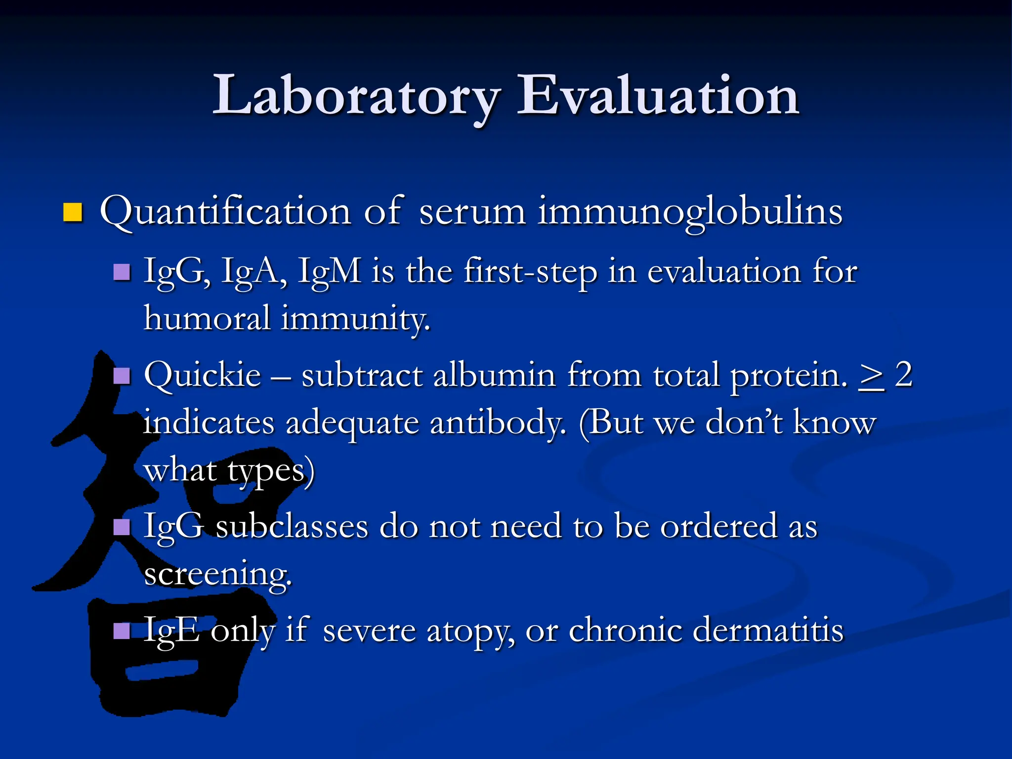
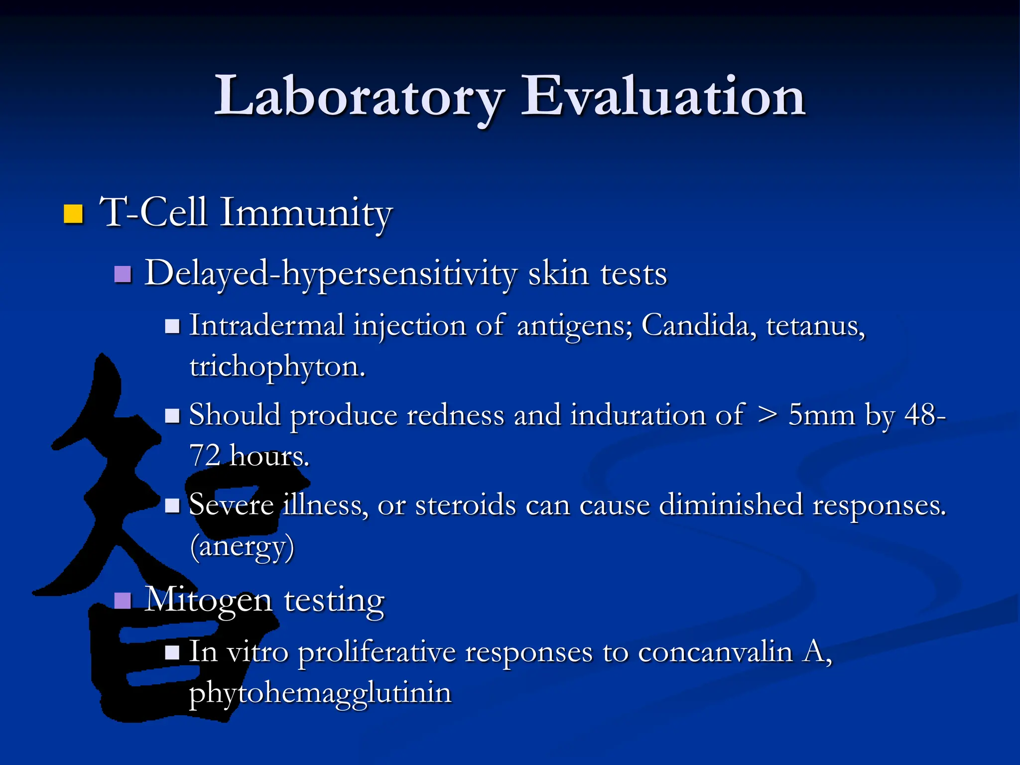
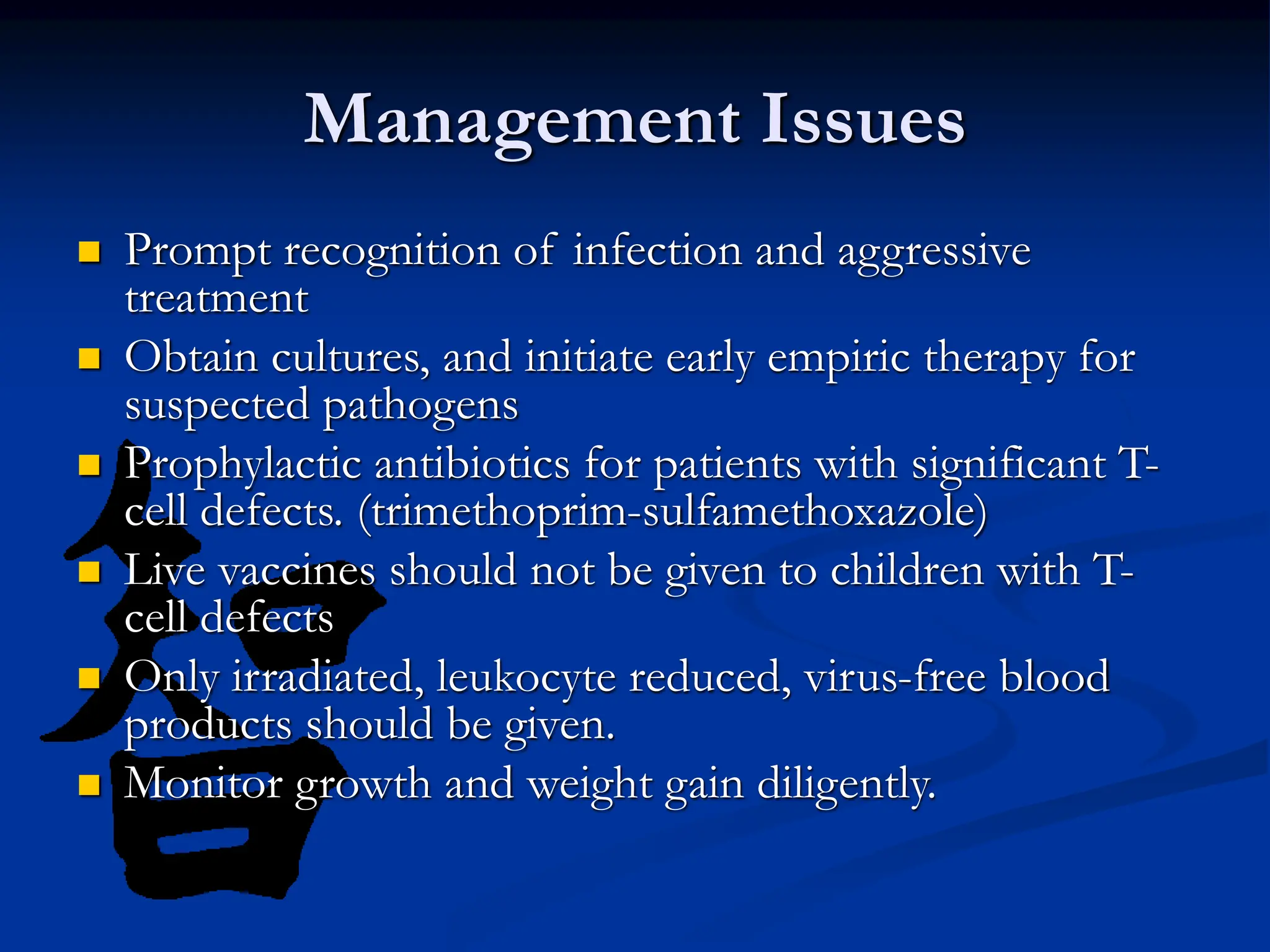
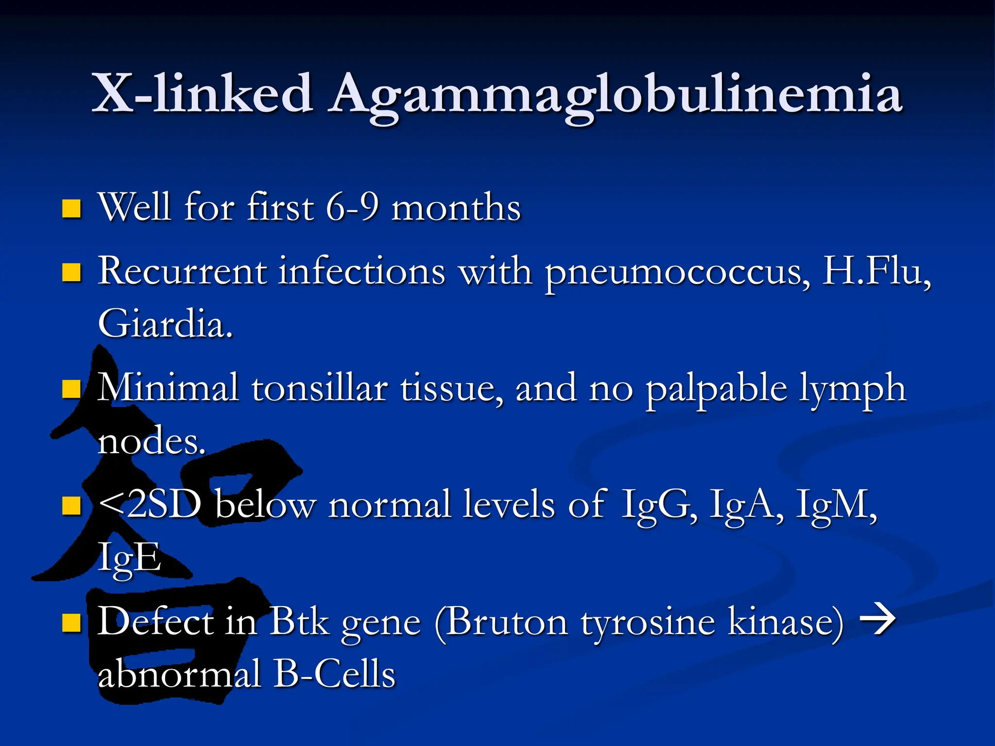
Primary Immune
Deficiencies:
Presentation, Diagnosis,
and Management
Or
Office Evaluation of Children with
Recurrent Infection
J. Aaron Henley, DO
Recurrent Infections Vs. Frequent
Infections
 Immune system not fully developed until at least
two years of age.
 Immuno-competent kids may have up to six
URIs per year. Ten URIs per year if they attend
daycare.
Recurrent Infections Vs. Frequent
Infections
 Common Risk Factors for Frequent Infections
 Day-care, school-aged siblings
 Second-hand smoke
 Atopy
 Anatomic abnormalities including ciliary defects
 Retained foreign body
 Gastroesophageal reflux
Recurrent Infections Vs. Frequent
Infections
 Frequent or recurrent sinopulmonary infections,
malabsorption, nasal polyps…
•Cystic Fibrosis
–1:2500
–More common than most of the
immunodeficiencies
Secondary Immunodeficiencies
 Two most common
 Malnutrition
 Vit. A deficiency - Infections of GI and resp. tract
 Zinc deficiency – Acrodermatitis enteropathica, SCID-like
syndrome
 B12 deficiency – impaired immunoglobulin production
 Protein and caloric deficiency – impaired immune
response
Secondary Immunodeficiency
 Two most common
 HIV infection – seventh leading cause of death in
children 1-4 years in the US.
 Third leading cause in black children 1-4 in the urban
northeastern US.
 Always think about it…
Primary Immunodeficiency
 A disorder in one or more components of the
immune system.
Components of Immunity
 Skin and mucosal barriers
 Innate immune system (nonspecific)
 Phagocytic cells, NK cells, complement
 Adaptive immune system (specific)
 T and B lymphocytes, antibodies
Classification of Immunodeficiency
1. Humoral (B-cell) – quantitative or qualitative
defects in antibody production account for
more than 50% of defects.
2. Cellular (T-cell) – usually combined with
humoral; account for 20-30%.
3. Phagocytic – defects in migration, or killing;
account for ~18%.
4. Complement – account for ~2%
Index of Suspicion
 >10 episodes acute otitis media per year (infants
and children).
 >2 episodes consolidated pneumonia per year.
 >2 life-threatening infections per lifetime.
 Two or more serious sinus infections within 1
year.
 Unusual organisms.
 Unusual response to organism.
Index of Suspicion
 Recurrent deep skin or organ abscesses.
 Two or more deep-seated infections such as meningitis,
osteomyelitis, cellulites or sepsis.
 Persistent oral thrush or candida infection elsewhere on the skin,
after age 1 year.
 Recurrent autoimmune phenomena.
 Dysmorphic features associated with recurrent infection.
 Infections worsening chronic disorders (asthma or seizure).
 Development of vaccine pathogen after vaccination (e.g., HiB
infection despite previous HiB vaccine).
 Family history of immunodeficiency or recurrent infection.
History Our Guide
 Important historical points
 Frequency, duration, severity, complications,
response to treatment
 Risk factors
 Family history
 Infection with low-virulence or unusual organisms
 Age of onset
History Our Guide
 Predominant B-Cell defects
 Onset after maternal antibodies diminish, usually
after 5-7 mos, later childhood to adulthood.
 Bacteria: strep, staph, H.flu; Campylobacter,
enteroviruses, giardia, cryptosporidia
 Recurrent sinopulmonary infections, chronic GI
symptoms, malabsorption, arthritis, viral
meningoencephalitis
 Autoimmunity, lymphoreticular malignancy;
thymoma, lymphoma
History Our Guide
 Predominant T-Cell Defects
 Early onset, usually 2-6 mos
 Bacteria, mycobacteria, viruses: CMV, EBV, varicella;
fungi, parasites, PCP, mycobacterium avium-
intracellulare
 FTT, protracted diarrhea, extensive mucocutaneous
candidiasis
 GVHD caused by maternal engrafment,
nonirradiated blood
 Hypocalcemic tetany in infancy
History Our Guide
 Granulocyte Defects
 Early onset, delayed separation of cord (>8 weeks),
poor wound healing
 Bacteria: staph, Pseudomonas, Serratia, Klebsiella;
Fungi: Candida, Nocardia, Aspergillus
 Dermatitis, impetigo, cellulitis, abscesses, supparative
lymphadenitis, periodontitis, osteomyelits
History Our Guide
 Complement Defects
 Late (C5-C9) – Neisserial infections: meningitidis,
septic arthritis from gonorrhoeae.
 Early (C1, C4, and C2) – autoimmune disease
 C3 deficiency – overwhelming sepsis, especially with
gram negative organisms
Physical Exam
 A benign physical exam does not rule out
immunodeficiency.
 Look for:
 General appearance, weight, overall health
 Hair, connective tissue
 Dysmorphic features
 Gingivitis, dental erosions, signs of sinusitis
 Tonsillar tissue, adenopathy, splenomegaly
 Arthritis, ataxia, neuro deficits
Disease Specific Skin Findings
 Eczema and petechiae – Wiskott-Aldrich
Syndrome
 Telangiectasia – Ataxia-Telangiectasia
 Oculocutaneous albinism – Chediak-Higashi
 Dermatomyositis-like rash – XLA
 Chronic dermatitis – Hyper-IgE
 Generalized molluscum, extensive warts,
candidiasis – T-Cell defects
Laboratory Evaluation
 CBC with differential
 Total WBC, ANC, ALC, AEC (age-appropriate
values)
 Lymphopenia = < 3,000 in infants, < 1500 in
children and adults
 Persistently high ANC occurs in LAD
 Hemolytic anemia, thrombocytopenia, leukopenia
occurs in some B-Cell deficiencies.
Laboratory Evaluation
 Quantification of serum immunoglobulins
 IgG, IgA, IgM is the first-step in evaluation for
humoral immunity.
 Quickie – subtract albumin from total protein. > 2
indicates adequate antibody. (But we don’t know
what types)
 IgG subclasses do not need to be ordered as
screening.
 IgE only if severe atopy, or chronic dermatitis
Laboratory Evaluation
 Qualitative Evaluation of Antibodies
 Isohemagglutins – Antibodies to ABO blood-group
determinants
 Antibodies to tetanus and diptheria glycoproteins
and pneumococcal polysaccharides.
 If low titers, give booster, then repeat titers 4 weeks later.
 Children younger than 2 can not be tested for
polysaccharide antigen antibody.
Laboratory Evaluation
 T-Cell Immunity
 Delayed-hypersensitivity skin tests
 Intradermal injection of antigens; Candida, tetanus,
trichophyton.
 Should produce redness and induration of > 5mm by 48-
72 hours.
 Severe illness, or steroids can cause diminished responses.
(anergy)
 Mitogen testing
 In vitro proliferative responses to concanvalin A,
phytohemagglutinin
Laboratory Evaluation
 Phagocytic Cell Function
 Adhesion antigens by flow cytometry (CD11/CD18)
– checks for adhesion defects
 Chemiluminescence – phagocytic killing power
Laboratory Evaluation
 Complement function
 Total hemolytic complement (CH50) – tests
functional integrity of classic complement pathway.
 AH50 – tests the functional integrity of alternate
pathway.
 The most common reason for an abnormal CH50 is
improper handling of specimen.
Treatment for Primary Immune
Deficiencies
 Bone marrow transplantation
 Immunoglobulin replacement
 Enzyme replacement
 Gene therapy
Management Issues
 Prompt recognition of infection and aggressive
treatment
 Obtain cultures, and initiate early empiric therapy for
suspected pathogens
 Prophylactic antibiotics for patients with significant T-
cell defects. (trimethoprim-sulfamethoxazole)
 Live vaccines should not be given to children with T-
cell defects
 Only irradiated, leukocyte reduced, virus-free blood
products should be given.
 Monitor growth and weight gain diligently.
Specific Diseases
 Well for first 6-9 months
 Recurrent infections with pneumococcus, H.Flu,
Giardia.
 Minimal tonsillar tissue, and no palpable lymph
nodes.
 <2SD below normal levels of IgG, IgA, IgM,
IgE
 Defect in Btk gene (Bruton tyrosine kinase) 
abnormal B-Cells
X-linked Agammaglobulinemia
 Well for first 6-9 months
 Recurrent infections with pneumococcus, H.Flu,
Giardia.
 Minimal tonsillar tissue, and no palpable lymph
nodes.
 <2SD below normal levels of IgG, IgA, IgM,
IgE
 Defect in Btk gene (Bruton tyrosine kinase) 
abnormal B-Cells
 Recurrent sinopulmonary infections with usual
pathogens.
 Age of onset 15-35 years. Equal male:female.
 Low IgG and poor antibody responses to
immunizations.
 Variable levels of IgM and IgA.
 Increased risk for autoimmune diseases and
malignancy.
 B-Cells phenotypically normal.
Common Variable
Immunodeficiency
 Recurrent sinopulmonary infections with usual
pathogens.
 Age of onset 15-35 years. Equal male:female.
 Low IgG and poor antibody responses to
immunizations.
 Variable levels of IgM and IgA.
 Increased risk for autoimmune diseases and
malignancy.
 B-Cells phenotypically normal.
 Recurrent sinopulmonary disease.
 Telangiectasias between 3-6 years.
 Ataxia soon after learning to walk, in wheelchair
by 10-12 years.
 Often low or absent IgA. Varaible depressions
of other immunoglobulins.
 Anergy and depressed mitogen studies.
 Risk for lymphoreticular malignancy.
Ataxia-Telangiectasia
 Recurrent sinopulmonary disease.
 Telangiectasias between 3-6 years.
 Ataxia soon after learning to walk, in wheelchair
by 10-12 years.
 Often low or absent IgA. Varaible depressions
of other immunoglobulins.
 Anergy and depressed mitogen studies.
 Risk for lymphoreticular malignancy.
 1:400 to 1:800
 Symptoms: GI, GU, RTI infections. Many
asymptomatic patients.
 Normal IgG and IgM response to pathogens
and vaccines.
 Role in diagnosis of Celiac disease.
 May evolve into CVID.
 Be very careful if pt. develops Kawasaki…
IgA Deficiency
 1:400 to 1:800
 Symptoms: GI, GU, RTI infections. Many
asymptomatic patients.
 Normal IgG and IgM response to pathogens
and vaccines.
 Role in diagnosis of Celiac disease.
 May evolve into CVID.
 Be very careful if pt. develops Kawasaki…
 Decreased IgG beyond 6 months.
 Normal IgA, and variable IgM.
 Able to synthesize antibodies to blood type,
diptheria, and tetanus antigens normally.
 May have increase otitis and sinusitis.
 Usually resolves by 4 years of age.
Transient Hypogammaglobulinemia
of Infancy
 Decreased IgG beyond 6 months.
 Normal IgA, and variable IgM.
 Able to synthesize antibodies to blood type,
diptheria, and tetanus antigens normally.
 May have increase otitis and sinusitis.
 Usually resolves by 4 years of age.
 Recurrent infections by three months. Can be
life-threatening.
 Candida, PCP, cryptosporidiosis, HSV, RSV,
rotavirus, adeno, entero, EBV, CMV.
 Absence of lyphoid tissue, lymphopenia, no
thymic shadow.
 Anergy, abnormal T-Cell proliferation, +/- B-
Cell dysfunction.
 Absence of adaptive immunity.
Severe Combined Immunodeficiency
 Recurrent infections by three months. Can be
life-threatening.
 Candida, PCP, cryptosporidiosis, HSV, RSV,
rotavirus, adeno, entero, EBV, CMV.
 Absence of lyphoid tissue, lymphopenia, no
thymic shadow.
 Anergy, abnormal T-Cell proliferation, +/- B-
Cell dysfunction.
 Absence of adaptive immunity.
 1:10 million
 Striking neutrophilia.
 Recurrent bacterial and fungal infections
without pus.
 Severe gingivitis, periodontitis, alveolar bone
loss.
 Decreased or absent CD18/CD11 by flow.
 Delayed separation of umbilical cord.
Leukocyte Adhesion Defect
 1:10 million
 Striking neutrophilia.
 Recurrent bacterial and fungal infections
without pus.
 Severe gingivitis, periodontitis, alveolar bone
loss.
 Decreased or absent CD18/CD11 by flow.
 Delayed separation of umbilical cord.
 Unsure of clinical relevance.
 May be evolving form of CVID.
 May be normal variation.
 Does not need treatment or evaluation unless
there is an impaired ability to form antibodies to
protein and polysaccharide antigens.
Selective IgG Subclass Deficiencies
 Unsure of clinical relevance.
 May be evolving form of CVID.
 May be normal variation.
 Does not need treatment or evaluation unless
there is an impaired ability to form antibodies to
protein and polysaccharide antigens.
 Variable hypoplasia of thymus and parathyroid.
 Hypocalcemia  seizures
 Susceptability to fungi, viruses, PCP.
 T-Cells variable in number, abnormal mitogen studies
 Normal to increased B-Cells, normal antibody levels.
 Microdeletion of 22q11.2
 Associated heart defects, facial anomalies, esophageal
atresia.
DiGeorge Anomaly
 Variable hypoplasia of thymus and parathyroid.
 Hypocalcemia  seizures
 Susceptability to fungi, viruses, PCP.
 T-Cells variable in number, abnormal mitogen studies
 Normal to increased B-Cells, normal antibody levels.
 Microdeletion of 22q11.2
 Associated heart defects, facial anomalies, esophageal
atresia.
 Initially asymptomatic.
 Defective response to EBV.
 Fulminant often fatal mono, lymphomas,
acquired hypogammalobulinemia.
 70% of boys die by 10.
 Impairment of antibody to EBNA
 Viral capsid antigen antibody titers may be
absent to exaggerated.
X-linked Lymphoproliferative
Syndrome
 Initially asymptomatic.
 Defective response to EBV.
 Fulminant often fatal mono, lymphomas,
acquired hypogammalobulinemia.
 70% of boys die by 10.
 Impairment of antibody to EBNA
 Viral capsid antigen antibody titers may be
absent to exaggerated.
 Recurrent abscesses, lymphadenitis, or
osteomyelitis at multiple sites.
 Unusual infections with catalase positive
organisms: Staph, Serratia, Aspergillus, Candida,
Salmonella, gram - enterics.
 Defect in NADPH oxidase enzyme  leading
to the inability to produce H2O2.
 No problem with streprococci.
 Chemiluminescence (DHR test)
Chronic Granulomatous Disease
 Recurrent abscesses, lymphadenitis, or
osteomyelitis at multiple sites.
 Unusual infections with catalase positive
organisms: Staph, Serratia, Aspergillus, Candida,
Salmonella, gram - enterics.
 Defect in NADPH oxidase enzyme  leading
to the inability to produce H2O2.
 No problem with streprococci.
 Chemiluminescence (DHR test)
 Regular, periodic oscillation in the number of
peripheral neutrophils.
 Neutropenia every 21+/- 3 days.
 May develop fever, stomatitis, pharyngitis,
pneumonia, occasionally sepsis and death.
 May spontaneously abate.
 Cycles become less noticeable with age.
Cyclic Neutropenia
 Regular, periodic oscillation in the number of
peripheral neutrophils.
 Neutropenia every 21+/- 3 days.
 May develop fever, stomatitis, pharyngitis,
pneumonia, occasionally sepsis and death.
 May spontaneously abate.
 Cycles become less noticeable with age.
 Chronic pruritic dermatitis.
 Recurrent staph infections of skin, lungs, joints,
and dental infections.
 Course facial features.
 Markedly elevated IgE and eosinophilia.
Hyper-IgE Syndrome
 Chronic pruritic dermatitis.
 Recurrent staph infections of skin, lungs, joints,
and dental infections.
 Course facial features.
 Markedly elevated IgE and eosinophilia.
 Partial oculocutaneous albinism.
 Frequent infections of skin, mucous
membranes, respiratory tract. Gram -, Gram +,
and fungi.
 Large inclusions in all nucleated blood cells.
 Accelerated lymphoma-like syndrome; non-
neoplastic infiltration of liver, spleen, and lymph
nodes associated with recurrent infections and
death.
Chediak-Higashi Syndrome
 Partial oculocutaneous albinism.
 Frequent infections of skin, mucous
membranes, respiratory tract. Gram -, Gram +,
and fungi.
 Large inclusions in all nucleated blood cells.
 Accelerated lymphoma-like syndrome; non-
neoplastic infiltration of liver, spleen, and lymph
nodes associated with recurrent infections and
death.
 1:4,000
 Most do not have increased infection rate.
 MPO important in producing hypochlorous
acid.
 Affected cells use an MPO-independent
pathway for killing pathogens.
 Occasionally, disseminated candidiasis.
 No specific treatment needed.
Myeloperoxidase Deficiency
 1:4,000
 Most do not have increased infection rate.
 MPO important in producing hypochlorous
acid.
 Affected cells use an MPO-independent
pathway for killing pathogens.
 Occasionally, disseminated candidiasis.
 No specific treatment needed.
 Atopic dermatitis
 Microcytic thrombocytopenia  bleeding
 Recurrent infections with encapsulated bacteria:
pneumococcus esp.
 Variable antibody levels. Often low IgM, high
IgA and IgE. Poor antibody finction.
 Low to low-normal T-Cells.
 WASPy boys.
Wiskott-Aldrich Syndrome
 Atopic dermatitis
 Microcytic thrombocytopenia  bleeding
 Recurrent infections with encapsulated bacteria:
pneumococcus esp.
 Variable antibody levels. Often low IgM, high
IgA and IgE. Poor antibody finction.
 Low to low-normal T-Cells.
 WASPy boys.
References
 Nelson Textbook of Pediatrics; 17th Edition. Primary Immune
Deficiency.
 Woroneika M. Primary Immune Deficiencies:Presentation,
Diagnosis, and Management. Ped Clinic North Am 47:6 2000
 Thatayatikom A. Chapter V.5. Immune Deficiency, Case Based
Pediatrics For Medical Students and Residents
Department of Pediatrics, University of Hawaii John A. Burns
School of Medicine 2003
 Bonilla F. Practice parameters for the diagnosis and management
of primary immunodeficiency. Ann All, Asth, Immun. 94:S1-
S63, 2005

Primary_Immune_Deficiencies_Presentations_Diagnosis_and Management.ppt

  • 1.
    Primary Immune Deficiencies: Presentation, Diagnosis, andManagement Or Office Evaluation of Children with Recurrent Infection J. Aaron Henley, DO
  • 2.
    Recurrent Infections Vs.Frequent Infections  Immune system not fully developed until at least two years of age.  Immuno-competent kids may have up to six URIs per year. Ten URIs per year if they attend daycare.
  • 3.
    Recurrent Infections Vs.Frequent Infections  Common Risk Factors for Frequent Infections  Day-care, school-aged siblings  Second-hand smoke  Atopy  Anatomic abnormalities including ciliary defects  Retained foreign body  Gastroesophageal reflux
  • 4.
    Recurrent Infections Vs.Frequent Infections  Frequent or recurrent sinopulmonary infections, malabsorption, nasal polyps… •Cystic Fibrosis –1:2500 –More common than most of the immunodeficiencies
  • 5.
    Secondary Immunodeficiencies  Twomost common  Malnutrition  Vit. A deficiency - Infections of GI and resp. tract  Zinc deficiency – Acrodermatitis enteropathica, SCID-like syndrome  B12 deficiency – impaired immunoglobulin production  Protein and caloric deficiency – impaired immune response
  • 6.
    Secondary Immunodeficiency  Twomost common  HIV infection – seventh leading cause of death in children 1-4 years in the US.  Third leading cause in black children 1-4 in the urban northeastern US.  Always think about it…
  • 7.
    Primary Immunodeficiency  Adisorder in one or more components of the immune system.
  • 8.
    Components of Immunity Skin and mucosal barriers  Innate immune system (nonspecific)  Phagocytic cells, NK cells, complement  Adaptive immune system (specific)  T and B lymphocytes, antibodies
  • 9.
    Classification of Immunodeficiency 1.Humoral (B-cell) – quantitative or qualitative defects in antibody production account for more than 50% of defects. 2. Cellular (T-cell) – usually combined with humoral; account for 20-30%. 3. Phagocytic – defects in migration, or killing; account for ~18%. 4. Complement – account for ~2%
  • 10.
    Index of Suspicion >10 episodes acute otitis media per year (infants and children).  >2 episodes consolidated pneumonia per year.  >2 life-threatening infections per lifetime.  Two or more serious sinus infections within 1 year.  Unusual organisms.  Unusual response to organism.
  • 11.
    Index of Suspicion Recurrent deep skin or organ abscesses.  Two or more deep-seated infections such as meningitis, osteomyelitis, cellulites or sepsis.  Persistent oral thrush or candida infection elsewhere on the skin, after age 1 year.  Recurrent autoimmune phenomena.  Dysmorphic features associated with recurrent infection.  Infections worsening chronic disorders (asthma or seizure).  Development of vaccine pathogen after vaccination (e.g., HiB infection despite previous HiB vaccine).  Family history of immunodeficiency or recurrent infection.
  • 12.
    History Our Guide Important historical points  Frequency, duration, severity, complications, response to treatment  Risk factors  Family history  Infection with low-virulence or unusual organisms  Age of onset
  • 13.
    History Our Guide Predominant B-Cell defects  Onset after maternal antibodies diminish, usually after 5-7 mos, later childhood to adulthood.  Bacteria: strep, staph, H.flu; Campylobacter, enteroviruses, giardia, cryptosporidia  Recurrent sinopulmonary infections, chronic GI symptoms, malabsorption, arthritis, viral meningoencephalitis  Autoimmunity, lymphoreticular malignancy; thymoma, lymphoma
  • 14.
    History Our Guide Predominant T-Cell Defects  Early onset, usually 2-6 mos  Bacteria, mycobacteria, viruses: CMV, EBV, varicella; fungi, parasites, PCP, mycobacterium avium- intracellulare  FTT, protracted diarrhea, extensive mucocutaneous candidiasis  GVHD caused by maternal engrafment, nonirradiated blood  Hypocalcemic tetany in infancy
  • 15.
    History Our Guide Granulocyte Defects  Early onset, delayed separation of cord (>8 weeks), poor wound healing  Bacteria: staph, Pseudomonas, Serratia, Klebsiella; Fungi: Candida, Nocardia, Aspergillus  Dermatitis, impetigo, cellulitis, abscesses, supparative lymphadenitis, periodontitis, osteomyelits
  • 16.
    History Our Guide Complement Defects  Late (C5-C9) – Neisserial infections: meningitidis, septic arthritis from gonorrhoeae.  Early (C1, C4, and C2) – autoimmune disease  C3 deficiency – overwhelming sepsis, especially with gram negative organisms
  • 17.
    Physical Exam  Abenign physical exam does not rule out immunodeficiency.  Look for:  General appearance, weight, overall health  Hair, connective tissue  Dysmorphic features  Gingivitis, dental erosions, signs of sinusitis  Tonsillar tissue, adenopathy, splenomegaly  Arthritis, ataxia, neuro deficits
  • 18.
    Disease Specific SkinFindings  Eczema and petechiae – Wiskott-Aldrich Syndrome  Telangiectasia – Ataxia-Telangiectasia  Oculocutaneous albinism – Chediak-Higashi  Dermatomyositis-like rash – XLA  Chronic dermatitis – Hyper-IgE  Generalized molluscum, extensive warts, candidiasis – T-Cell defects
  • 19.
    Laboratory Evaluation  CBCwith differential  Total WBC, ANC, ALC, AEC (age-appropriate values)  Lymphopenia = < 3,000 in infants, < 1500 in children and adults  Persistently high ANC occurs in LAD  Hemolytic anemia, thrombocytopenia, leukopenia occurs in some B-Cell deficiencies.
  • 20.
    Laboratory Evaluation  Quantificationof serum immunoglobulins  IgG, IgA, IgM is the first-step in evaluation for humoral immunity.  Quickie – subtract albumin from total protein. > 2 indicates adequate antibody. (But we don’t know what types)  IgG subclasses do not need to be ordered as screening.  IgE only if severe atopy, or chronic dermatitis
  • 21.
    Laboratory Evaluation  QualitativeEvaluation of Antibodies  Isohemagglutins – Antibodies to ABO blood-group determinants  Antibodies to tetanus and diptheria glycoproteins and pneumococcal polysaccharides.  If low titers, give booster, then repeat titers 4 weeks later.  Children younger than 2 can not be tested for polysaccharide antigen antibody.
  • 22.
    Laboratory Evaluation  T-CellImmunity  Delayed-hypersensitivity skin tests  Intradermal injection of antigens; Candida, tetanus, trichophyton.  Should produce redness and induration of > 5mm by 48- 72 hours.  Severe illness, or steroids can cause diminished responses. (anergy)  Mitogen testing  In vitro proliferative responses to concanvalin A, phytohemagglutinin
  • 23.
    Laboratory Evaluation  PhagocyticCell Function  Adhesion antigens by flow cytometry (CD11/CD18) – checks for adhesion defects  Chemiluminescence – phagocytic killing power
  • 24.
    Laboratory Evaluation  Complementfunction  Total hemolytic complement (CH50) – tests functional integrity of classic complement pathway.  AH50 – tests the functional integrity of alternate pathway.  The most common reason for an abnormal CH50 is improper handling of specimen.
  • 25.
    Treatment for PrimaryImmune Deficiencies  Bone marrow transplantation  Immunoglobulin replacement  Enzyme replacement  Gene therapy
  • 26.
    Management Issues  Promptrecognition of infection and aggressive treatment  Obtain cultures, and initiate early empiric therapy for suspected pathogens  Prophylactic antibiotics for patients with significant T- cell defects. (trimethoprim-sulfamethoxazole)  Live vaccines should not be given to children with T- cell defects  Only irradiated, leukocyte reduced, virus-free blood products should be given.  Monitor growth and weight gain diligently.
  • 27.
  • 28.
     Well forfirst 6-9 months  Recurrent infections with pneumococcus, H.Flu, Giardia.  Minimal tonsillar tissue, and no palpable lymph nodes.  <2SD below normal levels of IgG, IgA, IgM, IgE  Defect in Btk gene (Bruton tyrosine kinase)  abnormal B-Cells
  • 29.
    X-linked Agammaglobulinemia  Wellfor first 6-9 months  Recurrent infections with pneumococcus, H.Flu, Giardia.  Minimal tonsillar tissue, and no palpable lymph nodes.  <2SD below normal levels of IgG, IgA, IgM, IgE  Defect in Btk gene (Bruton tyrosine kinase)  abnormal B-Cells
  • 30.
     Recurrent sinopulmonaryinfections with usual pathogens.  Age of onset 15-35 years. Equal male:female.  Low IgG and poor antibody responses to immunizations.  Variable levels of IgM and IgA.  Increased risk for autoimmune diseases and malignancy.  B-Cells phenotypically normal.
  • 31.
    Common Variable Immunodeficiency  Recurrentsinopulmonary infections with usual pathogens.  Age of onset 15-35 years. Equal male:female.  Low IgG and poor antibody responses to immunizations.  Variable levels of IgM and IgA.  Increased risk for autoimmune diseases and malignancy.  B-Cells phenotypically normal.
  • 32.
     Recurrent sinopulmonarydisease.  Telangiectasias between 3-6 years.  Ataxia soon after learning to walk, in wheelchair by 10-12 years.  Often low or absent IgA. Varaible depressions of other immunoglobulins.  Anergy and depressed mitogen studies.  Risk for lymphoreticular malignancy.
  • 33.
    Ataxia-Telangiectasia  Recurrent sinopulmonarydisease.  Telangiectasias between 3-6 years.  Ataxia soon after learning to walk, in wheelchair by 10-12 years.  Often low or absent IgA. Varaible depressions of other immunoglobulins.  Anergy and depressed mitogen studies.  Risk for lymphoreticular malignancy.
  • 34.
     1:400 to1:800  Symptoms: GI, GU, RTI infections. Many asymptomatic patients.  Normal IgG and IgM response to pathogens and vaccines.  Role in diagnosis of Celiac disease.  May evolve into CVID.  Be very careful if pt. develops Kawasaki…
  • 35.
    IgA Deficiency  1:400to 1:800  Symptoms: GI, GU, RTI infections. Many asymptomatic patients.  Normal IgG and IgM response to pathogens and vaccines.  Role in diagnosis of Celiac disease.  May evolve into CVID.  Be very careful if pt. develops Kawasaki…
  • 36.
     Decreased IgGbeyond 6 months.  Normal IgA, and variable IgM.  Able to synthesize antibodies to blood type, diptheria, and tetanus antigens normally.  May have increase otitis and sinusitis.  Usually resolves by 4 years of age.
  • 37.
    Transient Hypogammaglobulinemia of Infancy Decreased IgG beyond 6 months.  Normal IgA, and variable IgM.  Able to synthesize antibodies to blood type, diptheria, and tetanus antigens normally.  May have increase otitis and sinusitis.  Usually resolves by 4 years of age.
  • 38.
     Recurrent infectionsby three months. Can be life-threatening.  Candida, PCP, cryptosporidiosis, HSV, RSV, rotavirus, adeno, entero, EBV, CMV.  Absence of lyphoid tissue, lymphopenia, no thymic shadow.  Anergy, abnormal T-Cell proliferation, +/- B- Cell dysfunction.  Absence of adaptive immunity.
  • 39.
    Severe Combined Immunodeficiency Recurrent infections by three months. Can be life-threatening.  Candida, PCP, cryptosporidiosis, HSV, RSV, rotavirus, adeno, entero, EBV, CMV.  Absence of lyphoid tissue, lymphopenia, no thymic shadow.  Anergy, abnormal T-Cell proliferation, +/- B- Cell dysfunction.  Absence of adaptive immunity.
  • 40.
     1:10 million Striking neutrophilia.  Recurrent bacterial and fungal infections without pus.  Severe gingivitis, periodontitis, alveolar bone loss.  Decreased or absent CD18/CD11 by flow.  Delayed separation of umbilical cord.
  • 41.
    Leukocyte Adhesion Defect 1:10 million  Striking neutrophilia.  Recurrent bacterial and fungal infections without pus.  Severe gingivitis, periodontitis, alveolar bone loss.  Decreased or absent CD18/CD11 by flow.  Delayed separation of umbilical cord.
  • 42.
     Unsure ofclinical relevance.  May be evolving form of CVID.  May be normal variation.  Does not need treatment or evaluation unless there is an impaired ability to form antibodies to protein and polysaccharide antigens.
  • 43.
    Selective IgG SubclassDeficiencies  Unsure of clinical relevance.  May be evolving form of CVID.  May be normal variation.  Does not need treatment or evaluation unless there is an impaired ability to form antibodies to protein and polysaccharide antigens.
  • 44.
     Variable hypoplasiaof thymus and parathyroid.  Hypocalcemia  seizures  Susceptability to fungi, viruses, PCP.  T-Cells variable in number, abnormal mitogen studies  Normal to increased B-Cells, normal antibody levels.  Microdeletion of 22q11.2  Associated heart defects, facial anomalies, esophageal atresia.
  • 45.
    DiGeorge Anomaly  Variablehypoplasia of thymus and parathyroid.  Hypocalcemia  seizures  Susceptability to fungi, viruses, PCP.  T-Cells variable in number, abnormal mitogen studies  Normal to increased B-Cells, normal antibody levels.  Microdeletion of 22q11.2  Associated heart defects, facial anomalies, esophageal atresia.
  • 46.
     Initially asymptomatic. Defective response to EBV.  Fulminant often fatal mono, lymphomas, acquired hypogammalobulinemia.  70% of boys die by 10.  Impairment of antibody to EBNA  Viral capsid antigen antibody titers may be absent to exaggerated.
  • 47.
    X-linked Lymphoproliferative Syndrome  Initiallyasymptomatic.  Defective response to EBV.  Fulminant often fatal mono, lymphomas, acquired hypogammalobulinemia.  70% of boys die by 10.  Impairment of antibody to EBNA  Viral capsid antigen antibody titers may be absent to exaggerated.
  • 48.
     Recurrent abscesses,lymphadenitis, or osteomyelitis at multiple sites.  Unusual infections with catalase positive organisms: Staph, Serratia, Aspergillus, Candida, Salmonella, gram - enterics.  Defect in NADPH oxidase enzyme  leading to the inability to produce H2O2.  No problem with streprococci.  Chemiluminescence (DHR test)
  • 49.
    Chronic Granulomatous Disease Recurrent abscesses, lymphadenitis, or osteomyelitis at multiple sites.  Unusual infections with catalase positive organisms: Staph, Serratia, Aspergillus, Candida, Salmonella, gram - enterics.  Defect in NADPH oxidase enzyme  leading to the inability to produce H2O2.  No problem with streprococci.  Chemiluminescence (DHR test)
  • 50.
     Regular, periodicoscillation in the number of peripheral neutrophils.  Neutropenia every 21+/- 3 days.  May develop fever, stomatitis, pharyngitis, pneumonia, occasionally sepsis and death.  May spontaneously abate.  Cycles become less noticeable with age.
  • 51.
    Cyclic Neutropenia  Regular,periodic oscillation in the number of peripheral neutrophils.  Neutropenia every 21+/- 3 days.  May develop fever, stomatitis, pharyngitis, pneumonia, occasionally sepsis and death.  May spontaneously abate.  Cycles become less noticeable with age.
  • 52.
     Chronic pruriticdermatitis.  Recurrent staph infections of skin, lungs, joints, and dental infections.  Course facial features.  Markedly elevated IgE and eosinophilia.
  • 53.
    Hyper-IgE Syndrome  Chronicpruritic dermatitis.  Recurrent staph infections of skin, lungs, joints, and dental infections.  Course facial features.  Markedly elevated IgE and eosinophilia.
  • 54.
     Partial oculocutaneousalbinism.  Frequent infections of skin, mucous membranes, respiratory tract. Gram -, Gram +, and fungi.  Large inclusions in all nucleated blood cells.  Accelerated lymphoma-like syndrome; non- neoplastic infiltration of liver, spleen, and lymph nodes associated with recurrent infections and death.
  • 55.
    Chediak-Higashi Syndrome  Partialoculocutaneous albinism.  Frequent infections of skin, mucous membranes, respiratory tract. Gram -, Gram +, and fungi.  Large inclusions in all nucleated blood cells.  Accelerated lymphoma-like syndrome; non- neoplastic infiltration of liver, spleen, and lymph nodes associated with recurrent infections and death.
  • 56.
     1:4,000  Mostdo not have increased infection rate.  MPO important in producing hypochlorous acid.  Affected cells use an MPO-independent pathway for killing pathogens.  Occasionally, disseminated candidiasis.  No specific treatment needed.
  • 57.
    Myeloperoxidase Deficiency  1:4,000 Most do not have increased infection rate.  MPO important in producing hypochlorous acid.  Affected cells use an MPO-independent pathway for killing pathogens.  Occasionally, disseminated candidiasis.  No specific treatment needed.
  • 58.
     Atopic dermatitis Microcytic thrombocytopenia  bleeding  Recurrent infections with encapsulated bacteria: pneumococcus esp.  Variable antibody levels. Often low IgM, high IgA and IgE. Poor antibody finction.  Low to low-normal T-Cells.  WASPy boys.
  • 59.
    Wiskott-Aldrich Syndrome  Atopicdermatitis  Microcytic thrombocytopenia  bleeding  Recurrent infections with encapsulated bacteria: pneumococcus esp.  Variable antibody levels. Often low IgM, high IgA and IgE. Poor antibody finction.  Low to low-normal T-Cells.  WASPy boys.
  • 60.
    References  Nelson Textbookof Pediatrics; 17th Edition. Primary Immune Deficiency.  Woroneika M. Primary Immune Deficiencies:Presentation, Diagnosis, and Management. Ped Clinic North Am 47:6 2000  Thatayatikom A. Chapter V.5. Immune Deficiency, Case Based Pediatrics For Medical Students and Residents Department of Pediatrics, University of Hawaii John A. Burns School of Medicine 2003  Bonilla F. Practice parameters for the diagnosis and management of primary immunodeficiency. Ann All, Asth, Immun. 94:S1- S63, 2005