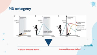
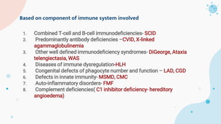
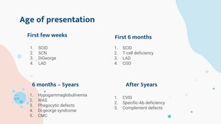
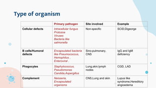
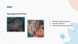
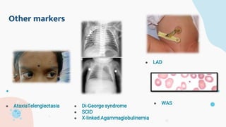
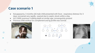
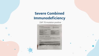
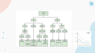
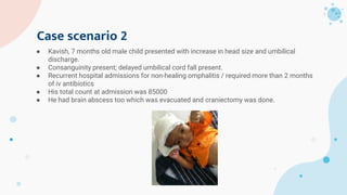
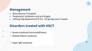
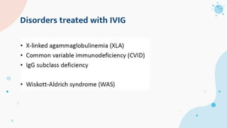
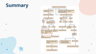
Approach
to PID
-P.T.Nanmaaran
Junior Resident
Pediatrics
Scope of this session
Background
01
Classification
02
Clinical markers
03
Case scenarios
04
Background
How common?
Introduction to Immunity
01
History
● The first PID was discovered 70 years ago —> CGD (around 1950)
● Then SCID—> WAS and Bruton’s agammaglobulinemia
● The introduction of immunoglobulin replacement (1952) and bone marrow
transplantation (1957
● Around 2010, PID discovered was around 150
● Now its more than 350 diseases described—> expansion in understanding immune basis
and genetic etiology.
Background- “Inborn Errors of Immunity”
 Primary immunodeficiency diseases (PIDs) are inherited disorders that
impair the immune response, leading to increased risk of infections,
immune dysregulation, autoimmune phenomena, inflammation, and
malignancy.
 >1 million cases in India; 6 million people —> PIDs worldwide, of which only
27,000–60,000 have been diagnosed
 Approximate incidence : 1 in 2000 to 1: 10,000
 For now more than 344 gene defects and 354 disease identified and it is
increasing….
Primary Immunodeficiency Disorders in India—A Situational ReviewAnkur Kumar Jindal, Rakesh Kumar Pilania, Amit Rawat and Surjit Singh*Allergy
Immunology Unit, Advanced Pediatrics Centre, Postgraduate Institute of Medical Education and Research, Chandigarh, India
Primary Immunodeficiency Disorders in India—A Situational ReviewAnkur Kumar Jindal, Rakesh Kumar Pilania, Amit Rawat and Surjit Singh*Allergy
Immunology Unit, Advanced Pediatrics Centre, Postgraduate Institute of Medical Education and Research, Chandigarh, India
Basics –Immunity
Innate Immunity Adaptive Immunity
Lag time Short Long
Active Always active Activated after antigen exposure
Specificity Non-specific Specific
Eg., Skin epithelium,GI and
Respiratory mucosa
Cells Neutrophils,Eosinophils,Basoph
ils,Monocytes and NK cells
Pattern recognition receptors
B and T lymphocytes,
Plasma cells—>antibody
Cells of adaptive immunity
B cells T cells
Produced in Bone Marrow Bone Marrow
Mature in Bone Marrow Thymus
Lymph node Follicles Para-follicular
Spleen Follicles Per—arteriolar region
Immune system Humoral —> antibody
mediated
Cell mediated—>
CD4/CD8 T cells
Cell mediated immunity
Humoral immunity
Jeffrey Model
foundation-10
warning signs
2000’s
 Unusual site
 Unusual organism
 Unusual severity
 Unusual response after
live vaccine
 Recurrence
 Failure to thrive
When to suspect PID?
10 warning signs…
● The overall significance of having any of the 10 warning signs to suspect
PID showed that
● When at least one warning sign was exhibited, the sensitivity was 100,
specificity was 26%.
● Meeting at least two criteria of the 10 warning signs led to a sensitivity
of 94% specificity of 64%.
● When at least three criteria of the 10 warning signs were fulfilled, the
sensitivity was 77% ,the specificity was 86%
The most common asymptomatic PID –
Selective IgA deficiency (1:223 to 1:1000)
The most common symptomatic PID –
Antibody deficiency
PID ontogeny
Cellular immune defect Humoral immune defect
12 months
General understanding
02
Classification
Based on component of immune system involved
1. Combined T-cell and B-cell immunodeficiencies- SCID
2. Predominantly antibody deficiencies –CVID, X-linked
agammaglobulinemia
3. Other well defined immunodeficiency syndromes- DiGeorge, Ataxia
telengiectasia, WAS
4. Diseases of immune dysregulation-HLH
5. Congenital defects of phagocyte number and function – LAD, CGD
6. Defects in innate immunity- MSMD, CMC
7. Auto-inflammatory disorders- FMF
8. Complement deficiencies( C1 inhibitor deficiency- hereditory
angioedema)
Approach to PID
Age of presentation
1. SCID
2. SCN
3. DiGeorge
4. LAD
First few weeks First 6 months
1. SCID
2. T-cell deficiency
3. LAD
4. CGD
1. Hypogammaglobulinemia
2. WAS
3. Phagocytic defects
4. Di-george syndrome
5. CMC
6 months – 5years After 5years
1. CVID
2. Specific-Ab deficiency
3. Complement defects
Type of organism
Primary pathogen Site involved Example
Cellular defects Intracellular fungus
Protozoa
Viruses
Bacteria like
salmonella
Non-specific SCID,Digeorge
B cells/Humoral
defects
Encapsulated bacteria
like Pneumococcus,
Hemophilus ,
Enteroviral .
Sino-pulmonary,
CNS
IgG and IgM
deficiency
Phagocytes Staphylococcus,
Pseudomonas
Candida,Aspergillus
Lung,skin,lymph
nodes
CGD, LAD
Complement Neisseria,
Encapsulated
organisms
CNS,Lung and skin Lupus like
syndrome,Hereditory
angioedema
Specific diseases
03
Clinical markers
Skin
● Wiskott Aldrich syndrome
● IPEX
● Hyper IgE syndrome
● Omenn syndrome
● Ig A deficiency
● CVID
1.Eczema
Skin
● CGD
● LAD
● Hyper Ig E syndrome
Recurrent skin infection, Periodontitis and
Gingivitis
Skin
● SCID
● CMC
● Hyper IgE syndrome
Recurrent mucosal candidiasis
Hair
● Chediak Higashi syndrome
● Griscelli syndrome
● Hermansky pudlack syndrome
Hypopigmented hair
Other markers
● AtaxiaTelengiectasia ● Di-George syndrome
● SCID
● X-linked Agammaglobulinemia
● WAS
● LAD
Specific diseases
04
Case Scenarios
Case scenario 1
● Karuppasamy, 3 months old male child presented with fever , respiratory distress for 3
days; worsened very rapidly ; expired due to septic shock within a day.
● ALC-3000; previous 3 sibling death at similar age; consanguinity present
● Previous child workup for complement and Ig profile was normal.
Severe Combined
Immunodeficiency
ZAP 70 mutation positive
SCID
● Usually present within first six months of life with failure to thrive, chronic
diarrhea, persistent oral thrush, skin rash, pneumonia, and sepsis
● Disseminated BCG infection is commonly seen in patients with SCID
● Lymphopenia is commonly seen with patients with SCID
● >22 groups of SCID is present. ADA deficiency, X-Linked SCID , Artemis
deficiency , PNP deficiency etc.,
● Less than 2 years + Less than 20% CD3 T cells/ALC<3000/mm3/one
defined mutation Diagnose SCID
SCID
Case scenario 2
● Kavish, 7 months old male child presented with increase in head size and umbilical
discharge.
● Consanguinity present; delayed umbilical cord fall present.
● Recurrent hospital admissions for non-healing omphalitis / required more than 2 months
of iv antibiotics
● His total count at admission was 85000
● He had brain abscess too which was evacuated and craniectomy was done.
LAD-1
Mutation proven positive
LAD
Leukocyte adhesion deficiency
● Recurrent infection + neutrophilia
● Delayed seperation of umbilical cord
● No pus formation
● No signs of inflammation
● LAD-1 : CD11/CD18 low
● LAD-2 : CD 15 /Normal CD11 and CD18
● LAD-2  associated with Bombay blood group
● LAD-3  Associated with Glanzman thrombasthenia like bleeding disorder
● HSCT can be tried in LAD 1 and LAD 3
Chronic granulomatous disease
Boy
Persistent
pneumonia
Persistent
lymphnode
swellling
Lung/Liver
abscess
Osteomyelitis
CGD
● Inflammatory granuloma present
● NBT/DHR
Case 3
● 18 months old boy; Recurrent otitis media and pneumonia since 8 months of
age
● Absent tonsils and Lymph node
● Consanguinity present
● IgA-16 mg/dL (40-200)
● IgG-184 mg/dL (490-1610)
● IgM- 9 mg/dL (50-299)
Agammaglobulinemia
● Absent B cells and Reduced Ig levels are
suggestive of Agammaglobulinemia
● X-Linked or AR inheritance
● 1952  Colonel Ogden Bruton found reduced
immunoglobulin levels in a boy with recurrent
infections .He was the first to give Ig (im)
● Basic defect: Pro B cell to mature B cell –
Btk gene expression defective
● More than 50% will have serious infection by 2
years of age
● Marked reduction in all types of
Immunoglobulin
X-LA vs CVID
B cell defects
● Reduced Ig with normal to low B cells with abnormal specific antibody responses 
common variable immunodeficiency (CVID)
● Markedly low IgG and IgA with normal to elevated IgM levels s/o Hyper IgM syndrome
● Antibody deficiency- best approach to diagnosis Serum specific antibody titers
(usually IgG) in response to vaccine antigens (tetanus toxoid and pneumococcus)
● Pre and post immunization levels will be diminished or absent
● In many PIDs, antibody responses to these antigens are absent.
Case 4
● 1 1/2 year old boy was admitted from Feb,12 to March
8,2021–> Presented with fever for 20 days; Multiple
swellings for 20 days;Not responding to oral antibiotics for 2
months—> worked up for Primary Immunodeficiency Ig
profile-Normal;Bone marrow-Paucity of myeloid series of
cells/No blasts;TBNK profile was normal.
● The child had issues 1.Left cervical abscess 2.Right
Otomycosis 3.Oral Candidiasis
● His ANC counts never raised >1000
Severe Congenital
Neutropenia
Severe congenital neutropenia
● Mutation in ELANE, HAX 1 and G6PC3
● More prone for Staphylococcus, E.coli and
Pseudomonal infections
● Cyclical neutropenia – once in 21 days
● Monocytosis is common during Neutropenic phase
● Treatment –G-CSF
● Defintite management HSCT
Some well known immunodeficiency syndromes
1. Wiscott Aldrich Syndrome
2. Ataxia Talengectasia
3. Di-george anomaly
4. Hyper IgE Syndromes(HIES)/AD (Jobs Syndrome)
● 4 year old boy Recurrent pneumonia and Otitis media ; eczema over
trunk and Thrombocytopenia
● PS was s/o Microplatelets
● Triad of Atopic dermatitis; Infections and Thrombocytopenia
● X-linked
● No autoimmunity
● WAS gene
Case 5
DiGeorge syndrome
● Hypocalcemic seizures
● Dysmorphic facies
● Decreased CD3 +T cells
● Diagnose by FISH
CBC in PID:
Neutrophil Normal Rules out LAD/
Neutropenia
Lymphocyte Normal Rules out T-cell defect
Platelet Normal Rules out WAS
Howel Jolly bodies Present Asplenia
CBC in PID:
Hypereosinophilia Present Omenn syndrome, Hyper
IgE syndrome,WAS
Rule of 2/3 rd’s
● For a child less than 3 years
o 2/3 of WBC should be lymphocytes
o 2/3 of lymphocytes should be T cells
o 2/3 of T cells should be CD4 cells
• For a child more than 3 years
o 2/3 of WBC should be neutrophils
o 2/3 of lymphocytes should be T cells
Screening tests for PID
B Cell defect Antibody levels(Ig)
Antibody titres to vaccination
(Protein and Polysaccharide)
T cell defect ALC
Flow cytometry (for naïve T cells)
Phagocytic defect ANC
Respiratory burst assay
Complement deficiency CH50
AH50
Approach
Disorders treated with HSCT
● Bone Marrow Transplant
● Prophylactic Antibiotics and anti-fungals
● Lifelong IVIg Replacement 0.4 - 0.6 gm/kg every 3 weeks
Management
Disorders treated with IVIG
Summary
Thank you…!

PID presentation.pptx

  • 1.
  • 2.
    Scope of thissession Background 01 Classification 02 Clinical markers 03 Case scenarios 04
  • 3.
  • 4.
    History ● The firstPID was discovered 70 years ago —> CGD (around 1950) ● Then SCID—> WAS and Bruton’s agammaglobulinemia ● The introduction of immunoglobulin replacement (1952) and bone marrow transplantation (1957 ● Around 2010, PID discovered was around 150 ● Now its more than 350 diseases described—> expansion in understanding immune basis and genetic etiology.
  • 5.
    Background- “Inborn Errorsof Immunity”  Primary immunodeficiency diseases (PIDs) are inherited disorders that impair the immune response, leading to increased risk of infections, immune dysregulation, autoimmune phenomena, inflammation, and malignancy.  >1 million cases in India; 6 million people —> PIDs worldwide, of which only 27,000–60,000 have been diagnosed  Approximate incidence : 1 in 2000 to 1: 10,000  For now more than 344 gene defects and 354 disease identified and it is increasing…. Primary Immunodeficiency Disorders in India—A Situational ReviewAnkur Kumar Jindal, Rakesh Kumar Pilania, Amit Rawat and Surjit Singh*Allergy Immunology Unit, Advanced Pediatrics Centre, Postgraduate Institute of Medical Education and Research, Chandigarh, India
  • 6.
    Primary Immunodeficiency Disordersin India—A Situational ReviewAnkur Kumar Jindal, Rakesh Kumar Pilania, Amit Rawat and Surjit Singh*Allergy Immunology Unit, Advanced Pediatrics Centre, Postgraduate Institute of Medical Education and Research, Chandigarh, India
  • 7.
    Basics –Immunity Innate ImmunityAdaptive Immunity Lag time Short Long Active Always active Activated after antigen exposure Specificity Non-specific Specific Eg., Skin epithelium,GI and Respiratory mucosa Cells Neutrophils,Eosinophils,Basoph ils,Monocytes and NK cells Pattern recognition receptors B and T lymphocytes, Plasma cells—>antibody
  • 8.
    Cells of adaptiveimmunity B cells T cells Produced in Bone Marrow Bone Marrow Mature in Bone Marrow Thymus Lymph node Follicles Para-follicular Spleen Follicles Per—arteriolar region Immune system Humoral —> antibody mediated Cell mediated—> CD4/CD8 T cells
  • 9.
  • 10.
  • 12.
    Jeffrey Model foundation-10 warning signs 2000’s Unusual site  Unusual organism  Unusual severity  Unusual response after live vaccine  Recurrence  Failure to thrive When to suspect PID?
  • 13.
    10 warning signs… ●The overall significance of having any of the 10 warning signs to suspect PID showed that ● When at least one warning sign was exhibited, the sensitivity was 100, specificity was 26%. ● Meeting at least two criteria of the 10 warning signs led to a sensitivity of 94% specificity of 64%. ● When at least three criteria of the 10 warning signs were fulfilled, the sensitivity was 77% ,the specificity was 86% The most common asymptomatic PID – Selective IgA deficiency (1:223 to 1:1000) The most common symptomatic PID – Antibody deficiency
  • 14.
    PID ontogeny Cellular immunedefect Humoral immune defect 12 months
  • 15.
  • 16.
    Based on componentof immune system involved 1. Combined T-cell and B-cell immunodeficiencies- SCID 2. Predominantly antibody deficiencies –CVID, X-linked agammaglobulinemia 3. Other well defined immunodeficiency syndromes- DiGeorge, Ataxia telengiectasia, WAS 4. Diseases of immune dysregulation-HLH 5. Congenital defects of phagocyte number and function – LAD, CGD 6. Defects in innate immunity- MSMD, CMC 7. Auto-inflammatory disorders- FMF 8. Complement deficiencies( C1 inhibitor deficiency- hereditory angioedema)
  • 17.
  • 18.
    Age of presentation 1.SCID 2. SCN 3. DiGeorge 4. LAD First few weeks First 6 months 1. SCID 2. T-cell deficiency 3. LAD 4. CGD 1. Hypogammaglobulinemia 2. WAS 3. Phagocytic defects 4. Di-george syndrome 5. CMC 6 months – 5years After 5years 1. CVID 2. Specific-Ab deficiency 3. Complement defects
  • 19.
    Type of organism Primarypathogen Site involved Example Cellular defects Intracellular fungus Protozoa Viruses Bacteria like salmonella Non-specific SCID,Digeorge B cells/Humoral defects Encapsulated bacteria like Pneumococcus, Hemophilus , Enteroviral . Sino-pulmonary, CNS IgG and IgM deficiency Phagocytes Staphylococcus, Pseudomonas Candida,Aspergillus Lung,skin,lymph nodes CGD, LAD Complement Neisseria, Encapsulated organisms CNS,Lung and skin Lupus like syndrome,Hereditory angioedema
  • 20.
  • 21.
    Skin ● Wiskott Aldrichsyndrome ● IPEX ● Hyper IgE syndrome ● Omenn syndrome ● Ig A deficiency ● CVID 1.Eczema
  • 22.
    Skin ● CGD ● LAD ●Hyper Ig E syndrome Recurrent skin infection, Periodontitis and Gingivitis
  • 23.
    Skin ● SCID ● CMC ●Hyper IgE syndrome Recurrent mucosal candidiasis
  • 24.
    Hair ● Chediak Higashisyndrome ● Griscelli syndrome ● Hermansky pudlack syndrome Hypopigmented hair
  • 25.
    Other markers ● AtaxiaTelengiectasia● Di-George syndrome ● SCID ● X-linked Agammaglobulinemia ● WAS ● LAD
  • 26.
  • 27.
    Case scenario 1 ●Karuppasamy, 3 months old male child presented with fever , respiratory distress for 3 days; worsened very rapidly ; expired due to septic shock within a day. ● ALC-3000; previous 3 sibling death at similar age; consanguinity present ● Previous child workup for complement and Ig profile was normal.
  • 28.
  • 29.
    SCID ● Usually presentwithin first six months of life with failure to thrive, chronic diarrhea, persistent oral thrush, skin rash, pneumonia, and sepsis ● Disseminated BCG infection is commonly seen in patients with SCID ● Lymphopenia is commonly seen with patients with SCID ● >22 groups of SCID is present. ADA deficiency, X-Linked SCID , Artemis deficiency , PNP deficiency etc., ● Less than 2 years + Less than 20% CD3 T cells/ALC<3000/mm3/one defined mutation Diagnose SCID
  • 30.
  • 32.
    Case scenario 2 ●Kavish, 7 months old male child presented with increase in head size and umbilical discharge. ● Consanguinity present; delayed umbilical cord fall present. ● Recurrent hospital admissions for non-healing omphalitis / required more than 2 months of iv antibiotics ● His total count at admission was 85000 ● He had brain abscess too which was evacuated and craniectomy was done.
  • 33.
  • 34.
  • 35.
    Leukocyte adhesion deficiency ●Recurrent infection + neutrophilia ● Delayed seperation of umbilical cord ● No pus formation ● No signs of inflammation ● LAD-1 : CD11/CD18 low ● LAD-2 : CD 15 /Normal CD11 and CD18 ● LAD-2  associated with Bombay blood group ● LAD-3  Associated with Glanzman thrombasthenia like bleeding disorder ● HSCT can be tried in LAD 1 and LAD 3
  • 36.
  • 37.
    CGD ● Inflammatory granulomapresent ● NBT/DHR
  • 38.
    Case 3 ● 18months old boy; Recurrent otitis media and pneumonia since 8 months of age ● Absent tonsils and Lymph node ● Consanguinity present ● IgA-16 mg/dL (40-200) ● IgG-184 mg/dL (490-1610) ● IgM- 9 mg/dL (50-299)
  • 39.
    Agammaglobulinemia ● Absent Bcells and Reduced Ig levels are suggestive of Agammaglobulinemia ● X-Linked or AR inheritance ● 1952  Colonel Ogden Bruton found reduced immunoglobulin levels in a boy with recurrent infections .He was the first to give Ig (im) ● Basic defect: Pro B cell to mature B cell – Btk gene expression defective ● More than 50% will have serious infection by 2 years of age ● Marked reduction in all types of Immunoglobulin
  • 40.
  • 41.
    B cell defects ●Reduced Ig with normal to low B cells with abnormal specific antibody responses  common variable immunodeficiency (CVID) ● Markedly low IgG and IgA with normal to elevated IgM levels s/o Hyper IgM syndrome ● Antibody deficiency- best approach to diagnosis Serum specific antibody titers (usually IgG) in response to vaccine antigens (tetanus toxoid and pneumococcus) ● Pre and post immunization levels will be diminished or absent ● In many PIDs, antibody responses to these antigens are absent.
  • 42.
    Case 4 ● 11/2 year old boy was admitted from Feb,12 to March 8,2021–> Presented with fever for 20 days; Multiple swellings for 20 days;Not responding to oral antibiotics for 2 months—> worked up for Primary Immunodeficiency Ig profile-Normal;Bone marrow-Paucity of myeloid series of cells/No blasts;TBNK profile was normal. ● The child had issues 1.Left cervical abscess 2.Right Otomycosis 3.Oral Candidiasis ● His ANC counts never raised >1000
  • 43.
  • 44.
    Severe congenital neutropenia ●Mutation in ELANE, HAX 1 and G6PC3 ● More prone for Staphylococcus, E.coli and Pseudomonal infections ● Cyclical neutropenia – once in 21 days ● Monocytosis is common during Neutropenic phase ● Treatment –G-CSF ● Defintite management HSCT
  • 45.
    Some well knownimmunodeficiency syndromes 1. Wiscott Aldrich Syndrome 2. Ataxia Talengectasia 3. Di-george anomaly 4. Hyper IgE Syndromes(HIES)/AD (Jobs Syndrome)
  • 46.
    ● 4 yearold boy Recurrent pneumonia and Otitis media ; eczema over trunk and Thrombocytopenia ● PS was s/o Microplatelets ● Triad of Atopic dermatitis; Infections and Thrombocytopenia ● X-linked ● No autoimmunity ● WAS gene Case 5
  • 47.
    DiGeorge syndrome ● Hypocalcemicseizures ● Dysmorphic facies ● Decreased CD3 +T cells ● Diagnose by FISH
  • 48.
    CBC in PID: NeutrophilNormal Rules out LAD/ Neutropenia Lymphocyte Normal Rules out T-cell defect Platelet Normal Rules out WAS Howel Jolly bodies Present Asplenia
  • 49.
    CBC in PID: HypereosinophiliaPresent Omenn syndrome, Hyper IgE syndrome,WAS
  • 50.
    Rule of 2/3rd’s ● For a child less than 3 years o 2/3 of WBC should be lymphocytes o 2/3 of lymphocytes should be T cells o 2/3 of T cells should be CD4 cells • For a child more than 3 years o 2/3 of WBC should be neutrophils o 2/3 of lymphocytes should be T cells
  • 51.
    Screening tests forPID B Cell defect Antibody levels(Ig) Antibody titres to vaccination (Protein and Polysaccharide) T cell defect ALC Flow cytometry (for naïve T cells) Phagocytic defect ANC Respiratory burst assay Complement deficiency CH50 AH50
  • 52.
  • 53.
    Disorders treated withHSCT ● Bone Marrow Transplant ● Prophylactic Antibiotics and anti-fungals ● Lifelong IVIg Replacement 0.4 - 0.6 gm/kg every 3 weeks Management
  • 54.
  • 55.
  • 56.