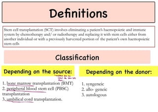
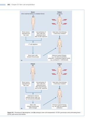
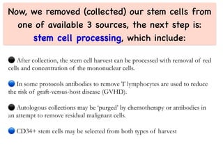
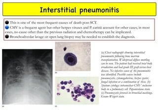
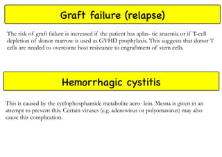
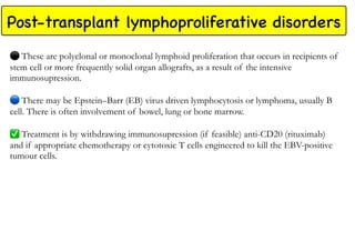
Bone marrow and
stem cell transplantation
Definitions
Stem cell transplantation (SCT) involves eliminating a patient’s haemopoietic and immune
system by chemotherapy and/ or radiotherapy and replacing it with stem cells either from
another individual or with a previously harvested portion of the patient’s own haemopoietic
stem cells
Depending on the source: Depending on the donor:
Classification
1. bone marrow transplantation (BMT)
2. peripheral blood stem cell (PBSC)
transplantation
3. umbilical cord transplantation.
1. syngeneic
2. allo- geneic
3. autologous
↳ Where You Can
Collect the stem cells
from. __ .
-
=
252 / Chapter 23: Stem cell transplantation
Figure 23.1 Procedures for (a) allogeneic, and (b) autologous stem cell transplantation. G-CSF, granulocyte colony-stimulating factor;
GVHD, graft-versus-host disease.
Donor
(a)
(HLA-matched sibling or unrelated donor)
Patient
(recipient)
High-dose chemotherapy
± total body irradiation
± T-cell depletion
Bone marrow
aspirated from
iliac crest
OR Leucopheresis of
stem cells from
peripheral blood
after G-CSF injections
Donor stem cells
infused intravenously
Intensive support therapy,
e.g. red cells and platelets,
antibiotics + prophylaxis against GVHD,
e.g. cyclosporin ± methotrexate
t
n
e
i
t
a
P
t
n
e
i
t
a
P
High-dose chemotherapy
± total body irradiation
± attempts to remove
residual tumour cells, e.g.
by monoclonal antibodies
Bone marrow
aspirated from
iliac crest
OR Leucopheresis of
stem cells from
peripheral blood
after chemotherapy
and G-CSF injections
Intensive support therapy,
e.g. red cells and platelets,
antibiotics
Stem cells
infused intravenously
(after storage)
(b)
o@g@qogtMK
the exact role of SCT in
the management of each
disease is complex and
depends on factors such
as disease severity and
subtype, remission status,
age and, for allogeneic
transplantation,
availability of a donor.
PBSC transplantation
This is now the preferred source of stem cells for both autolo- gous and allogeneic transplantation.
⚫
Peripheral blood stem cells (PBSCs) are taken using a cell-separator machine connected to the patient or donor via
peripheral cannulae.
Blood is taken through one cannula and pumped around the machine where mononuclear cells are collected by
centrifugation before the red cells are returned to the patient. This continuous process may take a few hours before
enough mononuclear cells are collected.
Peripheral blood normally contains too few
haemopoi- etic stem cells to allow collection of
sufficient numbers for transplantation.
Growth factors can increase the number by around
10–100 times. Granulocyte colony-stimulating factor
(G-CSF) is given to patients or donors as a course of
injections (typically 10 µg/kg/day for 4–6 days) until the
white cell count starts to rise. Plerixafor, an inhibitor of
stem cell adhesion in the bone marrow, is also given if
mobilisation is likely to be inadequate.
PBSC collections are then taken and, depending on
the efficiency of stem cell mobilization, repeated
collections may be needed for up to 3 days.
A typical out-patient protocol is G-CSF on days
1–4 and harvests on days 5 and 6.
The adequacy of the collection is assessed by CD34+
cell count. Generally greater than 2.0 × 10⁶/kg CD+
cells are needed for transplantation.
⚫
The donor is given a general
anaesthetic and 500–1200 mL of
marrow is harvested from the pelvis.
The marrow is anticoagulated and
a mononuclear cell count is taken to
assess the yield, which should be
approximately 2–4 × 10⁸ nucleated
cells/kg body weight of the recipient.
BM transplantation UC transplantation
Fetal blood is a rich source of
haemopoietic stem cells which may be
collected from cord blood.
Because of the relatively small numbers
of stem cells collected from a single cord,
they are most useful for children who do
not have a fully matching sibling or
unrelated donor.
Less stringent HLA-matching is needed.
Double cord donations may be needed
to obtain suf- ficient stem cells for adult
recipients.
Immune reconstitution is slower after
cord blood transplantation.
Now, we removed (collected) our stem cells from
one of available 3 sources, the next step is:
stem cell processing, which include:
After collection, the stem cell harvest can be processed with removal of red
cells and concentration of the mononuclear cells.
In some protocols antibodies to remove T lymphocytes are used to reduce
the risk of graft-versus-host disease (GVHD).
Autologous collections may be ‘purged’ by chemotherapy or antibodies in
an attempt to remove residual malignant cells.
CD34+ stem cells may be selected from both types of harvest
Then we have the process of conditioning:
⚫
Prior to infusion of haemopoietic stem cells, patients receive chemotherapy, sometimes in combination
with total body irradiation in a procedure called conditioning.
This is designed to eradicate the patient’s haemopoietic and immune system and, if present, malignancy.
In addition, in the setting of allogeneic SCT, by suppressing the host immune system, it helps to prevent
rejection of the ‘foreign’ stem cells.
Myeloablative
conditioning regimens
Non- Myeloablative
conditioning regimens
Myeloablative conditioning regimens
These irreversibly destroy the haemopoietic function of the bone marrow with high doses of
chemotherapy or radiotherapy.
TBI is usually used in patients with malignant disease and is usually administered over several days
(fractionated).
The most commonly used chemotherapy drug is cyclophosphamide but busulfan (preferably
intravenously), melphalan, or other drugs are given in some protocols. Treosulfan is a newer alkylating agent
in trials.
🔵
Before autologous transplantation, high-dose melphalan is used for myeloma and a combination regimen,
e.g. BEAM (carmustine, etoposide, cytarabine and melphalan) for lymphoma.
For benign disorders TBI is avoided and cyclophosphamide, busulfan and fludarabine with alemtuzumab
or ATG in vivo are employed in different protocols.
At least 36 hours are allowed for the elimination of the drugs from the circulation following the last
dose of chemotherapy before donor stem cells are infused.
Conditioning therapy is often complicated by mucositis and patients sometimes need parenteral
nutrition. T cell depletion of the donor stem cells in vitro reduces the risk of GVHD but increases
the risk of non-engraftment, infections and relapse.
Alternatively, T cell depletion of the host in vivo by antithymocyte globulin (ATG) or alemtuzumab
(anti-CD52) may be employed to reduce GVHD risk, but may increase risk of infection and relapse.
Non- Myeloablative conditioning regimens
These have been developed to reduce the morbidity and mortality of allogeneic
transplantation.
These do not completely destroy the host bone marrow. (Such regimes extend the
age range and increase the treatment indications for allogeneic transplantation).
These include agents such as fludarabine, low doses of busulfan or
cyclophosphamide, low-dose irradiation, ATG or other antibodies that delete T
cells.
The aim in these mini or low-intensity transplants is to use enough
immunosuppression to allow donor stem cells to engraft without completely
eradicating host marrow stem cells.
Donor leucocyte infusions (DLI) are commonly used at a later stage in order to
encourage complete donor engraftment and to enhance the graft-versus- leukaemia
or lymphoma effect.
Post-transplant engraftment and immunity
After a period of typically 1–3 weeks of severe pancytopenia, the first signs of successful
engraftment are monocytes and neutrophils in the blood with a subsequent increase in platelet count.
A reticulocytosis also begins.
G-CSF may be used to reduce the period of neutropenia.
Engraftment is usually quicker following PBSC transplantation compared with BMT.
➖
The marrow cellularity gradually returns to normal but the marrow reserve remains impaired for
1–2 years. There is profound immunodeficiency for 3–12 months with a low level of CD4 helper
cells.
Immune recovery is quicker after autologous and syngeneic SCT than following
allogeneic SCT.
The patient’s blood group changes to that of the donor and antigen-specific immunity
becomes that of the donor after approximately 60 days.
Autologous stem cell transplantation
This allows the delivery of a high dose of
chemotherapy, with or without radiotherapy, which
otherwise would result in prolonged bone marrow
aplasia.
⚫
Stem cells are harvested and stored before the
treatment is given and are then reinfused to rescue
the patient from the myeloablative effects of the
treatment.
A limitation of the procedure is that tumour cells
contaminating the stem cell harvest may be
reintroduced into the patient. The major problem
associated with autograft- ing is recurrence of the
original disease. GVHD is not an issue.
Nevertheless, autografting has a major role in the
treatment of haematological diseases such as
lymphoma and myeloma. Procedure-related
mortality is generally well below 5%.
The causes of death following autologous transplantation.
IPn, interstitial pneumonitis.
Allogeneic stem cell transplantation
⚫
In this procedure, stem cells
harvested from another person are
infused into the patient.
The procedure has a significant
morbid- ity and mortality and one of
the major reasons is the immunologi-
cal incompatibility between donor and
patient despite matching of the human
leucocyte antigens (HLA). This may
manifest as immunodeficiency, GVHD
or graft failure.
Paradoxically, there is also a graft-
versus-leukaemia (GVL) effect which
probably underlies much of the success
of the procedure.
Complications
(Relapse)
The overall rate from the procedure itself is lowest (less than 5%) for
autologous SCT and highest in unrelated and haploidentical SCT.
Let’s talk about some complications in more details:
(manifest as jaundice,
hepatomegaly and
ascites or weight gain)
Due to conditioning regimen,
especially high doses of
cyclophosphamide and
previous chemotherapy to
the heart
The use of prophylactic co-trimoxazole and oral aciclovir for 3–6 months
reduces the risk of Pneumocystis and herpes infections, respectively.
restrictive pneumonitis and bronchiolitis obliterans
myasthenia, rheumatoid arthritis, anaemia,
thrombocytopenia or neutro- penia.
especially non-Hodgkin lymphoma
↳
Affecting Respiratory system
↳ Prophylaxis: Peneatin
especial
[ s
other endocrinePatties, growthfailure, hypothyroidism,
Impaired sexual
ofthi Development
• Hemolysis due to ABO Inampabilit , MAHA
Graft-versus-host disease (GVHD)
This is caused by donor-derived immune cells, particularly T lymphocytes,
reacting against recipient tissues.
Its incidence is increased with:
1. increasing age of donor and recipient
2. if there is any degree of HLA mismatch between them.
3. Donor alloimmunization, e.g. a female who has had multiple pregnancies
4. in the recipient, viral infection (e.g. CMV), liver, inflammatory bowel or
rheumatological disease.
GVHD prophylaxis is usually given as ciclosporin (or tacrolimus), with
methotrexate (or sirolimus or mycophenolate mofetil).
Acute GVHD Chronic GVHD
Acute GVHD
In acute pattern GVHD, usually occurring in the first 100 days, the skin, gastrointestinal tract
or liver are affected.
The skin rash typically affects the face, palms, soles and ears but may, in severe cases, affect the
whole body.
Diarrhoea may lead to fluid and electrolyte depletion.
Typically, bilirubin and alkaline phosphatase are raised but the other hepatic enzymes are relatively
normal.
Acute GVHD is usually treated by high doses of corticosteroids which are effective in the
majority of cases.
Chronic GVHD
In chronic pattern GVHD, which usually occurs after 100 days and may
evolve from acute GVHD, these tissues are involved, but also the joints and
other serosal surfaces, the oral mucosa and lacrimal glands. Features of
autoimmune disease with scleroderma, Sjögren’s syndrome and lichen planus may
develop.
🔵
The immune system is impaired (including hyposplenism) with risk of
infection.
Malabsorption and pulmonary abnormalities are frequent.
Corticosteroids are tried with second-line drugs, including ciclosporin,
rituximab, sirolimus, mycophenolate mofetil and extracorporeal
photopheresis. The response may be poor.
Infections
Time sequence for development of different types of infection
following allogeneic stem cell transplantation
In the early post-transplant period, bacterial or fungal infections are frequent.
Prophylactic therapy with aciclovir, antifungal agents and oral antibiotics is often given. If a fever or
other evidence of an infection occurs, broad-spectrum intravenous antibiotics are commenced
immediately after blood cultures and other appropriate microbiological specimens have been taken.
🔵
Failure of response to antibacterial agents is usually an indication to commence systemic antifungal
therapy with amphotericin B, caspofungin or voriconazole. Fungal infections, especially Candida and
Aspergillus species are a particular problem because of the prolonged neutropenia.
Viral infections, particularly with the herpes group of
viruses, are frequent with herpes simplex,
cytomegalovirus (CMV) and varicella zoster virus (VZV)
occurring at dif- ferent peak intervals.
CMV presents a particular threat and is associated
with a potentially fatal interstitial pneumonitis as well as
with hepatitis and falling blood counts.
🔵
Ganciclovir, foscarnet, cidofovir (a newer powerful
but nephrotoxic agent) and CMV immunoglobulin may
be tried for established CMV infection.
Interstitial pneumonitis
This is one of the most frequent causes of death post-SCT.
🔵
CMV is a frequent agent but other herpes viruses and P. carinii account for other cases; in most
cases, no cause other than the previous radiation and chemotherapy can be implicated.
Bronchoalveolar lavage or open lung biopsy may be needed to establish the diagnosis.
(a) Chest radiograph showing interstitial
pneumonitis following bone marrow
transplantation. Widespread diffuse mottling
can be seen. The patient had received total body
irradiation and had grade III graft-versus-host
disease. No infective cause of the pneumonitis
was identified. Possible causes include
pneumocystis, cytomegalovirus, herpes zoster,
fungal infection or a combination of these. (b)
Sputum cytology: intranuclear CMV inclusion
body in a pulmonary cell. Papanicolaou stain.
(c) Pneumocystis jirovecii in bronchial washings,
Gram–Weigert stain.
Graft failure (relapse)
The risk of graft failure is increased if the patient has aplas- tic anaemia or if T-cell
depletion of donor marrow is used as GVHD prophylaxis. This suggests that donor T
cells are needed to overcome host resistance to engraftment of stem cells.
Hemorrhagic cystitis
This is caused by the cyclophosphamide metabolite acro- lein. Mesna is given in an
attempt to prevent this. Certain viruses (e.g. adenovirus or polyomavirus) may also
cause this complication.
Post-transplant lymphoproliferative disorders
These are polyclonal or monoclonal lymphoid proliferation that occurs in recipients of
stem cell or more frequently solid organ allografts, as a result of the intensive
immunosupression.
There may be Epstein–Barr (EB) virus driven lymphocytosis or lymphoma, usually B
cell. There is often involvement of bowel, lung or bone marrow.
✅
Treatment is by withdrawing immunosupression (if feasible) anti-CD20 (rituximab)
and if appropriate chemotherapy or cytotoxic T cells engineered to kill the EBV-positive
tumour cells.
Graft-versus-leukaemia effect
and donor leucocyte infusion
Just take a look at it
After allogeneic transplantation the donor immune system helps to eradicate the patient’s leukaemia, a
phenomenon known as the graft-versus-leukaemia (GVL) effect.
Evidence includes the decreased relapse rate in patients with GVHD, the increased relapse rate in identical
twins and, most convincingly, the ability of donor leucocyte infusions (DLI) to cure relapsed leukaemia in
some patients.
Graft-versus-lymphoma and -myeloma effects also exist.
The principle of DLI is that peripheral blood mononuclear cells are collected from the original allograft
donor and directly infused into the patient at the time of leukaemia relapse.
There is a large difference in the outcome of different diseases treated by DLI. Chronic myeloid leukaemia
(CML) is most sensitive whereas acute lymphoblastic leukaemia rarely responds. In CML the response to
DLI is better in cases of early relapse.
PCR is used to monitor serial blood samples for evidence of recurrence of the BCR-ABL1 transcript
before karyotypic or clinical relapse occurs.
DLI can then be used in cases of molecular relapse. The response to DLI may take several weeks but
usually results in a permanent cure. The mechanism is unclear but a T-cell-mediated alloreactive immune
response is likely to be a major component.
Positron emission tomography (PET) scans can be used to detect residual disease in cases of lymphoma and
to guide the requirement for DLI and determining the disease response
References:
Prepared by:
Bijar .A. Muhammed
6th year medical student

12.1. Stem Cell Transplantation.pdf

  • 1.
    Bone marrow and stemcell transplantation
  • 2.
    Definitions Stem cell transplantation(SCT) involves eliminating a patient’s haemopoietic and immune system by chemotherapy and/ or radiotherapy and replacing it with stem cells either from another individual or with a previously harvested portion of the patient’s own haemopoietic stem cells Depending on the source: Depending on the donor: Classification 1. bone marrow transplantation (BMT) 2. peripheral blood stem cell (PBSC) transplantation 3. umbilical cord transplantation. 1. syngeneic 2. allo- geneic 3. autologous ↳ Where You Can Collect the stem cells from. __ . - =
  • 3.
    252 / Chapter23: Stem cell transplantation Figure 23.1 Procedures for (a) allogeneic, and (b) autologous stem cell transplantation. G-CSF, granulocyte colony-stimulating factor; GVHD, graft-versus-host disease. Donor (a) (HLA-matched sibling or unrelated donor) Patient (recipient) High-dose chemotherapy ± total body irradiation ± T-cell depletion Bone marrow aspirated from iliac crest OR Leucopheresis of stem cells from peripheral blood after G-CSF injections Donor stem cells infused intravenously Intensive support therapy, e.g. red cells and platelets, antibiotics + prophylaxis against GVHD, e.g. cyclosporin ± methotrexate t n e i t a P t n e i t a P High-dose chemotherapy ± total body irradiation ± attempts to remove residual tumour cells, e.g. by monoclonal antibodies Bone marrow aspirated from iliac crest OR Leucopheresis of stem cells from peripheral blood after chemotherapy and G-CSF injections Intensive support therapy, e.g. red cells and platelets, antibiotics Stem cells infused intravenously (after storage) (b)
  • 4.
  • 5.
    the exact roleof SCT in the management of each disease is complex and depends on factors such as disease severity and subtype, remission status, age and, for allogeneic transplantation, availability of a donor.
  • 6.
    PBSC transplantation This isnow the preferred source of stem cells for both autolo- gous and allogeneic transplantation. ⚫ Peripheral blood stem cells (PBSCs) are taken using a cell-separator machine connected to the patient or donor via peripheral cannulae. Blood is taken through one cannula and pumped around the machine where mononuclear cells are collected by centrifugation before the red cells are returned to the patient. This continuous process may take a few hours before enough mononuclear cells are collected. Peripheral blood normally contains too few haemopoi- etic stem cells to allow collection of sufficient numbers for transplantation. Growth factors can increase the number by around 10–100 times. Granulocyte colony-stimulating factor (G-CSF) is given to patients or donors as a course of injections (typically 10 µg/kg/day for 4–6 days) until the white cell count starts to rise. Plerixafor, an inhibitor of stem cell adhesion in the bone marrow, is also given if mobilisation is likely to be inadequate. PBSC collections are then taken and, depending on the efficiency of stem cell mobilization, repeated collections may be needed for up to 3 days. A typical out-patient protocol is G-CSF on days 1–4 and harvests on days 5 and 6. The adequacy of the collection is assessed by CD34+ cell count. Generally greater than 2.0 × 10⁶/kg CD+ cells are needed for transplantation.
  • 7.
    ⚫ The donor isgiven a general anaesthetic and 500–1200 mL of marrow is harvested from the pelvis. The marrow is anticoagulated and a mononuclear cell count is taken to assess the yield, which should be approximately 2–4 × 10⁸ nucleated cells/kg body weight of the recipient. BM transplantation UC transplantation Fetal blood is a rich source of haemopoietic stem cells which may be collected from cord blood. Because of the relatively small numbers of stem cells collected from a single cord, they are most useful for children who do not have a fully matching sibling or unrelated donor. Less stringent HLA-matching is needed. Double cord donations may be needed to obtain suf- ficient stem cells for adult recipients. Immune reconstitution is slower after cord blood transplantation.
  • 8.
    Now, we removed(collected) our stem cells from one of available 3 sources, the next step is: stem cell processing, which include: After collection, the stem cell harvest can be processed with removal of red cells and concentration of the mononuclear cells. In some protocols antibodies to remove T lymphocytes are used to reduce the risk of graft-versus-host disease (GVHD). Autologous collections may be ‘purged’ by chemotherapy or antibodies in an attempt to remove residual malignant cells. CD34+ stem cells may be selected from both types of harvest
  • 9.
    Then we havethe process of conditioning: ⚫ Prior to infusion of haemopoietic stem cells, patients receive chemotherapy, sometimes in combination with total body irradiation in a procedure called conditioning. This is designed to eradicate the patient’s haemopoietic and immune system and, if present, malignancy. In addition, in the setting of allogeneic SCT, by suppressing the host immune system, it helps to prevent rejection of the ‘foreign’ stem cells. Myeloablative conditioning regimens Non- Myeloablative conditioning regimens
  • 10.
    Myeloablative conditioning regimens Theseirreversibly destroy the haemopoietic function of the bone marrow with high doses of chemotherapy or radiotherapy. TBI is usually used in patients with malignant disease and is usually administered over several days (fractionated). The most commonly used chemotherapy drug is cyclophosphamide but busulfan (preferably intravenously), melphalan, or other drugs are given in some protocols. Treosulfan is a newer alkylating agent in trials. 🔵 Before autologous transplantation, high-dose melphalan is used for myeloma and a combination regimen, e.g. BEAM (carmustine, etoposide, cytarabine and melphalan) for lymphoma. For benign disorders TBI is avoided and cyclophosphamide, busulfan and fludarabine with alemtuzumab or ATG in vivo are employed in different protocols. At least 36 hours are allowed for the elimination of the drugs from the circulation following the last dose of chemotherapy before donor stem cells are infused. Conditioning therapy is often complicated by mucositis and patients sometimes need parenteral nutrition. T cell depletion of the donor stem cells in vitro reduces the risk of GVHD but increases the risk of non-engraftment, infections and relapse. Alternatively, T cell depletion of the host in vivo by antithymocyte globulin (ATG) or alemtuzumab (anti-CD52) may be employed to reduce GVHD risk, but may increase risk of infection and relapse.
  • 11.
    Non- Myeloablative conditioningregimens These have been developed to reduce the morbidity and mortality of allogeneic transplantation. These do not completely destroy the host bone marrow. (Such regimes extend the age range and increase the treatment indications for allogeneic transplantation). These include agents such as fludarabine, low doses of busulfan or cyclophosphamide, low-dose irradiation, ATG or other antibodies that delete T cells. The aim in these mini or low-intensity transplants is to use enough immunosuppression to allow donor stem cells to engraft without completely eradicating host marrow stem cells. Donor leucocyte infusions (DLI) are commonly used at a later stage in order to encourage complete donor engraftment and to enhance the graft-versus- leukaemia or lymphoma effect.
  • 12.
    Post-transplant engraftment andimmunity After a period of typically 1–3 weeks of severe pancytopenia, the first signs of successful engraftment are monocytes and neutrophils in the blood with a subsequent increase in platelet count. A reticulocytosis also begins. G-CSF may be used to reduce the period of neutropenia. Engraftment is usually quicker following PBSC transplantation compared with BMT. ➖ The marrow cellularity gradually returns to normal but the marrow reserve remains impaired for 1–2 years. There is profound immunodeficiency for 3–12 months with a low level of CD4 helper cells. Immune recovery is quicker after autologous and syngeneic SCT than following allogeneic SCT. The patient’s blood group changes to that of the donor and antigen-specific immunity becomes that of the donor after approximately 60 days.
  • 14.
    Autologous stem celltransplantation This allows the delivery of a high dose of chemotherapy, with or without radiotherapy, which otherwise would result in prolonged bone marrow aplasia. ⚫ Stem cells are harvested and stored before the treatment is given and are then reinfused to rescue the patient from the myeloablative effects of the treatment. A limitation of the procedure is that tumour cells contaminating the stem cell harvest may be reintroduced into the patient. The major problem associated with autograft- ing is recurrence of the original disease. GVHD is not an issue. Nevertheless, autografting has a major role in the treatment of haematological diseases such as lymphoma and myeloma. Procedure-related mortality is generally well below 5%. The causes of death following autologous transplantation. IPn, interstitial pneumonitis.
  • 15.
    Allogeneic stem celltransplantation ⚫ In this procedure, stem cells harvested from another person are infused into the patient. The procedure has a significant morbid- ity and mortality and one of the major reasons is the immunologi- cal incompatibility between donor and patient despite matching of the human leucocyte antigens (HLA). This may manifest as immunodeficiency, GVHD or graft failure. Paradoxically, there is also a graft- versus-leukaemia (GVL) effect which probably underlies much of the success of the procedure.
  • 16.
  • 17.
    (Relapse) The overall ratefrom the procedure itself is lowest (less than 5%) for autologous SCT and highest in unrelated and haploidentical SCT. Let’s talk about some complications in more details: (manifest as jaundice, hepatomegaly and ascites or weight gain) Due to conditioning regimen, especially high doses of cyclophosphamide and previous chemotherapy to the heart The use of prophylactic co-trimoxazole and oral aciclovir for 3–6 months reduces the risk of Pneumocystis and herpes infections, respectively. restrictive pneumonitis and bronchiolitis obliterans myasthenia, rheumatoid arthritis, anaemia, thrombocytopenia or neutro- penia. especially non-Hodgkin lymphoma ↳ Affecting Respiratory system ↳ Prophylaxis: Peneatin especial [ s other endocrinePatties, growthfailure, hypothyroidism, Impaired sexual ofthi Development • Hemolysis due to ABO Inampabilit , MAHA
  • 18.
    Graft-versus-host disease (GVHD) Thisis caused by donor-derived immune cells, particularly T lymphocytes, reacting against recipient tissues. Its incidence is increased with: 1. increasing age of donor and recipient 2. if there is any degree of HLA mismatch between them. 3. Donor alloimmunization, e.g. a female who has had multiple pregnancies 4. in the recipient, viral infection (e.g. CMV), liver, inflammatory bowel or rheumatological disease. GVHD prophylaxis is usually given as ciclosporin (or tacrolimus), with methotrexate (or sirolimus or mycophenolate mofetil). Acute GVHD Chronic GVHD
  • 19.
    Acute GVHD In acutepattern GVHD, usually occurring in the first 100 days, the skin, gastrointestinal tract or liver are affected. The skin rash typically affects the face, palms, soles and ears but may, in severe cases, affect the whole body. Diarrhoea may lead to fluid and electrolyte depletion. Typically, bilirubin and alkaline phosphatase are raised but the other hepatic enzymes are relatively normal. Acute GVHD is usually treated by high doses of corticosteroids which are effective in the majority of cases.
  • 20.
    Chronic GVHD In chronicpattern GVHD, which usually occurs after 100 days and may evolve from acute GVHD, these tissues are involved, but also the joints and other serosal surfaces, the oral mucosa and lacrimal glands. Features of autoimmune disease with scleroderma, Sjögren’s syndrome and lichen planus may develop. 🔵 The immune system is impaired (including hyposplenism) with risk of infection. Malabsorption and pulmonary abnormalities are frequent. Corticosteroids are tried with second-line drugs, including ciclosporin, rituximab, sirolimus, mycophenolate mofetil and extracorporeal photopheresis. The response may be poor.
  • 21.
    Infections Time sequence fordevelopment of different types of infection following allogeneic stem cell transplantation In the early post-transplant period, bacterial or fungal infections are frequent. Prophylactic therapy with aciclovir, antifungal agents and oral antibiotics is often given. If a fever or other evidence of an infection occurs, broad-spectrum intravenous antibiotics are commenced immediately after blood cultures and other appropriate microbiological specimens have been taken. 🔵 Failure of response to antibacterial agents is usually an indication to commence systemic antifungal therapy with amphotericin B, caspofungin or voriconazole. Fungal infections, especially Candida and Aspergillus species are a particular problem because of the prolonged neutropenia. Viral infections, particularly with the herpes group of viruses, are frequent with herpes simplex, cytomegalovirus (CMV) and varicella zoster virus (VZV) occurring at dif- ferent peak intervals. CMV presents a particular threat and is associated with a potentially fatal interstitial pneumonitis as well as with hepatitis and falling blood counts. 🔵 Ganciclovir, foscarnet, cidofovir (a newer powerful but nephrotoxic agent) and CMV immunoglobulin may be tried for established CMV infection.
  • 22.
    Interstitial pneumonitis This isone of the most frequent causes of death post-SCT. 🔵 CMV is a frequent agent but other herpes viruses and P. carinii account for other cases; in most cases, no cause other than the previous radiation and chemotherapy can be implicated. Bronchoalveolar lavage or open lung biopsy may be needed to establish the diagnosis. (a) Chest radiograph showing interstitial pneumonitis following bone marrow transplantation. Widespread diffuse mottling can be seen. The patient had received total body irradiation and had grade III graft-versus-host disease. No infective cause of the pneumonitis was identified. Possible causes include pneumocystis, cytomegalovirus, herpes zoster, fungal infection or a combination of these. (b) Sputum cytology: intranuclear CMV inclusion body in a pulmonary cell. Papanicolaou stain. (c) Pneumocystis jirovecii in bronchial washings, Gram–Weigert stain.
  • 23.
    Graft failure (relapse) Therisk of graft failure is increased if the patient has aplas- tic anaemia or if T-cell depletion of donor marrow is used as GVHD prophylaxis. This suggests that donor T cells are needed to overcome host resistance to engraftment of stem cells. Hemorrhagic cystitis This is caused by the cyclophosphamide metabolite acro- lein. Mesna is given in an attempt to prevent this. Certain viruses (e.g. adenovirus or polyomavirus) may also cause this complication.
  • 24.
    Post-transplant lymphoproliferative disorders Theseare polyclonal or monoclonal lymphoid proliferation that occurs in recipients of stem cell or more frequently solid organ allografts, as a result of the intensive immunosupression. There may be Epstein–Barr (EB) virus driven lymphocytosis or lymphoma, usually B cell. There is often involvement of bowel, lung or bone marrow. ✅ Treatment is by withdrawing immunosupression (if feasible) anti-CD20 (rituximab) and if appropriate chemotherapy or cytotoxic T cells engineered to kill the EBV-positive tumour cells.
  • 25.
    Graft-versus-leukaemia effect and donorleucocyte infusion Just take a look at it
  • 26.
    After allogeneic transplantationthe donor immune system helps to eradicate the patient’s leukaemia, a phenomenon known as the graft-versus-leukaemia (GVL) effect. Evidence includes the decreased relapse rate in patients with GVHD, the increased relapse rate in identical twins and, most convincingly, the ability of donor leucocyte infusions (DLI) to cure relapsed leukaemia in some patients. Graft-versus-lymphoma and -myeloma effects also exist. The principle of DLI is that peripheral blood mononuclear cells are collected from the original allograft donor and directly infused into the patient at the time of leukaemia relapse. There is a large difference in the outcome of different diseases treated by DLI. Chronic myeloid leukaemia (CML) is most sensitive whereas acute lymphoblastic leukaemia rarely responds. In CML the response to DLI is better in cases of early relapse. PCR is used to monitor serial blood samples for evidence of recurrence of the BCR-ABL1 transcript before karyotypic or clinical relapse occurs. DLI can then be used in cases of molecular relapse. The response to DLI may take several weeks but usually results in a permanent cure. The mechanism is unclear but a T-cell-mediated alloreactive immune response is likely to be a major component. Positron emission tomography (PET) scans can be used to detect residual disease in cases of lymphoma and to guide the requirement for DLI and determining the disease response
  • 28.
    References: Prepared by: Bijar .A.Muhammed 6th year medical student