Downloaded 124 times
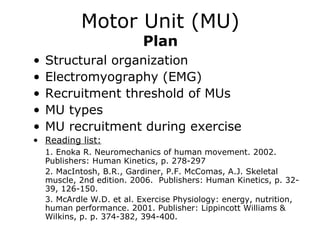
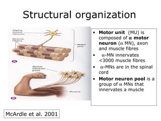
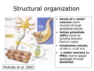
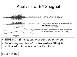
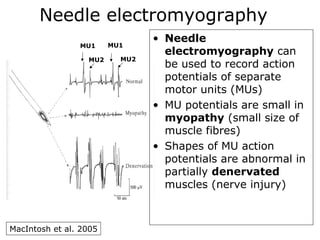
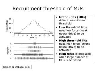
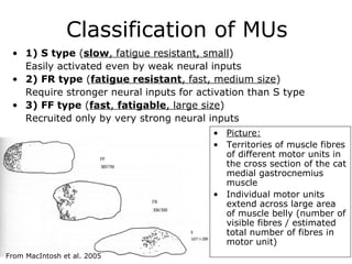
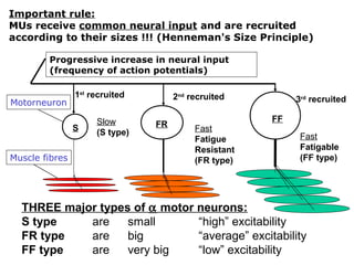
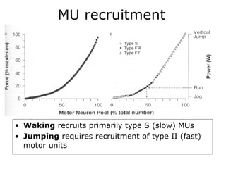
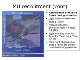


A motor unit is composed of an α motor neuron, its axon, and the muscle fibers it innervates. There are three main types of motor units - S, FR, and FF - that are recruited in order of size according to the Henneman size principle, with S units recruited first and FF units recruited only during high force generation. Electromyography can be used to study individual motor units and their recruitment patterns during different levels of exercise.


