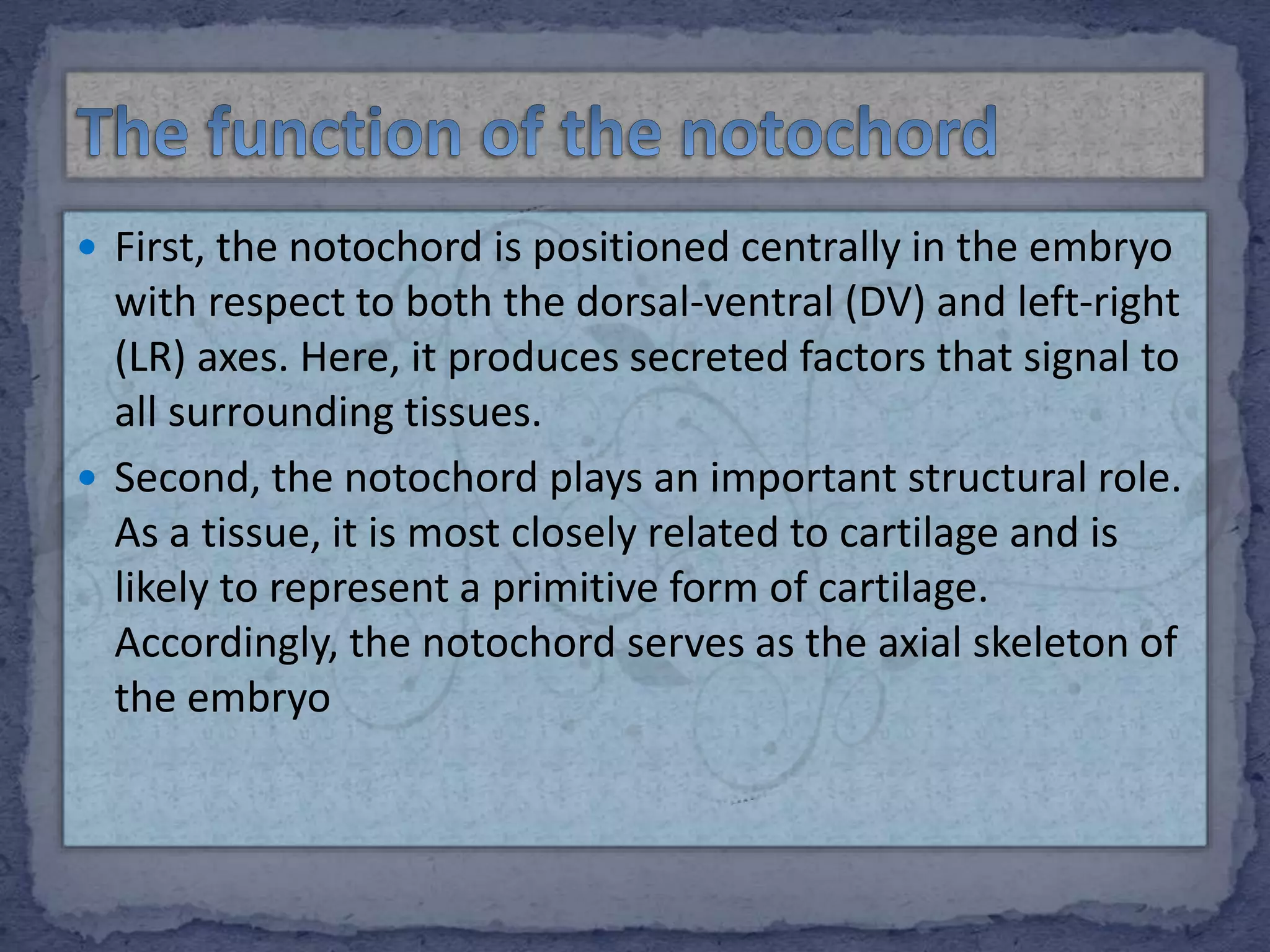
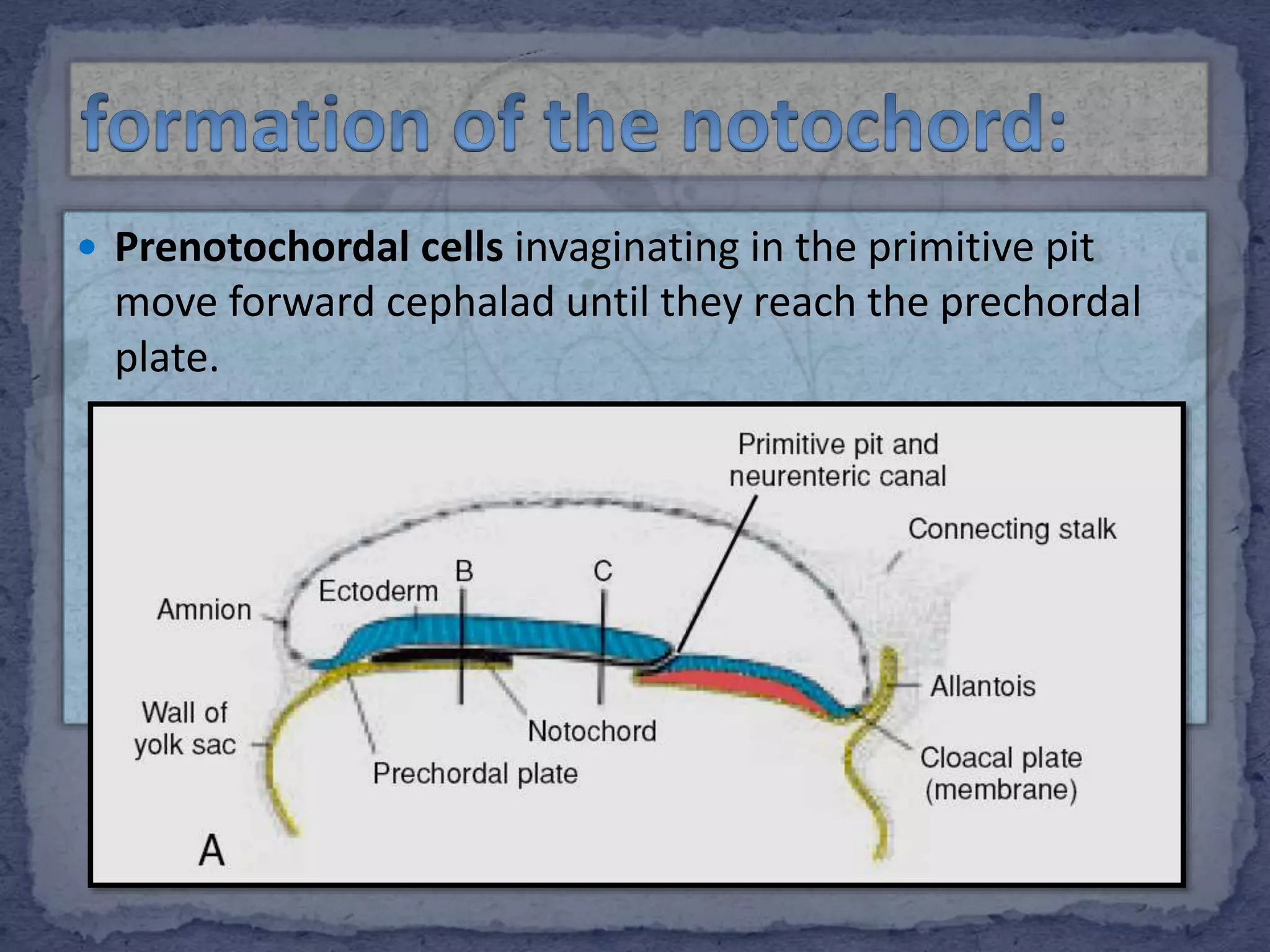
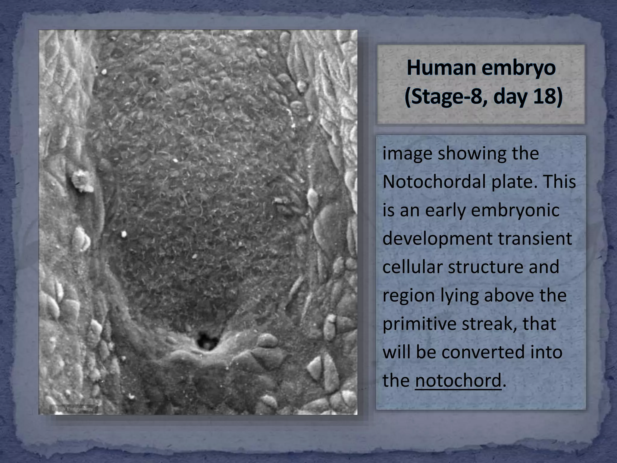
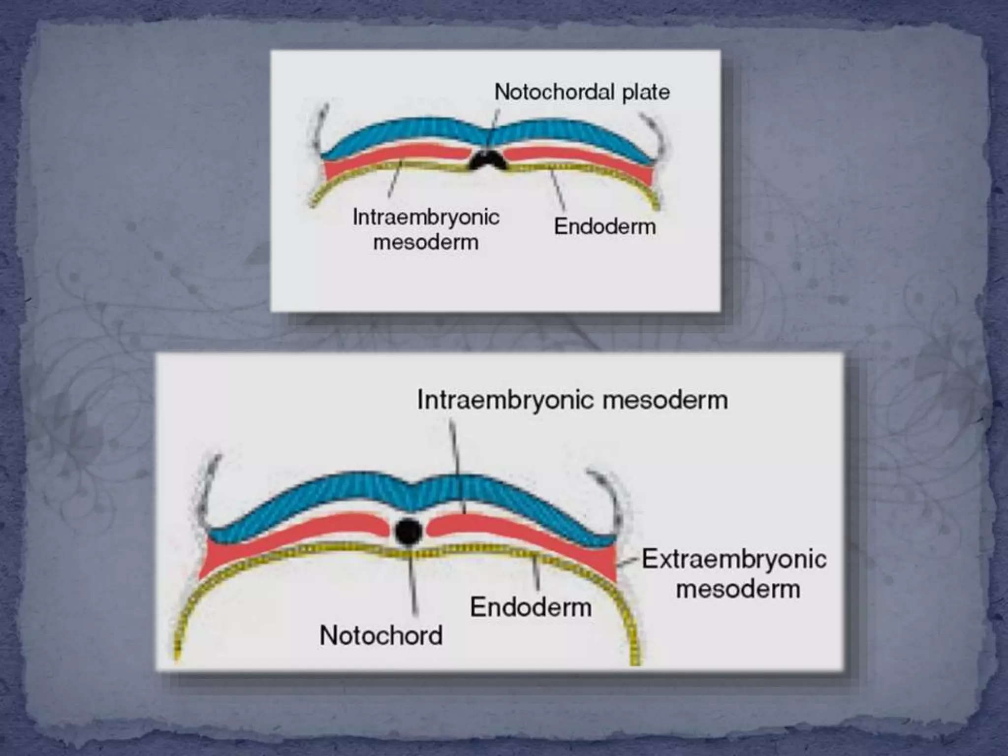
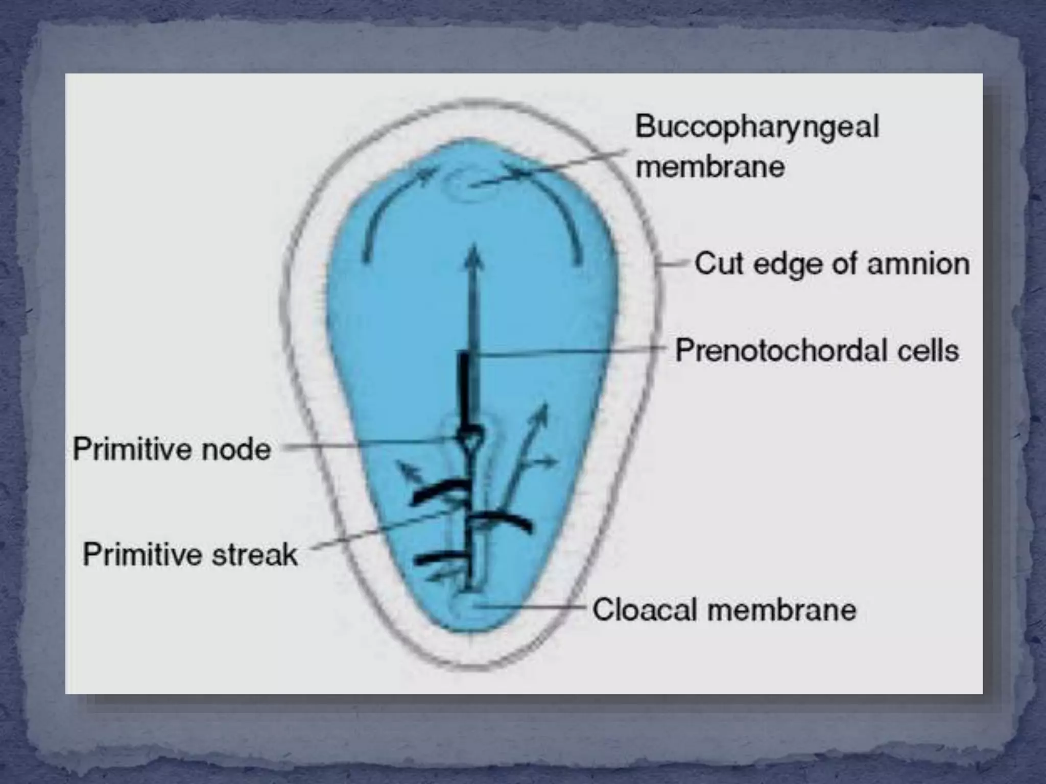
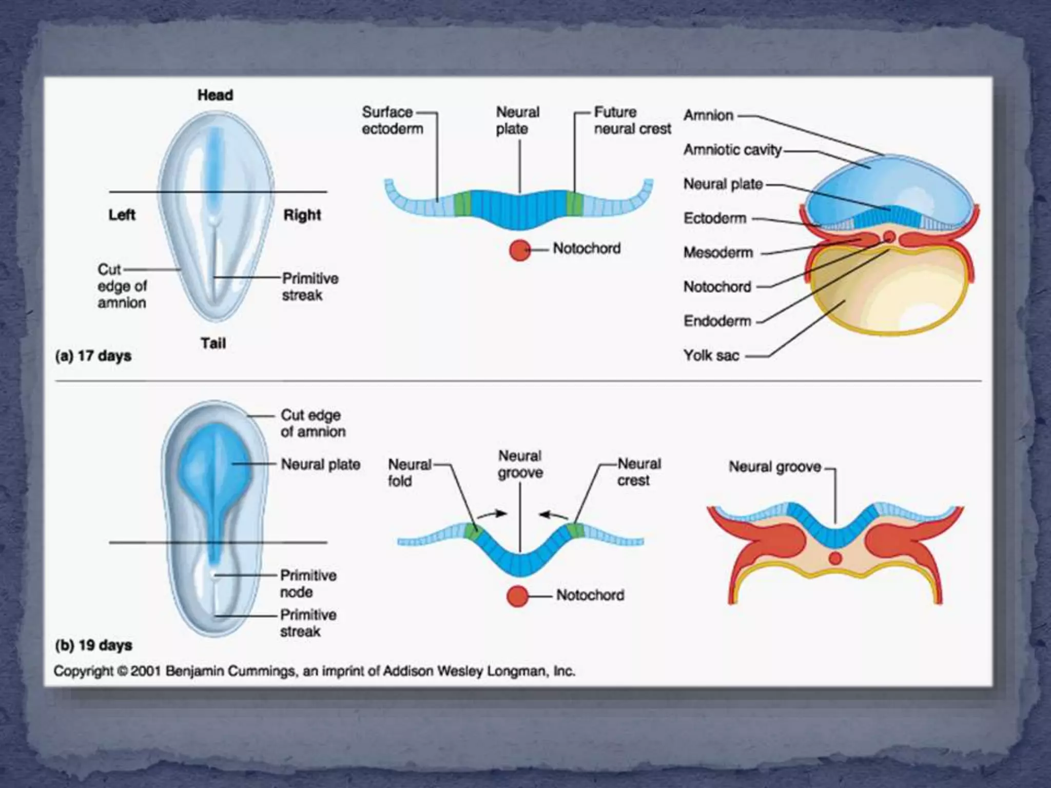
2. The notochord is an embryonic midline structure
common to all members of the phylum Chordata. In
higher vertebrates, the notochord exists transiently
and has at least two important functions .
3.
4. First, the notochord is positioned centrally in the embryo
with respect to both the dorsal-ventral (DV) and left-right
(LR) axes. Here, it produces secreted factors that signal to
all surrounding tissues.
Second, the notochord plays an important structural role.
As a tissue, it is most closely related to cartilage and is
likely to represent a primitive form of cartilage.
Accordingly, the notochord serves as the axial skeleton of
the embryo
5. Prenotochordal cells invaginating in the primitive pit
move forward cephalad until they reach the prechordal
plate.
6. image showing the
Notochordal plate. This
is an early embryonic
development transient
cellular structure and
region lying above the
primitive streak, that
will be converted into
the notochord.
7. These Prenotochordal cells become intercalated in the
hypoblast so that, for a short time, the midline of the
embryo consists of two cell layers that form the
Notochordal plate.
As the hypoblast is replaced by endoderm cells moving in
at the streak, cells of the notochordal plate proliferate
and detach from the endoderm. They then form a solid
cord of cells, the definitive notochord. which underlies
the neural tube and serves as the basis for the axial
skeleton.
8.
9. Because elongation of the notochord is a dynamic
process, the cranial end forms first, and caudal regions
are added as the primitive streak assumes a more caudal
position. The notochord and prenotochordal cells extend
cranially to the Prechordal plate (an area just caudal to
the buccopharyngeal membrane) and caudally to the
primitive pit. At the point where the pit forms an
indentation in the epiblast, the neurenteric canal
temporarily connects the amniotic. and yolk sac cavities.
10.
11. It extends throughout the entire length of the future
vertebral column, and reaches as far as the anterior end
of the midbrain, where it ends in a hook-like extremity in
the region of the future dorsum sellæ of the sphenoid
bone.
Initially it exists between the neural tube and the
endoderm of the yolk-sac, but soon becomes separated
from them by the mesoderm, which grows medially and
surrounds it. From the mesoderm surrounding the neural
tube and notochord, the skull, vertebral column, and the
membranes of the brain and medulla spinales are
developed.
12.
13. The cloacal membrane is formed at the caudal end of the
embryonic disc. This membrane, which is similar in
structure to the buccopharyngeal membrane, consists of
tightly adherent ectoderm and endoderm cells with no
intervening mesoderm.
when the cloacal membrane appears the posterior wall
of the yolk sac forms a small diverticulum that extends
into the connecting stalk. This diverticulum, the
“allantoenteric diverticulum”, or “allantois”, appears
around the 16th day of development. Although in some
lower vertebrates the allantois serves as a reservoir for
excretion products of the renal system, in humans it
remains rudimentary but may be involved in
abnormalities of bladder development.
14.
15. The notochord is the defining structure of the
chordates, and has essential roles in
vertebrate development. It serves as a source
of midline signals that pattern surrounding
tissues and as a major skeletal element of the
developing embryo. Genetic and
embryological studies over the past decade
have informed us about the development and
function of the notochord.
18. is frequently associated with vertebral
anomalies. Complete cleft of the vertebral column is
associated with gastrointestinal tract and central nervous
system anomalies.
It is an extremely rare form of spinal dysraphism.
Management of SNS associated with dorsal enteric fistula
varies from case to case, depends upon the associated
anomalies and system involved; therefore, management
must be individualized. It is also considered that staging
procedure is needed for proper correction of these
anomalies