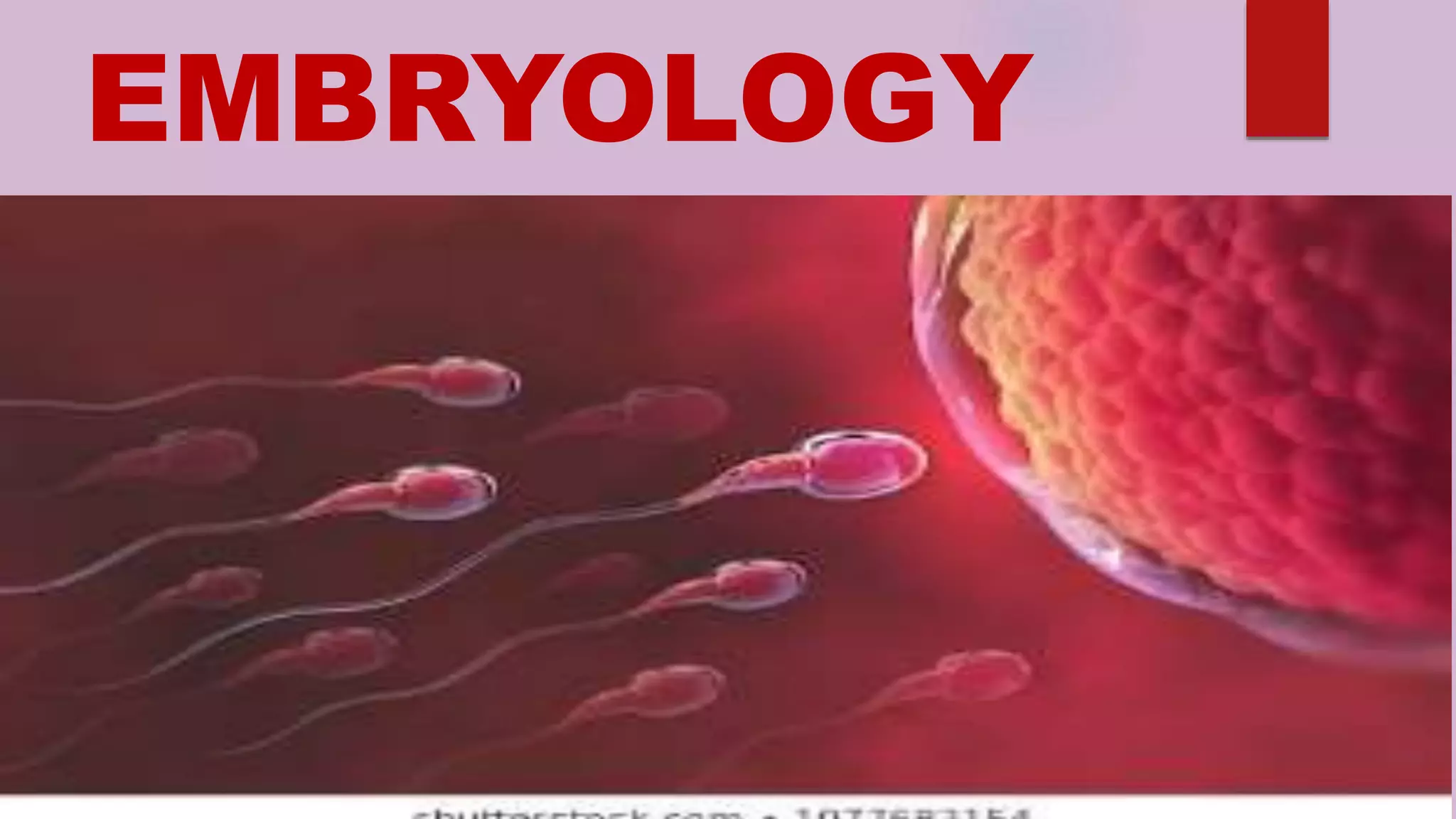
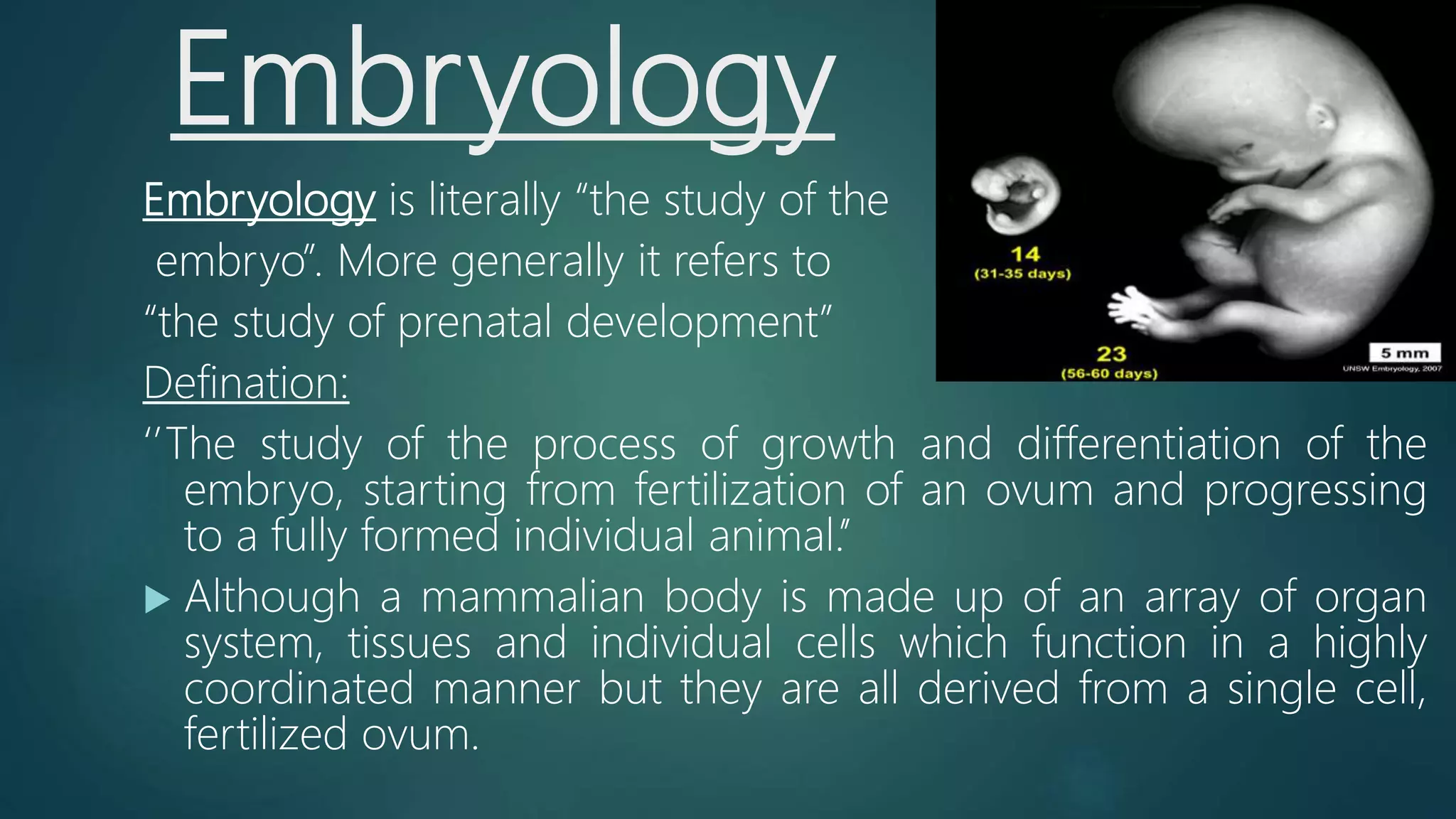
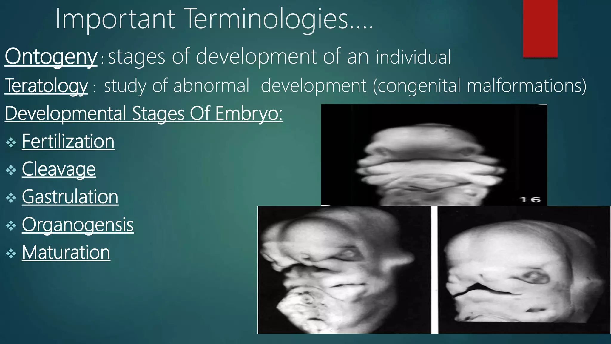
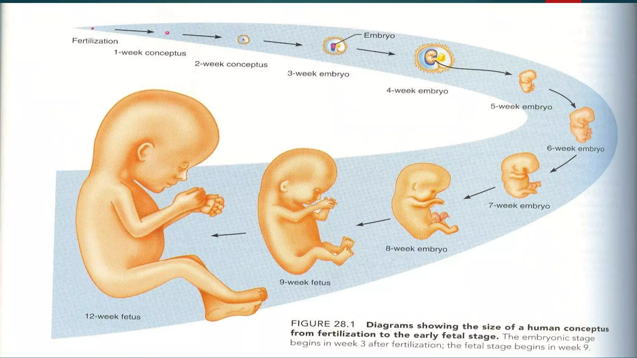
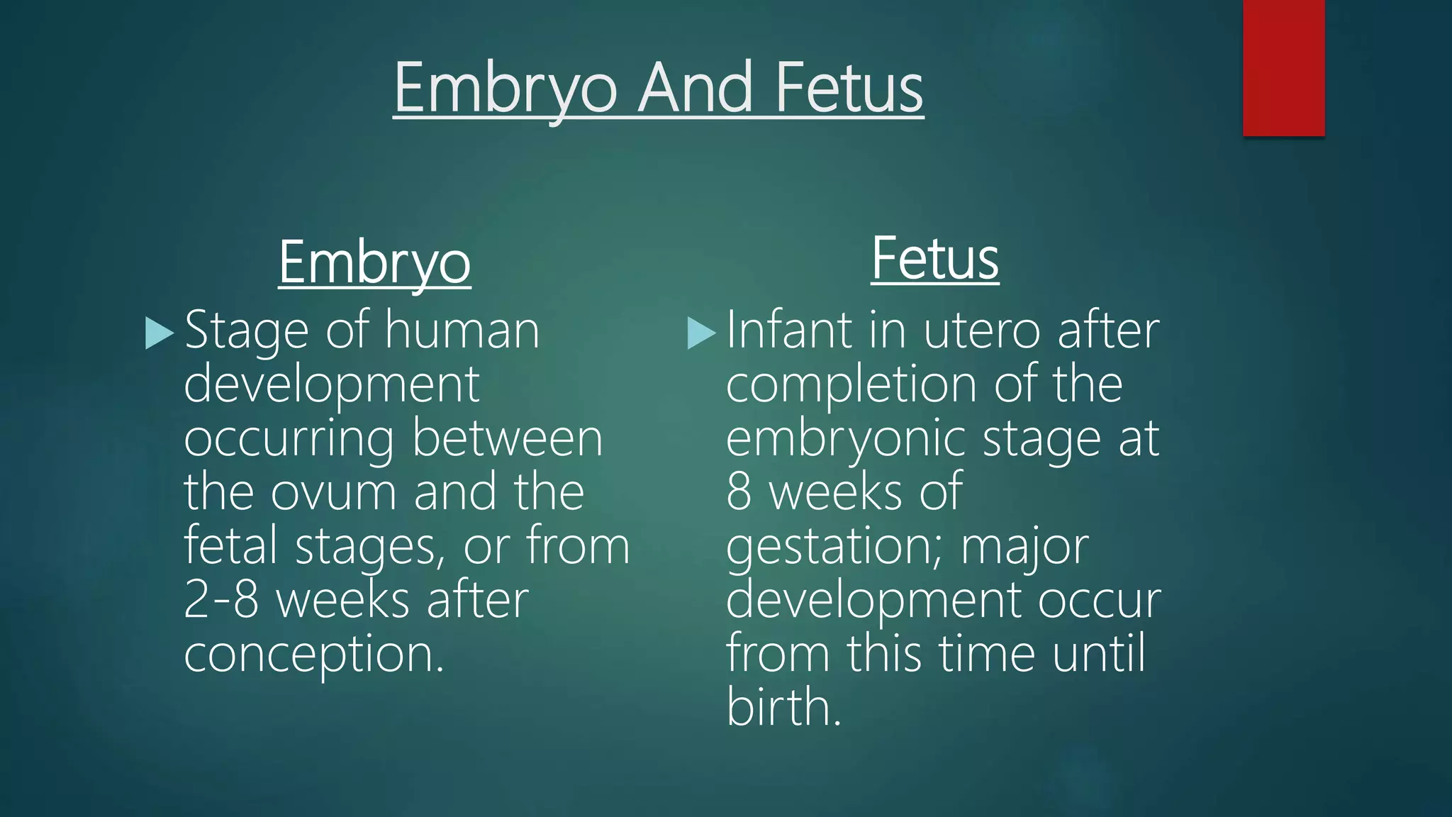
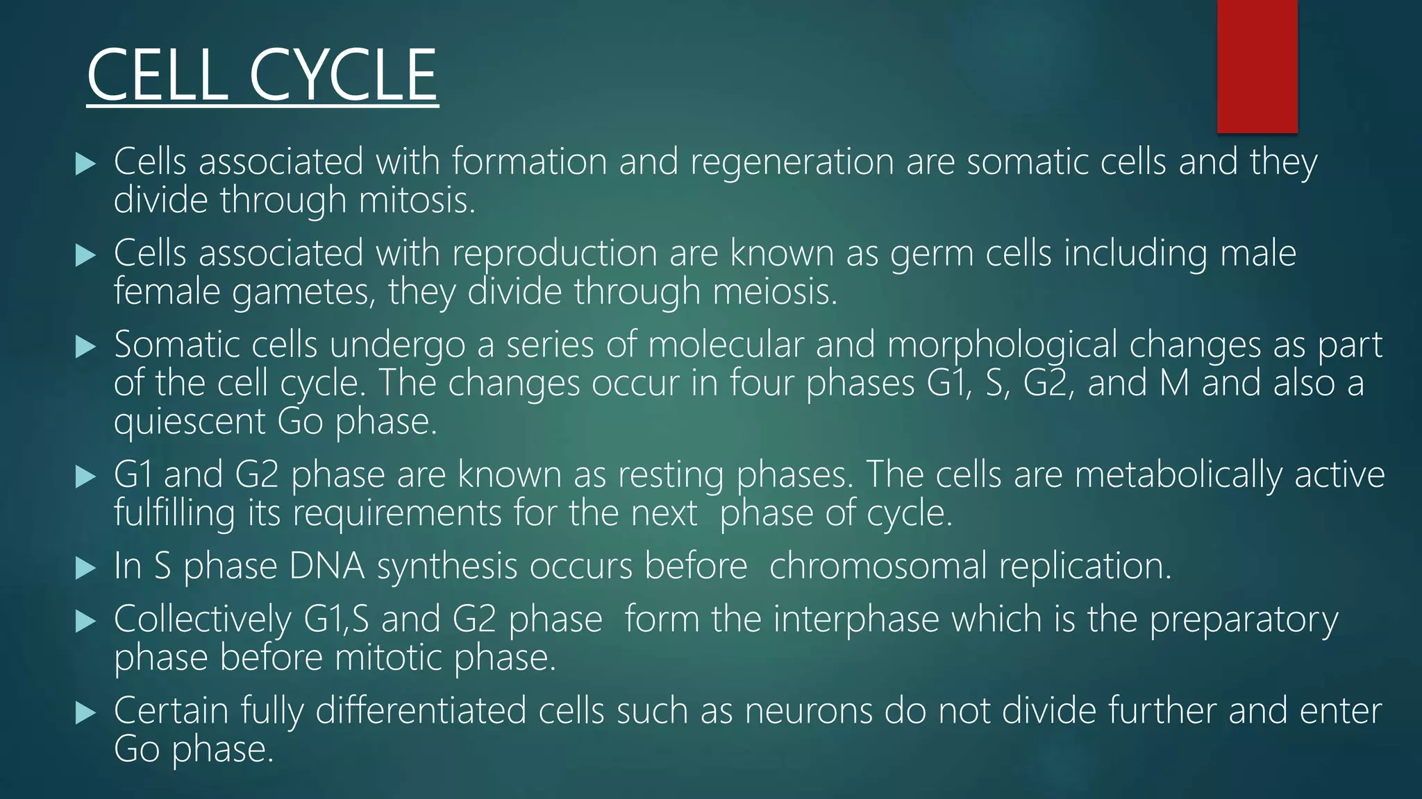
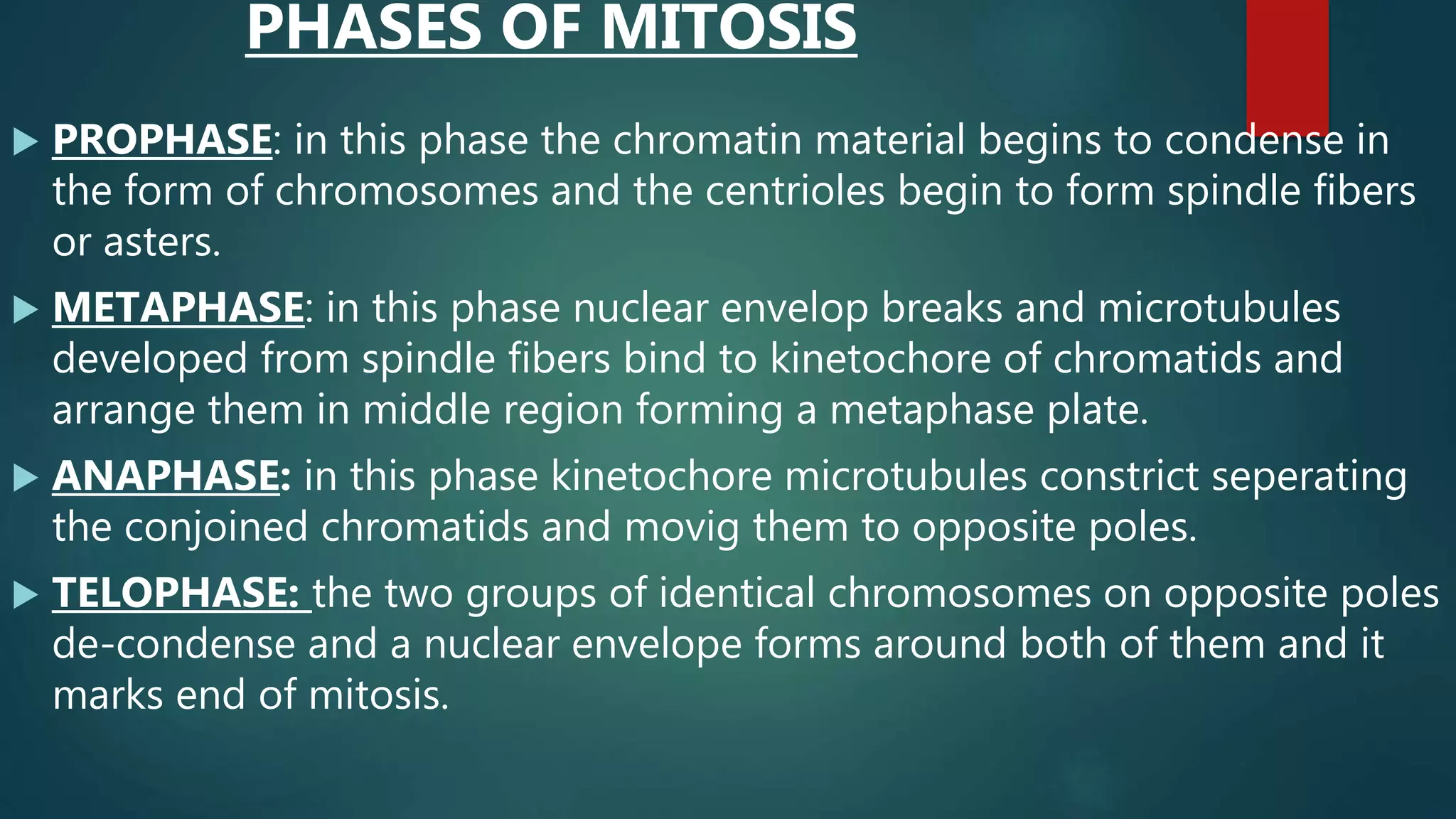
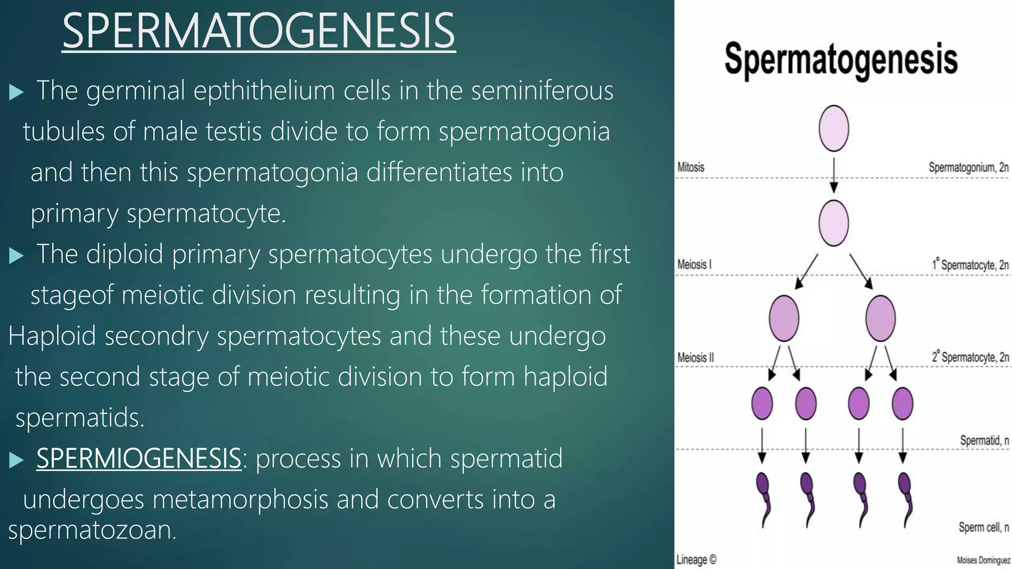
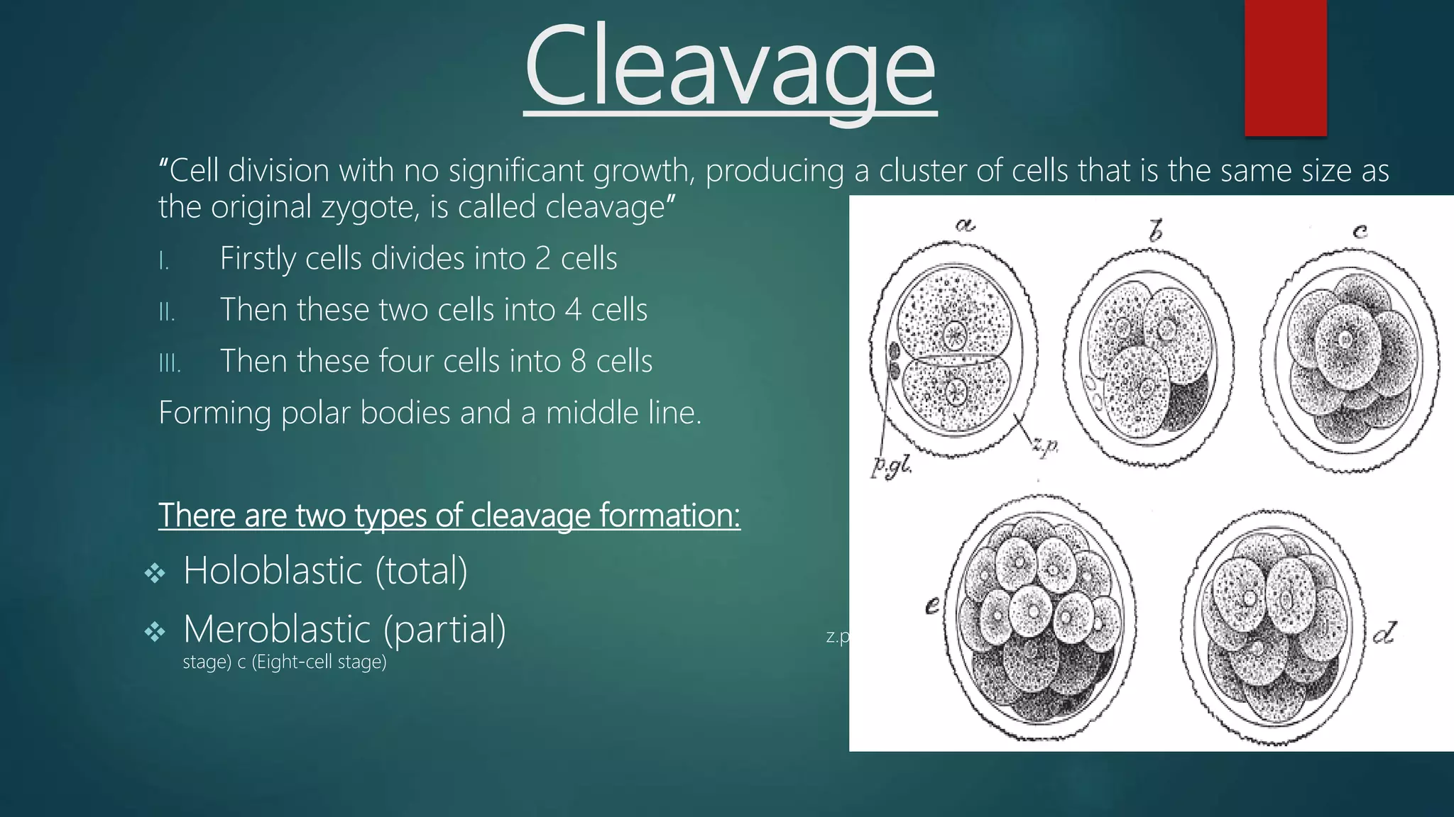
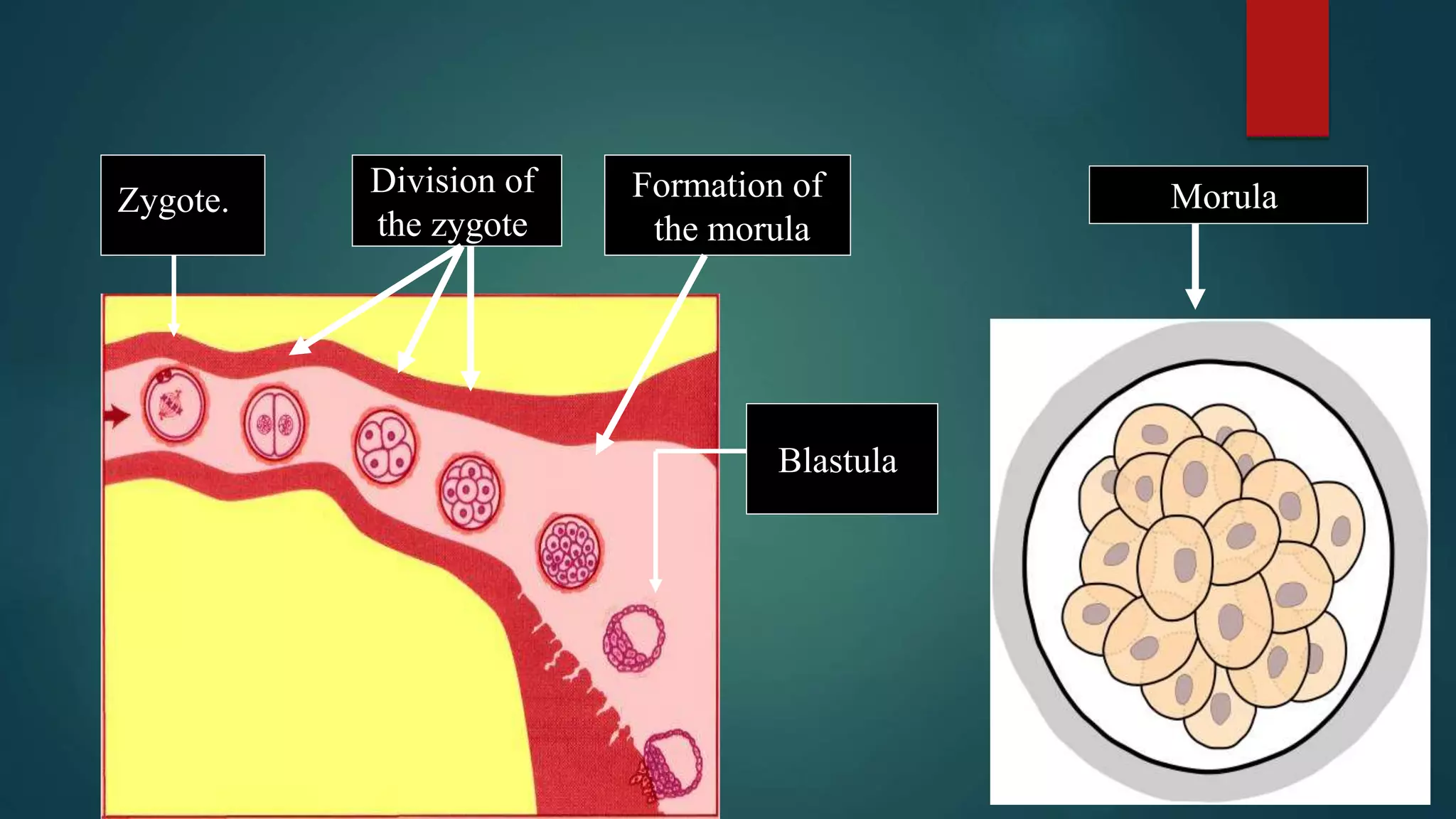
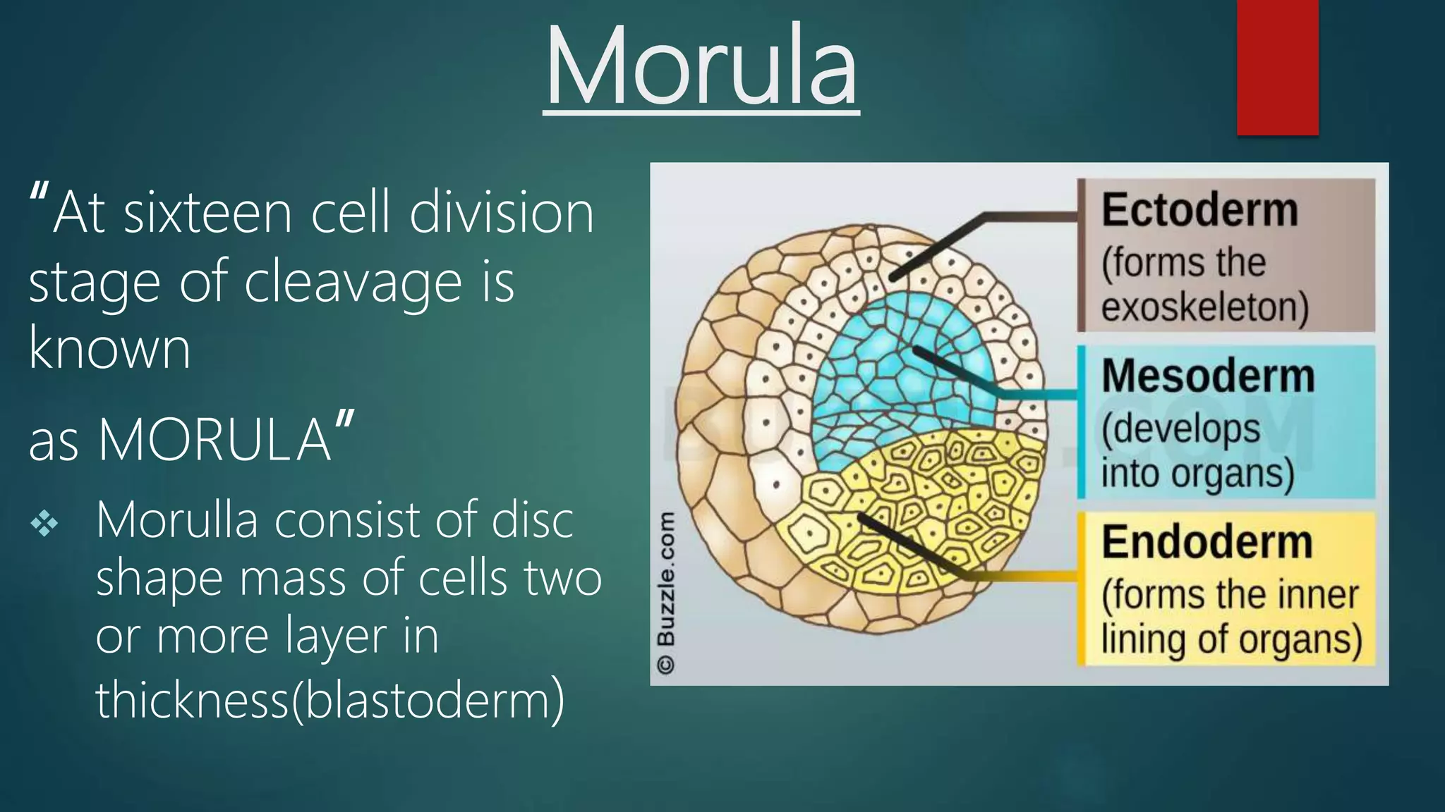
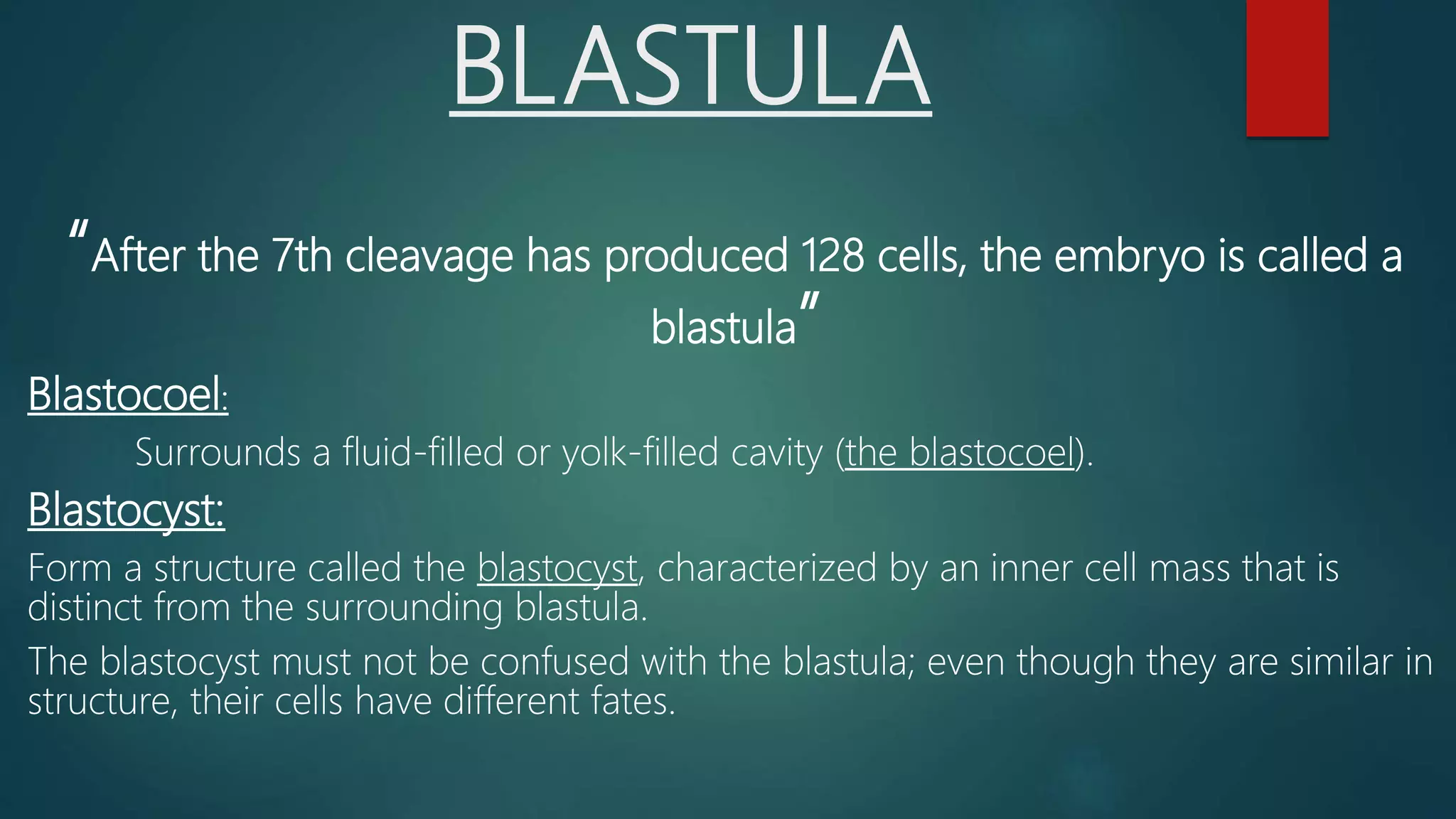
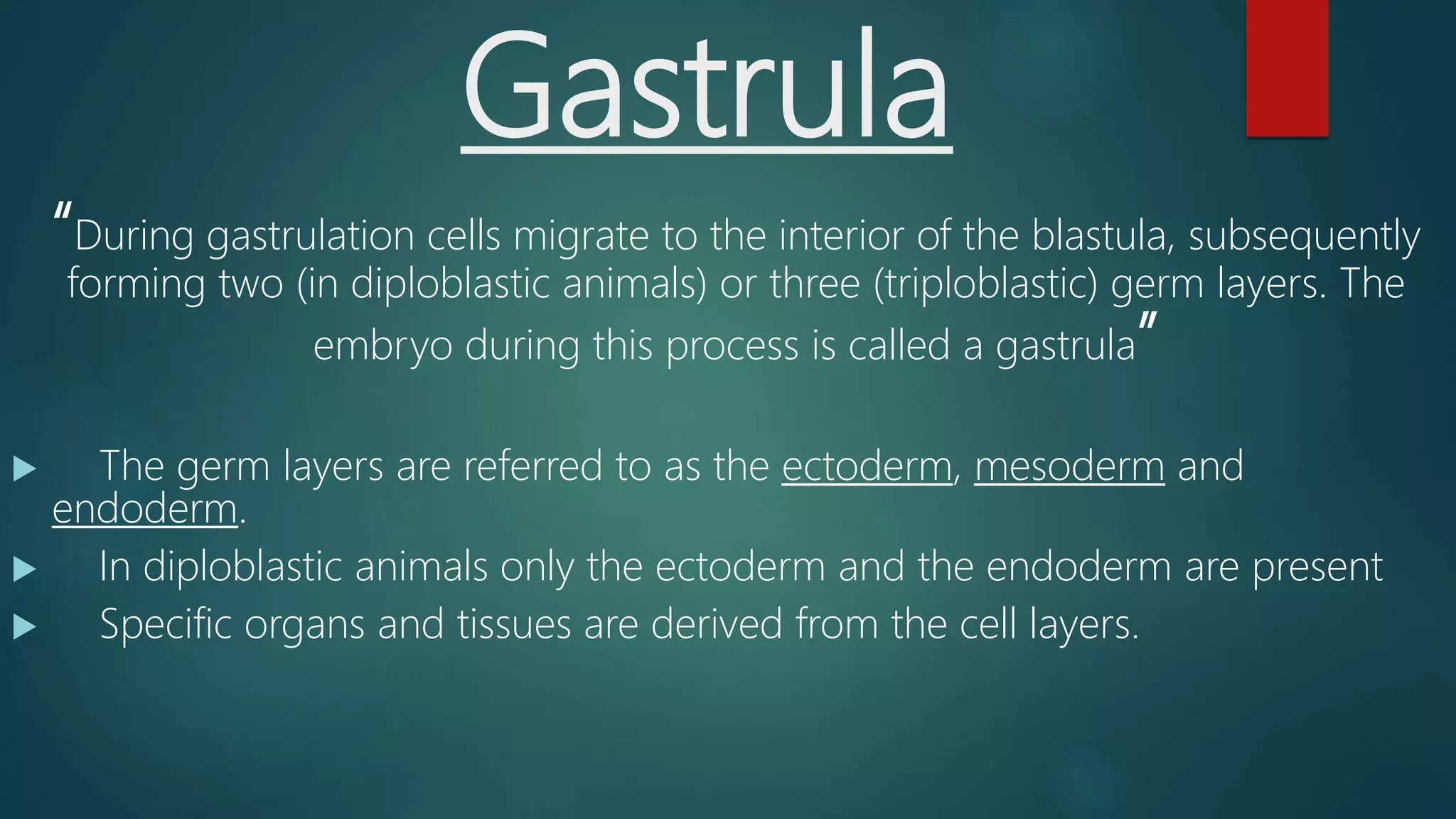
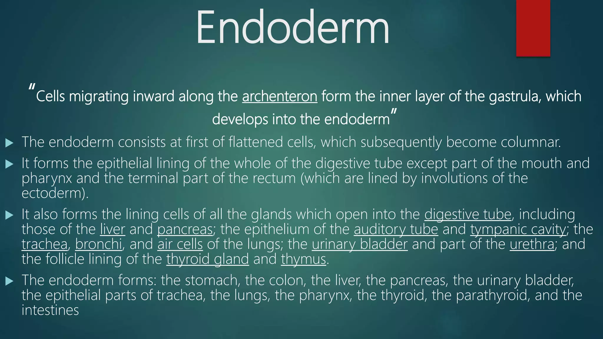
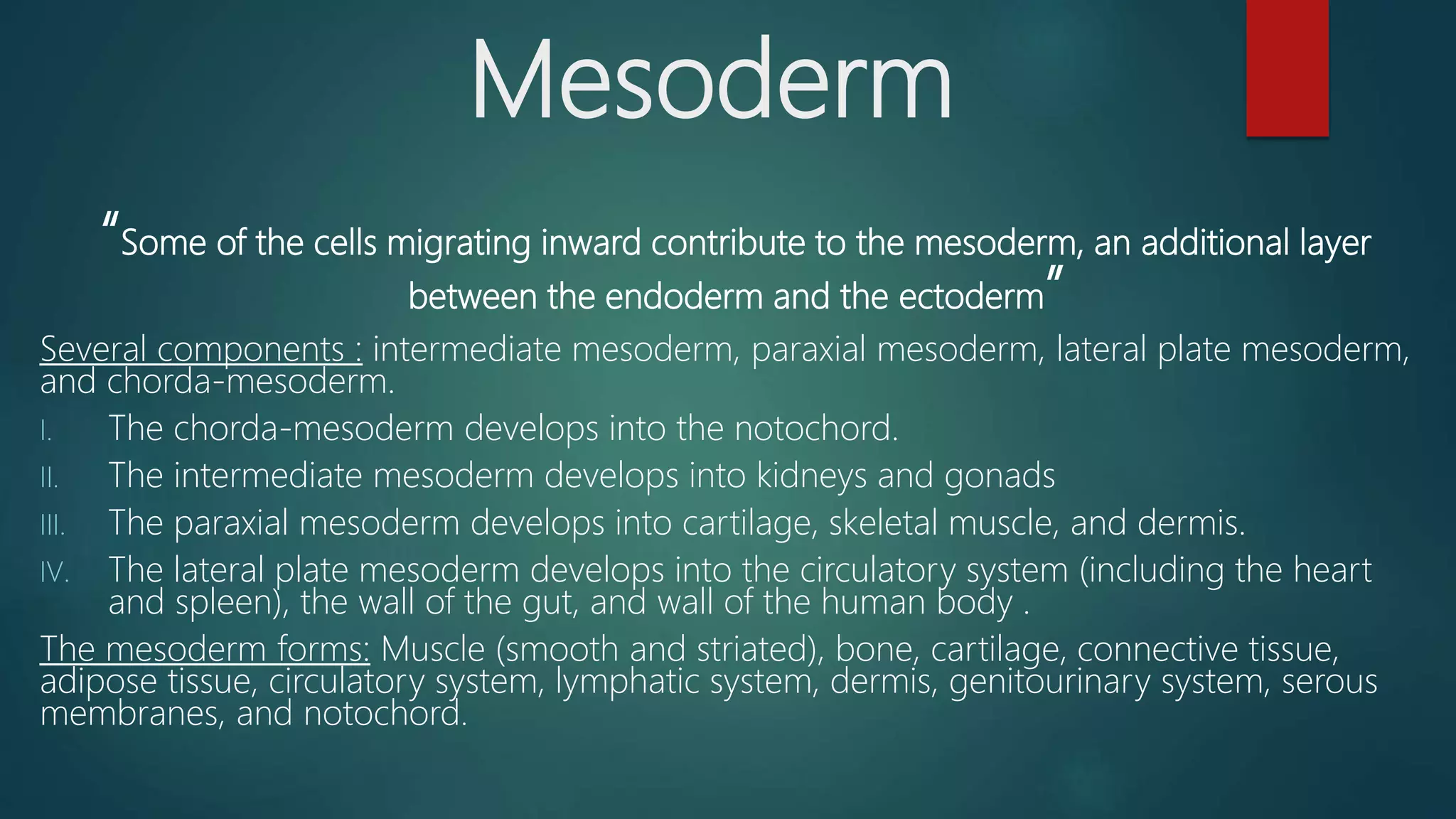
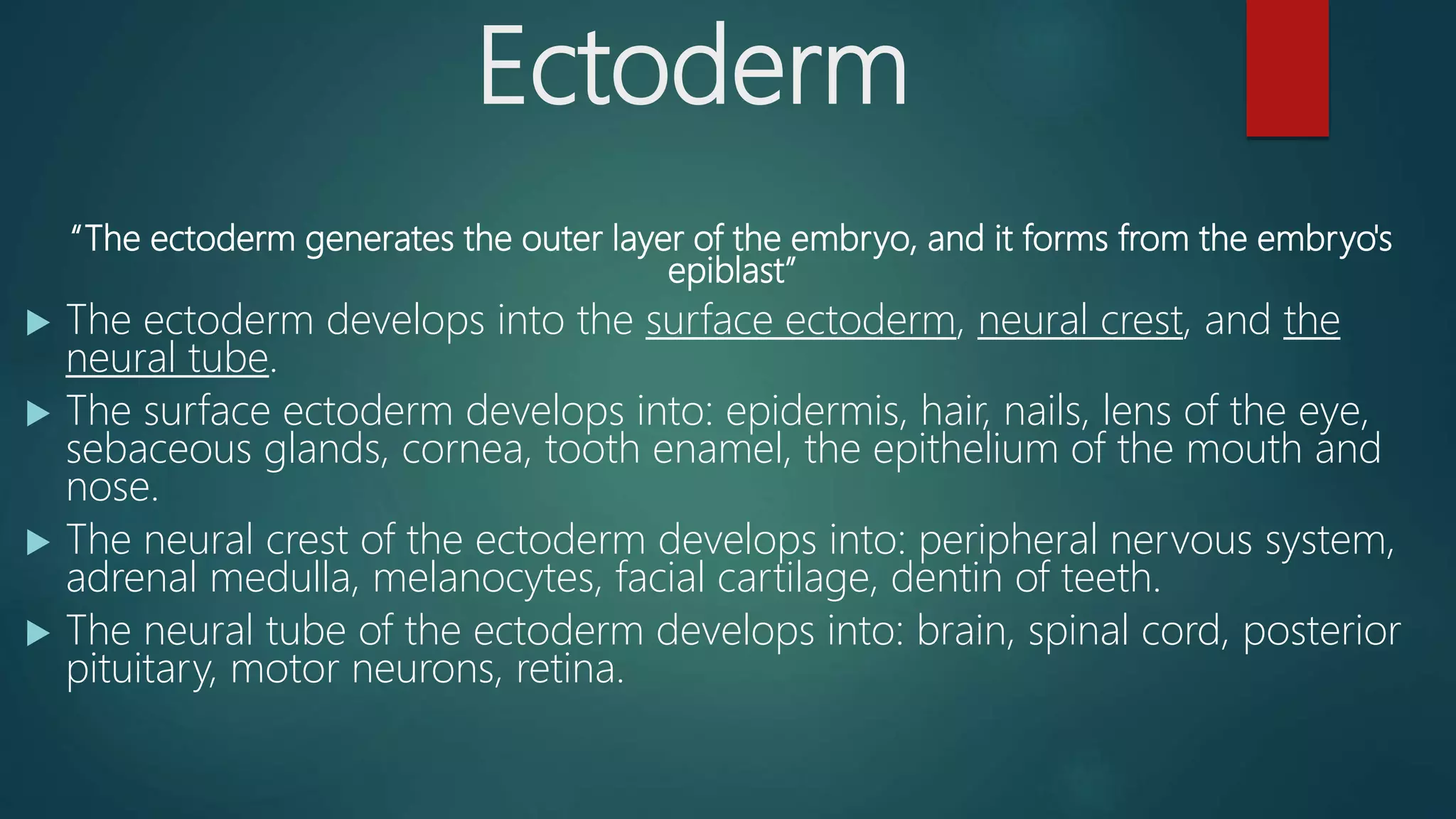
EMBRYOLOGY
Submitted To:
Dr. Riaz Hussain Pasha
Submitted By:
Haseeb Mazhar (19-Arid-2285)
Ehtasham ul Haq (19-Arid-2280)
Muhammad Sharjeel
Haider Balouch (19-Arid-2312)
Embryology
Embryology is literally “the study of the
embryo”. More generally it refers to
“the study of prenatal development”
Defination:
‘’The study of the process of growth and differentiation of the
embryo, starting from fertilization of an ovum and progressing
to a fully formed individual animal.’’
 Although a mammalian body is made up of an array of organ
system, tissues and individual cells which function in a highly
coordinated manner but they are all derived from a single cell,
fertilized ovum.
Ontogeny : stages of development of an individual
Teratology : study of abnormal development (congenital malformations)
Developmental Stages Of Embryo:
 Fertilization
 Cleavage
 Gastrulation
 Organogensis
 Maturation
Important Terminologies….
Embryo And Fetus
Embryo
Stage of human
development
occurring between
the ovum and the
fetal stages, or from
2-8 weeks after
conception.
Fetus
Infant in utero after
completion of the
embryonic stage at
8 weeks of
gestation; major
development occur
from this time until
birth.
CELL CYCLE
 Cells associated with formation and regeneration are somatic cells and they
divide through mitosis.
 Cells associated with reproduction are known as germ cells including male
female gametes, they divide through meiosis.
 Somatic cells undergo a series of molecular and morphological changes as part
of the cell cycle. The changes occur in four phases G1, S, G2, and M and also a
quiescent Go phase.
 G1 and G2 phase are known as resting phases. The cells are metabolically active
fulfilling its requirements for the next phase of cycle.
 In S phase DNA synthesis occurs before chromosomal replication.
 Collectively G1,S and G2 phase form the interphase which is the preparatory
phase before mitotic phase.
 Certain fully differentiated cells such as neurons do not divide further and enter
Go phase.
PHASES OF MITOSIS
 PROPHASE: in this phase the chromatin material begins to condense in
the form of chromosomes and the centrioles begin to form spindle fibers
or asters.
 METAPHASE: in this phase nuclear envelop breaks and microtubules
developed from spindle fibers bind to kinetochore of chromatids and
arrange them in middle region forming a metaphase plate.
 ANAPHASE: in this phase kinetochore microtubules constrict seperating
the conjoined chromatids and movig them to opposite poles.
 TELOPHASE: the two groups of identical chromosomes on opposite poles
de-condense and a nuclear envelope forms around both of them and it
marks end of mitosis.
PHASES OF MEIOSIS
 Meiosis consists of two phases MEIOSIS 1 and MEISOSIS 2 each consisting of
further sub phases.
 MEIOSIS 2 is same as mitosis but meiosis 1 has different steps.
 PROPHASE1: during this phase non sister chromatids combine with each other
at specific points(chiasmata formation) and exchange their segments(crossing
over). This helps in genetic variation in next generation.
 METAPHASE1: in this phase homologous chromosomes arrange them selfs in
the from of a metaphase plate.
 ANAPHASE1: in this phase the tetrads split into dyads and move towards
opposite poles.
 TELOPHASE1: in this phase nuclear envelop develops around nucleus and it is
followed by cytokinesis.
Gametogenesis
 Gametogenesis is a process of formation and development of
male(sperm) and female(egg) gametes.
 During this process reduction of chromosome takes place e.g. in case of
human chromosome number is reduced from 46 to 23 during
gametogenesis. It also involves the chromosomal and cytoplasmic
changes.
 The large rounded primary male germ cells lose most of the cytoplasm
and develops head tail and neck to assist the movement of sperms.
 The rounded female germ cells become larger as a result of increase in
their amount of cytoplasm and called ovum.
SPERMATOGENESIS
 The germinal epthithelium cells in the seminiferous
tubules of male testis divide to form spermatogonia
and then this spermatogonia differentiates into
primary spermatocyte.
 The diploid primary spermatocytes undergo the first
stageof meiotic division resulting in the formation of
Haploid secondry spermatocytes and these undergo
the second stage of meiotic division to form haploid
spermatids.
 SPERMIOGENESIS: process in which spermatid
undergoes metamorphosis and converts into a
spermatozoan.
OOGENESIS
 A pair of ovaries lies with in the female body cavity which produces
many oogonia which mitotically divide to form primary oocytes.
 These primary oocytes meiotically divide into the haploid secondary
oocyte and first polar body.
 Second meiotic division in the oocyte proceeds as far as metaphase
but is not completed until the oocyte is fertilized by the sperm.
 The discharge of ovum from the ovary is called as ovulation.
Fertilization
 Embryogenesis Starts with the fertilization of the egg cell ovum
by a sperm cell Spermatozoon
 Once fertilized, the ovum Is known as ZYGOTE ( a single diploid
cell).
 Depending on the specie fertilization can be :
a. Inside body
b. Outside of the body
 The zygote, which contains 46 chromosomes, rapidly divide by
mitosis to form mass of cells as 2,4, 8, 16, 32….
 This mass of cells continue dividing in order to give the different
tissues then organs of the human body.
Cleavage
“Cell division with no significant growth, producing a cluster of cells that is the same size as
the original zygote, is called cleavage”
I. Firstly cells divides into 2 cells
II. Then these two cells into 4 cells
III. Then these four cells into 8 cells
Forming polar bodies and a middle line.
There are two types of cleavage formation:
 Holoblastic (total)
 Meroblastic (partial) z.p( Zona pellucida) p.gl (Polar bodies) a (Two-cell stage) b (Four-cell
stage) c (Eight-cell stage)
Continue….
 Holoblastic cleavage:
Occurs in animals with little yolk in their eggs, such
as humans and other mammals who receive nourishment as embryos
from the mother, via the placenta or milk.
 Meroblastic cleavage:
Occurs in animals whose eggs have more yolk; i.e.
birds and reptiles. Because cleavage is impeded in the vegetal pole,
there is a very uneven distribution and size of cells, being more
numerous and smaller at the animal pole of the zygote
Zygote.
Division of
the zygote
Formation of
the morula
Morula
Blastula
Morula
“At sixteen cell division
stage of cleavage is
known
as MORULA”
 Morulla consist of disc
shape mass of cells two
or more layer in
thickness(blastoderm)
BLASTULA
“After the 7th cleavage has produced 128 cells, the embryo is called a
blastula”
Blastocoel:
Surrounds a fluid-filled or yolk-filled cavity (the blastocoel).
Blastocyst:
Form a structure called the blastocyst, characterized by an inner cell mass that is
distinct from the surrounding blastula.
The blastocyst must not be confused with the blastula; even though they are similar in
structure, their cells have different fates.
Gastrula
“During gastrulation cells migrate to the interior of the blastula, subsequently
forming two (in diploblastic animals) or three (triploblastic) germ layers. The
embryo during this process is called a gastrula”
 The germ layers are referred to as the ectoderm, mesoderm and
endoderm.
 In diploblastic animals only the ectoderm and the endoderm are present
 Specific organs and tissues are derived from the cell layers.
NOTOCHORD AND MESODERM
FORMATION
primitive streak: The mesodermal cells medially
and caudally migrate towards the center forming
a mid thick line called primitive streak.
primitive ridges: The continuous migration of
cells forms a groove along the length of the
primitive streak which is marked by the thickened
margins known as primitive ridges.
Neural plate: will fold to form neural groove
Neural groove: fuses to form neural tube
Neural tube: Gives rise to brain and spinal cord
Differentiation of
Germinal Layers
Germ Layer
“A germ layer, is a primary layer of cells that form during
embryogenesis”
The three germ layers in vertebrates are particularly
pronounced:
 Ectoderm
 Mesoderm
 Endoderm
Endoderm
“Cells migrating inward along the archenteron form the inner layer of the gastrula, which
develops into the endoderm”
 The endoderm consists at first of flattened cells, which subsequently become columnar.
 It forms the epithelial lining of the whole of the digestive tube except part of the mouth and
pharynx and the terminal part of the rectum (which are lined by involutions of the
ectoderm).
 It also forms the lining cells of all the glands which open into the digestive tube, including
those of the liver and pancreas; the epithelium of the auditory tube and tympanic cavity; the
trachea, bronchi, and air cells of the lungs; the urinary bladder and part of the urethra; and
the follicle lining of the thyroid gland and thymus.
 The endoderm forms: the stomach, the colon, the liver, the pancreas, the urinary bladder,
the epithelial parts of trachea, the lungs, the pharynx, the thyroid, the parathyroid, and the
intestines
Mesoderm
“Some of the cells migrating inward contribute to the mesoderm, an additional layer
between the endoderm and the ectoderm”
Several components : intermediate mesoderm, paraxial mesoderm, lateral plate mesoderm,
and chorda-mesoderm.
I. The chorda-mesoderm develops into the notochord.
II. The intermediate mesoderm develops into kidneys and gonads
III. The paraxial mesoderm develops into cartilage, skeletal muscle, and dermis.
IV. The lateral plate mesoderm develops into the circulatory system (including the heart
and spleen), the wall of the gut, and wall of the human body .
The mesoderm forms: Muscle (smooth and striated), bone, cartilage, connective tissue,
adipose tissue, circulatory system, lymphatic system, dermis, genitourinary system, serous
membranes, and notochord.
Ectoderm
“The ectoderm generates the outer layer of the embryo, and it forms from the embryo's
epiblast”
 The ectoderm develops into the surface ectoderm, neural crest, and the
neural tube.
 The surface ectoderm develops into: epidermis, hair, nails, lens of the eye,
sebaceous glands, cornea, tooth enamel, the epithelium of the mouth and
nose.
 The neural crest of the ectoderm develops into: peripheral nervous system,
adrenal medulla, melanocytes, facial cartilage, dentin of teeth.
 The neural tube of the ectoderm develops into: brain, spinal cord, posterior
pituitary, motor neurons, retina.
Overview
Developmental stages of embryo

Developmental stages of embryo

  • 1.
  • 2.
    Submitted To: Dr. RiazHussain Pasha Submitted By: Haseeb Mazhar (19-Arid-2285) Ehtasham ul Haq (19-Arid-2280) Muhammad Sharjeel Haider Balouch (19-Arid-2312)
  • 3.
    Embryology Embryology is literally“the study of the embryo”. More generally it refers to “the study of prenatal development” Defination: ‘’The study of the process of growth and differentiation of the embryo, starting from fertilization of an ovum and progressing to a fully formed individual animal.’’  Although a mammalian body is made up of an array of organ system, tissues and individual cells which function in a highly coordinated manner but they are all derived from a single cell, fertilized ovum.
  • 4.
    Ontogeny : stagesof development of an individual Teratology : study of abnormal development (congenital malformations) Developmental Stages Of Embryo:  Fertilization  Cleavage  Gastrulation  Organogensis  Maturation Important Terminologies….
  • 6.
    Embryo And Fetus Embryo Stageof human development occurring between the ovum and the fetal stages, or from 2-8 weeks after conception. Fetus Infant in utero after completion of the embryonic stage at 8 weeks of gestation; major development occur from this time until birth.
  • 7.
    CELL CYCLE  Cellsassociated with formation and regeneration are somatic cells and they divide through mitosis.  Cells associated with reproduction are known as germ cells including male female gametes, they divide through meiosis.  Somatic cells undergo a series of molecular and morphological changes as part of the cell cycle. The changes occur in four phases G1, S, G2, and M and also a quiescent Go phase.  G1 and G2 phase are known as resting phases. The cells are metabolically active fulfilling its requirements for the next phase of cycle.  In S phase DNA synthesis occurs before chromosomal replication.  Collectively G1,S and G2 phase form the interphase which is the preparatory phase before mitotic phase.  Certain fully differentiated cells such as neurons do not divide further and enter Go phase.
  • 8.
    PHASES OF MITOSIS PROPHASE: in this phase the chromatin material begins to condense in the form of chromosomes and the centrioles begin to form spindle fibers or asters.  METAPHASE: in this phase nuclear envelop breaks and microtubules developed from spindle fibers bind to kinetochore of chromatids and arrange them in middle region forming a metaphase plate.  ANAPHASE: in this phase kinetochore microtubules constrict seperating the conjoined chromatids and movig them to opposite poles.  TELOPHASE: the two groups of identical chromosomes on opposite poles de-condense and a nuclear envelope forms around both of them and it marks end of mitosis.
  • 9.
    PHASES OF MEIOSIS Meiosis consists of two phases MEIOSIS 1 and MEISOSIS 2 each consisting of further sub phases.  MEIOSIS 2 is same as mitosis but meiosis 1 has different steps.  PROPHASE1: during this phase non sister chromatids combine with each other at specific points(chiasmata formation) and exchange their segments(crossing over). This helps in genetic variation in next generation.  METAPHASE1: in this phase homologous chromosomes arrange them selfs in the from of a metaphase plate.  ANAPHASE1: in this phase the tetrads split into dyads and move towards opposite poles.  TELOPHASE1: in this phase nuclear envelop develops around nucleus and it is followed by cytokinesis.
  • 11.
    Gametogenesis  Gametogenesis isa process of formation and development of male(sperm) and female(egg) gametes.  During this process reduction of chromosome takes place e.g. in case of human chromosome number is reduced from 46 to 23 during gametogenesis. It also involves the chromosomal and cytoplasmic changes.  The large rounded primary male germ cells lose most of the cytoplasm and develops head tail and neck to assist the movement of sperms.  The rounded female germ cells become larger as a result of increase in their amount of cytoplasm and called ovum.
  • 13.
    SPERMATOGENESIS  The germinalepthithelium cells in the seminiferous tubules of male testis divide to form spermatogonia and then this spermatogonia differentiates into primary spermatocyte.  The diploid primary spermatocytes undergo the first stageof meiotic division resulting in the formation of Haploid secondry spermatocytes and these undergo the second stage of meiotic division to form haploid spermatids.  SPERMIOGENESIS: process in which spermatid undergoes metamorphosis and converts into a spermatozoan.
  • 14.
    OOGENESIS  A pairof ovaries lies with in the female body cavity which produces many oogonia which mitotically divide to form primary oocytes.  These primary oocytes meiotically divide into the haploid secondary oocyte and first polar body.  Second meiotic division in the oocyte proceeds as far as metaphase but is not completed until the oocyte is fertilized by the sperm.  The discharge of ovum from the ovary is called as ovulation.
  • 15.
    Fertilization  Embryogenesis Startswith the fertilization of the egg cell ovum by a sperm cell Spermatozoon  Once fertilized, the ovum Is known as ZYGOTE ( a single diploid cell).  Depending on the specie fertilization can be : a. Inside body b. Outside of the body  The zygote, which contains 46 chromosomes, rapidly divide by mitosis to form mass of cells as 2,4, 8, 16, 32….  This mass of cells continue dividing in order to give the different tissues then organs of the human body.
  • 16.
    Cleavage “Cell division withno significant growth, producing a cluster of cells that is the same size as the original zygote, is called cleavage” I. Firstly cells divides into 2 cells II. Then these two cells into 4 cells III. Then these four cells into 8 cells Forming polar bodies and a middle line. There are two types of cleavage formation:  Holoblastic (total)  Meroblastic (partial) z.p( Zona pellucida) p.gl (Polar bodies) a (Two-cell stage) b (Four-cell stage) c (Eight-cell stage)
  • 17.
    Continue….  Holoblastic cleavage: Occursin animals with little yolk in their eggs, such as humans and other mammals who receive nourishment as embryos from the mother, via the placenta or milk.  Meroblastic cleavage: Occurs in animals whose eggs have more yolk; i.e. birds and reptiles. Because cleavage is impeded in the vegetal pole, there is a very uneven distribution and size of cells, being more numerous and smaller at the animal pole of the zygote
  • 18.
    Zygote. Division of the zygote Formationof the morula Morula Blastula
  • 19.
    Morula “At sixteen celldivision stage of cleavage is known as MORULA”  Morulla consist of disc shape mass of cells two or more layer in thickness(blastoderm)
  • 20.
    BLASTULA “After the 7thcleavage has produced 128 cells, the embryo is called a blastula” Blastocoel: Surrounds a fluid-filled or yolk-filled cavity (the blastocoel). Blastocyst: Form a structure called the blastocyst, characterized by an inner cell mass that is distinct from the surrounding blastula. The blastocyst must not be confused with the blastula; even though they are similar in structure, their cells have different fates.
  • 21.
    Gastrula “During gastrulation cellsmigrate to the interior of the blastula, subsequently forming two (in diploblastic animals) or three (triploblastic) germ layers. The embryo during this process is called a gastrula”  The germ layers are referred to as the ectoderm, mesoderm and endoderm.  In diploblastic animals only the ectoderm and the endoderm are present  Specific organs and tissues are derived from the cell layers.
  • 23.
    NOTOCHORD AND MESODERM FORMATION primitivestreak: The mesodermal cells medially and caudally migrate towards the center forming a mid thick line called primitive streak. primitive ridges: The continuous migration of cells forms a groove along the length of the primitive streak which is marked by the thickened margins known as primitive ridges. Neural plate: will fold to form neural groove Neural groove: fuses to form neural tube Neural tube: Gives rise to brain and spinal cord
  • 24.
  • 25.
    Germ Layer “A germlayer, is a primary layer of cells that form during embryogenesis” The three germ layers in vertebrates are particularly pronounced:  Ectoderm  Mesoderm  Endoderm
  • 26.
    Endoderm “Cells migrating inwardalong the archenteron form the inner layer of the gastrula, which develops into the endoderm”  The endoderm consists at first of flattened cells, which subsequently become columnar.  It forms the epithelial lining of the whole of the digestive tube except part of the mouth and pharynx and the terminal part of the rectum (which are lined by involutions of the ectoderm).  It also forms the lining cells of all the glands which open into the digestive tube, including those of the liver and pancreas; the epithelium of the auditory tube and tympanic cavity; the trachea, bronchi, and air cells of the lungs; the urinary bladder and part of the urethra; and the follicle lining of the thyroid gland and thymus.  The endoderm forms: the stomach, the colon, the liver, the pancreas, the urinary bladder, the epithelial parts of trachea, the lungs, the pharynx, the thyroid, the parathyroid, and the intestines
  • 27.
    Mesoderm “Some of thecells migrating inward contribute to the mesoderm, an additional layer between the endoderm and the ectoderm” Several components : intermediate mesoderm, paraxial mesoderm, lateral plate mesoderm, and chorda-mesoderm. I. The chorda-mesoderm develops into the notochord. II. The intermediate mesoderm develops into kidneys and gonads III. The paraxial mesoderm develops into cartilage, skeletal muscle, and dermis. IV. The lateral plate mesoderm develops into the circulatory system (including the heart and spleen), the wall of the gut, and wall of the human body . The mesoderm forms: Muscle (smooth and striated), bone, cartilage, connective tissue, adipose tissue, circulatory system, lymphatic system, dermis, genitourinary system, serous membranes, and notochord.
  • 28.
    Ectoderm “The ectoderm generatesthe outer layer of the embryo, and it forms from the embryo's epiblast”  The ectoderm develops into the surface ectoderm, neural crest, and the neural tube.  The surface ectoderm develops into: epidermis, hair, nails, lens of the eye, sebaceous glands, cornea, tooth enamel, the epithelium of the mouth and nose.  The neural crest of the ectoderm develops into: peripheral nervous system, adrenal medulla, melanocytes, facial cartilage, dentin of teeth.  The neural tube of the ectoderm develops into: brain, spinal cord, posterior pituitary, motor neurons, retina.
  • 29.

Editor's Notes

  • #24 The central region of the ectoderm forms a columnar epithelium described as the neural plate. This epithelium will fold dorsally to form the neural groove, which then fuses to form an initially open at either end hollow neural tube. The neural tube forms the central nervous system (brain and spinal cord).