This document provides an overview of the anatomy of the ankle and foot complex. It discusses the bones, joints, ligaments, and arches of the foot. Key points include:
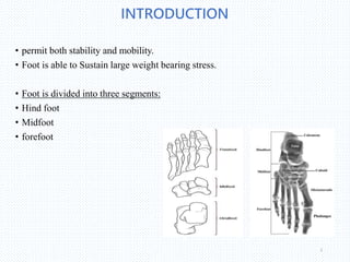
- The foot is divided into the hindfoot, midfoot, and forefoot and permits both stability and mobility while sustaining large weight-bearing stresses.
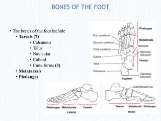
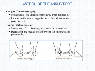
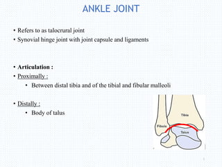
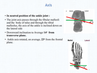
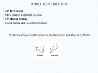
- Major bones include the tarsals, metatarsals, and phalanges. The talocrural joint allows dorsiflexion, plantar flexion, inversion, and eversion.
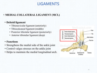
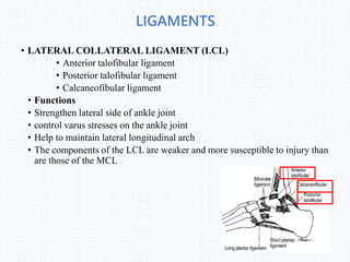
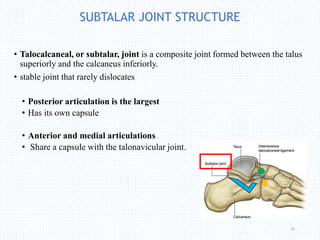
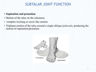
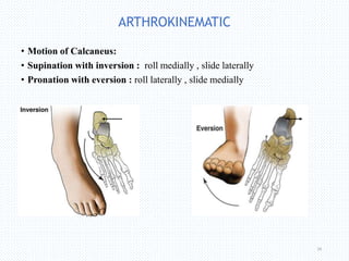
- Ligaments like the deltoid and collateral ligaments reinforce and support the ankle joint. The subtalar joint permits complex supination and pronation motions.