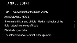
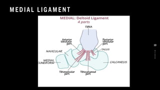
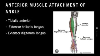
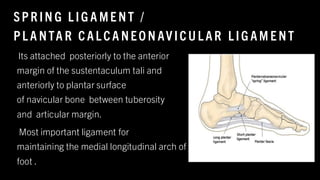
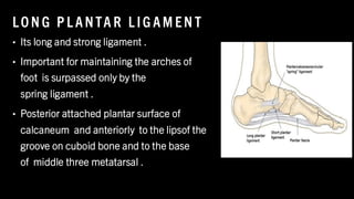
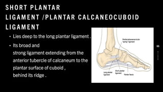
The document provides a comprehensive overview of the ankle and foot complex, detailing its anatomy, joint types, movements, and stability mechanisms. It describes the various ligaments, muscle attachments, and functional roles of the ankle joint, including its contributions to posture and mobility during gait. Additionally, the document includes descriptions of clinical tests for assessing ankle integrity and identifying potential injuries.


![T BIA L IS A NT ER IO R
• ORIGIN :- Lateral condyle and
superior 2/3 of anterolateral surface of tibia .
• INSERTION :- Medial and inferior surface of
medial cuneiform and base of 1st metatarsal
.
• NERVE SUPPLY :- Peroneal nerve { L4 , L5 ]
• ACTION : - Dorsiflexion of ankle .
8
/
1
/
2
0
2
4
16](https://image.slidesharecdn.com/anklejoint21-250204080450-e548fec2/85/ANKLE-JOINT-2-1-pdf-ppt-physiotherapist-16-320.jpg)
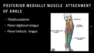
![T IBIA L IS PO ST E R IO R
• ORIGIN :- Posterior superior face of
tibia inferior to soleal line , posterior
surface of fibula .
• INSERTION :- Tuberosity of navicular ,
cuneiform , cuboid and
Sustanticulum Tali of calcaneus , base of
2nd , 3rd and 4th MT
• NERVE SUPPLY :- Tibial nerve [ L4 , L5 ]
• ACTION :- Plantar flexion
8
/
1
/
2
0
2
4
20](https://image.slidesharecdn.com/anklejoint21-250204080450-e548fec2/85/ANKLE-JOINT-2-1-pdf-ppt-physiotherapist-20-320.jpg)
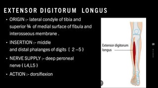
![F E X LO R DIG ITO R U M L O NG US
• ORIGIN :- Medial part of posterior
surface of tibia inferior to soleal line .
• INSERTION :- Base
of distal phalanges of digits 2- 5
• NERVE SUPPLY :- Tibial nerve [ L5
, S1 & S2 ]
• ACTION l:- Plantar flexion
8
/
1
/
2
0
2
4
21](https://image.slidesharecdn.com/anklejoint21-250204080450-e548fec2/85/ANKLE-JOINT-2-1-pdf-ppt-physiotherapist-21-320.jpg)
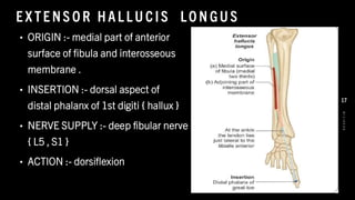
![F L E XO R H AL L U CIS LO N GU S
• ORIGIN :- Inferior 2/3 of
posterior surface of fibula and
interosseous membrane .
• INSERTION :- base of distal phalanx of
digit 1 [ hallux ]
• NERVE SUPPLY :- Tibial nerve [ L4,
L5 ,S1,S2 & S3 ]
• ACTION :- Plantar flexion
8
/
1
/
2
0
2
4
22](https://image.slidesharecdn.com/anklejoint21-250204080450-e548fec2/85/ANKLE-JOINT-2-1-pdf-ppt-physiotherapist-22-320.jpg)
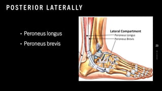
![PE RO N E US [ F IBU L A R IS ] LO N GU S
• ORGIN :- Head and superior 2/3 of
lateral surface of tibia .
• INSERTION :- Base of 1st
metatarsal and medial cuneiform .
• NERVE SUPPLY :- peroneal
nerve [ L5 , S1 ]
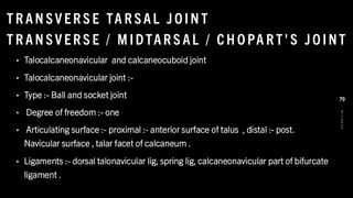
• ACTION : - Plantar flexion
8
/
1
/
2
0
2
4
24](https://image.slidesharecdn.com/anklejoint21-250204080450-e548fec2/85/ANKLE-JOINT-2-1-pdf-ppt-physiotherapist-24-320.jpg)
![PE RO N E US [ F IB UL A R IS ] B R E V IS
• ORIGIN :- inferior 2/3 of lateral surface of
fibula
• INSERTION :- dorsal surface of
tuberosity on lateral side of base of
5th metatarsal .
• NERVE SUPPLY :- superficial peroneal
nerve
• ACTION :- plantar flexion of ankle
8
/
1
/
2
0
2
4
25](https://image.slidesharecdn.com/anklejoint21-250204080450-e548fec2/85/ANKLE-JOINT-2-1-pdf-ppt-physiotherapist-25-320.jpg)


![TA L AR T ILT TE ST [ KL E IG E R ]
• Patient :- sits with leg hanging freely over
exam table .
• Examiner :- stabilize the leg medially with
one hand and cup the hind foot with other
hand . Inversion force applied to the hind foot
• Positive :-- tilt occurs at tibiotalar joint .
• Consistent with :- sprain or tear of the
anterior talofibular ligament
and calcaneofibular ligament
8
/
1
/
2
0
2
4
45](https://image.slidesharecdn.com/anklejoint21-250204080450-e548fec2/85/ANKLE-JOINT-2-1-pdf-ppt-physiotherapist-45-320.jpg)

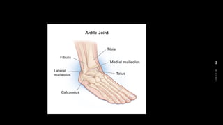
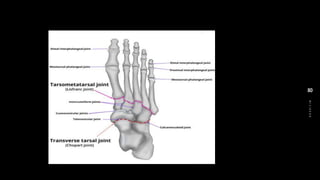
![PAR TS O F F O OT
• Hindfoot [ rear foot ] [ talus , calcaneus }
• Midfoot [ mid tarsal joint ] [ cuboid , navicular
, three cuneiform bone ]
• Forefoot [metatarsals , phalanges ]
8
/
1
/
2
0
2
4
49](https://image.slidesharecdn.com/anklejoint21-250204080450-e548fec2/85/ANKLE-JOINT-2-1-pdf-ppt-physiotherapist-49-320.jpg)
![F L E X IO N AN D E X TE NS IO N O F M TP J O INT
• Lumbricals
• Flexor hallucis brevis
• Extensor
digitorum longus { digits 2
–5 ]
• Extensor digitorum brevis
• Extensor hallucis longus {
digit 1 }
8
/
1
/
2
0
2
4
50](https://image.slidesharecdn.com/anklejoint21-250204080450-e548fec2/85/ANKLE-JOINT-2-1-pdf-ppt-physiotherapist-50-320.jpg)
![F L E X IO N AN D E X TE NS IO N O F PIP JO INT
• Flexor digitorum longus [ digits
2- 5 ]
• Flexor digitorum brevis [digits
2 –5 ]
• Flexor hallucis longus [ digit 1]
Extensor digitorum longus [
digits 2- 5 ]
Extensor digitorum brevis
Extensor hallucis longus [
digit 1]
8
/
1
/
2
0
2
4
51](https://image.slidesharecdn.com/anklejoint21-250204080450-e548fec2/85/ANKLE-JOINT-2-1-pdf-ppt-physiotherapist-51-320.jpg)
![F L E X IO N AN D E X TE NS IO N O F DIP JO IN T
• Flexor digitorum longus {
digits 2 – 5}
• Extensor digitorum longus
{ digits 2 –5 ]
• Extensor digitorum brevis
8
/
1
/
2
0
2
4
52](https://image.slidesharecdn.com/anklejoint21-250204080450-e548fec2/85/ANKLE-JOINT-2-1-pdf-ppt-physiotherapist-52-320.jpg)
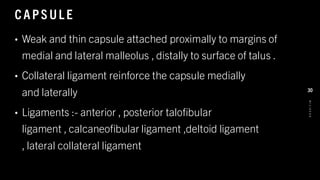
![STAB IL IT Y
• Joint design :- excessive mobility is prevented
due to alternating concave - convex facet .
• Capsule :- a loose capsule enclosing the entire joint ,
which thickness to form medial , lateral and
posterior talocalcaneal ligament that stabilize the capsule .
• Ligaments :- talocalcaneal [ all three
parts] talocalcaneal interosseous , cervical ligament .
8
/
1
/
2
0
2
4
68](https://image.slidesharecdn.com/anklejoint21-250204080450-e548fec2/85/ANKLE-JOINT-2-1-pdf-ppt-physiotherapist-68-320.jpg)
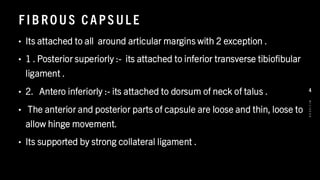
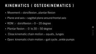
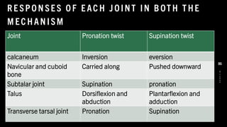
![KIN EM ATIC S [ OST EO KIN EM ATIC S ]
• Movement available :- supination :- inversion + adduction + plantarflexion
• Pronation :- eversion + abduction + dorsiflexion
• Plane and axis :- Triplanar movements occurring around a unique axis which is
obliquely oriented { 42 degree from transverse plane , 16 degree from sagittal plane }
• Inversion :- sagittal axis , frontal plane , ROM { 0 -- 30 degree }
• Eversion :- sagittal axis , frontal plane , ROM { 0 –10 degree }
• Abduction :- vertical axis , transverse plane , ROM { 0 –30 degree }
• Adduction :- vertical axis , frontal plane , ROM { 0 –1 0 degree }
8
/
1
/
2
0
2
4
70](https://image.slidesharecdn.com/anklejoint21-250204080450-e548fec2/85/ANKLE-JOINT-2-1-pdf-ppt-physiotherapist-70-320.jpg)
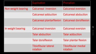

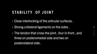
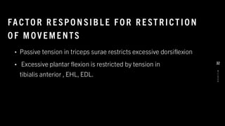
![SU PINAT IO N A ND P RO NAT IO N O F F O OT
• These are really component of the movement of inversion and
eversion .
• In pronation and supination of fore foot [ tarsus and
metatarsus ] moves on the calcaneum and talus .
• The medial border or fore foot elevated in supination [ part of
inversion ] and lateral border elevated in pronation .
8
/
1
/
2
0
2
4
75](https://image.slidesharecdn.com/anklejoint21-250204080450-e548fec2/85/ANKLE-JOINT-2-1-pdf-ppt-physiotherapist-75-320.jpg)


![ankle and foot biomechanics[1].pptx............................](https://cdn.slidesharecdn.com/ss_thumbnails/null1-260117154635-fe1ee6a7-thumbnail.jpg?width=640&height=640&fit=bounds)


![flat foot.ppt [pes planus ] #physio.# rehabilitation](https://cdn.slidesharecdn.com/ss_thumbnails/flatfoot-240310065759-517e9bef-thumbnail.jpg?width=640&height=640&fit=bounds)


