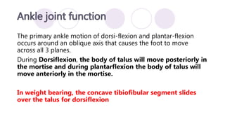
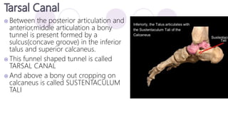
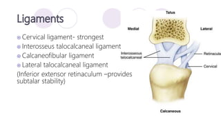
Vaibhavi Rathod
 Stability
• Provide stable base of support in variety of weight bearing
postures
• Acts as a rigid lever for effective push off during gait
 Mobility
• Absorbs stress & shock – pliability of foot
• Propulsion of body in walking
 Prehensile
 26 bones & 2 sesamoid bones;
divided into 3 functional segments –
 Forefoot – (Anterior)
• Metatarsals 5 ,Phalanges 14
 Midfoot – (Middle)
• Navicular, Cuboid, Cuneiform 3
 Hindfoot – (Posterior)
• Talus ,Calcaneus
25 component joints
 Proximal & distal tibiofibular joints
 Ankle (Talo-crural ) joint
 Talo-calcaneal (subtalar) joint
 Talo-navicular joint
 Calcaneo-cuboidal joint
 5 tarso-metatarsal joints
 5 metatarso-phalangeal joints
 9 interphalangeal joints
The term ankle specifically refers to :
• Talocrural joint-The formation of the mortise (a hole) by the
medial malleoli (Tibia) and lateral malleoli (fibula) with the talus
lying in between them makes up the talocrural joint.
• The ankle is a synovial Hinge joint with joint capsule and
associated ligaments.
• It is generally considered to have a single oblique axis with 1°of
freedom.
PROXIMAL ARTICULAR STRUCTURE
• The proximal segment of ankle is composed of concave surface
of distal tibia and of tibial and fibular malleoli.
• The structure of distal tibia and the two malleoli is referred to as
a MORITSE.
• The mortise of the ankle is adjustable, relying on the proximal
and distal tibio-fibular joints.
TALUS
3 Articular Surfaces
• Larger lateral facet-triangular shaped
• Smaller medial facet-comma shaped
• Superior facet- TROCHLEAR
It is a plane synovial joint formed by articulation of head of fibula
with the postero- lateral aspect of tibia.
•It is between the convex tibial facet and concave fibular facet
• Each proximal tibiofibular joint is surrounded by a joint capsule
that is reinforced by anterior and posterior tibiofibular ligaments.
It is a syndesmosis or fibrous union.
• It is in between the concave facet of tibia and convex facet of
fibula.
• Tibia and fibula do not come into contact with each other at this
point but are separated by fibro-adipose tissue.
• The ligaments of distal tibio-fibular joint are primarily
responsible for maintaining a stable mortise.
NO JOINT CAPSULE
Medial aspect
 Deltoid ligament complex
 Anterior tibiotalar ligament
 Posterior tibiotalar ligament
 Tibiocalcaneal ligament
 Tibionavicular ligament
Lateral aspect
 Anterior talofibular ligament
 Posterior talofibular ligament
 Calcaneofibular ligament
(supported by peroneal retinaculum)
Medial collateral ligaments
Lateral collateral ligaments
The primary ankle motion of dorsi-flexion and plantar-flexion
occurs around an oblique axis that causes the foot to move
across all 3 planes.
During Dorsiflexion, the body of talus will move posteriorly in
the mortise and during plantarflexion the body of talus will
move anteriorly in the mortise.
In weight bearing, the concave tibiofibular segment slides
over the talus for dorsiflexion
Dorsiflexors
 Tibialis anterior
 Extensor digitorum longus
 Extensor hallucis longus
 Peroneus tertius
Plantarflexors
 Gastrocnemius
 Soleus
The talocalcaneal or subtalar joint is a composite joint formed by
three separate plane articulations between the talus superiorly
and calcaneus inferiorly.
Provides a triplanar movement around a single joint axis.
Posterior Articulation-
 Largest concave facet-undersurface of body of talus
 convex facet- calcaneus
Anterior and medial articulation – Reverse of above
 Between the posterior articulation and
anterior,middle articulation a bony
tunnel is present formed by a
sulcus(concave groove) in the inferior
talus and superior calcaneus.
 This funnel shaped tunnel is called
TARSAL CANAL
 And above a bony out cropping on
calcaneus is called SUSTENTACULUM
TALI
 Cervical ligament- strongest
 Interosseus talocalcaneal ligament
 Calcaneofibular ligament
 Lateral talocalcaneal ligament
(Inferior extensor retinaculum –provides
subtalar stability)
 Complex twisting or screw like motion
 Subtalar axis lies halfway between between longitudinal and
vertical axis
 Pronation and supination includes equal magnitudes of
eversion/ inversion and abduction/ adduction.
NON WEIGHT BEARING WEIGHT BEARING
SUPINATION Calcaneal inversion
Calcaneal adduction
Calcaneal plantarflexion
Calcaneal inversion
Talar abduction
Talar dorsiflexion
Tibiofibular lateral rotation
PRONATION Calcaneal eversion
Calcaneal abduction
Calcaneal dorsiflexion
Calcaneal eversion
Talar adduction
Talar plantarflexion
Tibiofibular medial rotation
 The transverse tarsal joint, also called the midtarsal or Chopart
joint
 It is a compound joint formed by the
1.Talonavicular joint
2.calcaneocuboid joints .
 The two joints together present an S- shaped joint line that
transects the foot horizontally, dividing the hindfoot from the
midfoot and forefoot.
 The navicular and the cuboid bones are considered, immobile in
the weight-bearing foot.
 The proximal portion of the talonavicular articulation is formed
by the anterior portion of the head of the talus, and the
distal portion of the articulation, by the concave posterior
aspect of the navicular bone.
Ligaments
• Inferior aspect of the joint capsule- plantar
calcaneo-navicular ligament/spring ligament
• Medially- deltoid ligament
• Laterally-bifurcate ligament
 It is formed proximally by the anterior calcaneus and distally
by the posterior cuboid bone
Ligaments
 Laterally - lateral band of the bifurcate ligament (also known as
the calcaneocuboid ligament)
 Dorsally –dorsal calcaneocuboid ligament,
 Inferiorly -plantar calcaneocuboid (short plantar) and the long
plantar ligaments
 Any weight-bearing subtalar motion includes talar
abduction/adduction- dorsiflexion/plantarflexion that also
causes motion at the talonavicular joint
 Calcaneal inversion/eversion that causes motion at the
calcaneocuboid joint.
 As the subtalar joint supinates, its linkage to the transverse
tarsal joint causes both the talonavicular joint and the
calcaneocuboid joint to begin to supinate also.(CLOSE PACKED
POSITION)
 When the subtalar joint is pronated and loose-packed, the
transverse tarsal joint is also mobile and LOOSE PACKED
 The transverse tarsal joint is the transitional link between the
hindfoot and the forefoot, serving to (1)add to the
supination/pronation range of the subtalar joint and (2)
compensate the forefoot for hindfoot position.
Weight-Bearing Hindfoot Pronation and Transverse Tarsal Joint
Motion
In the weight-bearing position, medial rotation of the tibia for
example-pivoting on a fixed foot • Weight-Bearing Hindfoot
Supination and Transverse Tarsal Joint Motion • A lateral
force on the leg will create • subtalar supination in the weight-
bearing subtalar joint with a relative pronation of the transverse
tarsal joint (opposite motion of the forefoot segment) to
appropriate weight-bearing on a level surface Supination of the
subtalar joint, however, can proceed to only a certain point
the transverse tarsal joint also begins to supinate.
 The Tarsometatarsal TMT joints are plane synovial joints
formed by the distal row of tarsal bones (posteriorly) and the
bases of the metatarsals.
 LIGAMENT Deep transverse metatarsal ligament • This spans
the heads of the metatarsals on the plantar surface and is
similar to that found in the hand. • Contribute to stability of
proximal located TMT joints by preventing excessive motion
and splaying of metatarsal heads.
 FUNCTION • In
weightbearing,the TMT joints
function primarily to augment
the function of the transverse
tarsal joint; that is, the TMT
joints attempt to regulate
position of the metatarsals and
phalanges (the forefoot) in
relation to the weight-bearing
surface.
 When the hind foot pronates substantially in wt-bearing
position TTJ Joint will supinate to counter rotate the forefoot
to keep the plantar aspect of the foot in contact with the
ground.
 TMT –medial forefoot will press the ground lateral foot will lift
off the ground 1st and 2nd ray -dorsiflexion ,4th and 5th ray-
plantarflexion
 The entire forefoot undergoes an inversion rotation around a
hypothetical axis at the second ray.
When the hind foot and TTJ are locked in supination ,the
adjustment of forefoot position will be left entirely to TMT Joint
hence it will pronate.
 TMT-forefoot medial –lift off the ground
lateral-press to the ground 1st and 2nd ray-plantarflex 4th
and 5th –dorsiflexion and eversion accompanies
 A ray is defined as a functional unit formed by a metatarsal and (for
the first through third rays) its associated cuneiform bone.
 The axis of the first ray is inclined in such as way that dorsiflexion of
the first ray also includes inversion and adduction, whereas
plantarflexion is accompanied by eversion and abduction.
 Movements of the fifth ray around its axis are more restricted and
occur with the opposite arrangement of components. Dorsiflexion is
accompanied by eversion and abduction, and plantarflexion is
accompanied by inversion and adduction.
 The axis for the third ray nearly coincides with a coronal axis; the
predominant motion is dorsiflexion/plantarflexion.
 Longitudinal arch is formed anteriorly by the metatarsal heads
and posteriorly by calcaneus
 Talus is the keystone of the arch
 Transverse arch is formed by the anterior tarsals and the
middle cuneiform
Medial longitudinal ligament
 Spring ligament
 Interosseous talocalcaneal lig
 Deltoid ligament
 Plantar aponeurosis
Lateral longitudinal ligament
 Long and short plantar ligament
ankleandfootcomplex-190730140126 (1).pdf

ankleandfootcomplex-190730140126 (1).pdf

  • 1.
  • 2.
     Stability • Providestable base of support in variety of weight bearing postures • Acts as a rigid lever for effective push off during gait  Mobility • Absorbs stress & shock – pliability of foot • Propulsion of body in walking  Prehensile
  • 3.
     26 bones& 2 sesamoid bones; divided into 3 functional segments –  Forefoot – (Anterior) • Metatarsals 5 ,Phalanges 14  Midfoot – (Middle) • Navicular, Cuboid, Cuneiform 3  Hindfoot – (Posterior) • Talus ,Calcaneus
  • 4.
    25 component joints Proximal & distal tibiofibular joints  Ankle (Talo-crural ) joint  Talo-calcaneal (subtalar) joint  Talo-navicular joint  Calcaneo-cuboidal joint  5 tarso-metatarsal joints  5 metatarso-phalangeal joints  9 interphalangeal joints
  • 5.
    The term anklespecifically refers to : • Talocrural joint-The formation of the mortise (a hole) by the medial malleoli (Tibia) and lateral malleoli (fibula) with the talus lying in between them makes up the talocrural joint. • The ankle is a synovial Hinge joint with joint capsule and associated ligaments. • It is generally considered to have a single oblique axis with 1°of freedom.
  • 6.
    PROXIMAL ARTICULAR STRUCTURE •The proximal segment of ankle is composed of concave surface of distal tibia and of tibial and fibular malleoli. • The structure of distal tibia and the two malleoli is referred to as a MORITSE. • The mortise of the ankle is adjustable, relying on the proximal and distal tibio-fibular joints.
  • 8.
    TALUS 3 Articular Surfaces •Larger lateral facet-triangular shaped • Smaller medial facet-comma shaped • Superior facet- TROCHLEAR
  • 9.
    It is aplane synovial joint formed by articulation of head of fibula with the postero- lateral aspect of tibia. •It is between the convex tibial facet and concave fibular facet • Each proximal tibiofibular joint is surrounded by a joint capsule that is reinforced by anterior and posterior tibiofibular ligaments.
  • 10.
    It is asyndesmosis or fibrous union. • It is in between the concave facet of tibia and convex facet of fibula. • Tibia and fibula do not come into contact with each other at this point but are separated by fibro-adipose tissue. • The ligaments of distal tibio-fibular joint are primarily responsible for maintaining a stable mortise. NO JOINT CAPSULE
  • 12.
    Medial aspect  Deltoidligament complex  Anterior tibiotalar ligament  Posterior tibiotalar ligament  Tibiocalcaneal ligament  Tibionavicular ligament Lateral aspect  Anterior talofibular ligament  Posterior talofibular ligament  Calcaneofibular ligament (supported by peroneal retinaculum)
  • 13.
  • 14.
    The primary anklemotion of dorsi-flexion and plantar-flexion occurs around an oblique axis that causes the foot to move across all 3 planes. During Dorsiflexion, the body of talus will move posteriorly in the mortise and during plantarflexion the body of talus will move anteriorly in the mortise. In weight bearing, the concave tibiofibular segment slides over the talus for dorsiflexion
  • 15.
    Dorsiflexors  Tibialis anterior Extensor digitorum longus  Extensor hallucis longus  Peroneus tertius Plantarflexors  Gastrocnemius  Soleus
  • 16.
    The talocalcaneal orsubtalar joint is a composite joint formed by three separate plane articulations between the talus superiorly and calcaneus inferiorly. Provides a triplanar movement around a single joint axis. Posterior Articulation-  Largest concave facet-undersurface of body of talus  convex facet- calcaneus Anterior and medial articulation – Reverse of above
  • 18.
     Between theposterior articulation and anterior,middle articulation a bony tunnel is present formed by a sulcus(concave groove) in the inferior talus and superior calcaneus.  This funnel shaped tunnel is called TARSAL CANAL  And above a bony out cropping on calcaneus is called SUSTENTACULUM TALI
  • 19.
     Cervical ligament-strongest  Interosseus talocalcaneal ligament  Calcaneofibular ligament  Lateral talocalcaneal ligament (Inferior extensor retinaculum –provides subtalar stability)
  • 20.
     Complex twistingor screw like motion  Subtalar axis lies halfway between between longitudinal and vertical axis  Pronation and supination includes equal magnitudes of eversion/ inversion and abduction/ adduction.
  • 21.
    NON WEIGHT BEARINGWEIGHT BEARING SUPINATION Calcaneal inversion Calcaneal adduction Calcaneal plantarflexion Calcaneal inversion Talar abduction Talar dorsiflexion Tibiofibular lateral rotation PRONATION Calcaneal eversion Calcaneal abduction Calcaneal dorsiflexion Calcaneal eversion Talar adduction Talar plantarflexion Tibiofibular medial rotation
  • 22.
     The transversetarsal joint, also called the midtarsal or Chopart joint  It is a compound joint formed by the 1.Talonavicular joint 2.calcaneocuboid joints .  The two joints together present an S- shaped joint line that transects the foot horizontally, dividing the hindfoot from the midfoot and forefoot.  The navicular and the cuboid bones are considered, immobile in the weight-bearing foot.
  • 24.
     The proximalportion of the talonavicular articulation is formed by the anterior portion of the head of the talus, and the distal portion of the articulation, by the concave posterior aspect of the navicular bone. Ligaments • Inferior aspect of the joint capsule- plantar calcaneo-navicular ligament/spring ligament • Medially- deltoid ligament • Laterally-bifurcate ligament
  • 25.
     It isformed proximally by the anterior calcaneus and distally by the posterior cuboid bone Ligaments  Laterally - lateral band of the bifurcate ligament (also known as the calcaneocuboid ligament)  Dorsally –dorsal calcaneocuboid ligament,  Inferiorly -plantar calcaneocuboid (short plantar) and the long plantar ligaments
  • 26.
     Any weight-bearingsubtalar motion includes talar abduction/adduction- dorsiflexion/plantarflexion that also causes motion at the talonavicular joint  Calcaneal inversion/eversion that causes motion at the calcaneocuboid joint.  As the subtalar joint supinates, its linkage to the transverse tarsal joint causes both the talonavicular joint and the calcaneocuboid joint to begin to supinate also.(CLOSE PACKED POSITION)
  • 27.
     When thesubtalar joint is pronated and loose-packed, the transverse tarsal joint is also mobile and LOOSE PACKED  The transverse tarsal joint is the transitional link between the hindfoot and the forefoot, serving to (1)add to the supination/pronation range of the subtalar joint and (2) compensate the forefoot for hindfoot position.
  • 29.
    Weight-Bearing Hindfoot Pronationand Transverse Tarsal Joint Motion In the weight-bearing position, medial rotation of the tibia for example-pivoting on a fixed foot • Weight-Bearing Hindfoot Supination and Transverse Tarsal Joint Motion • A lateral force on the leg will create • subtalar supination in the weight- bearing subtalar joint with a relative pronation of the transverse tarsal joint (opposite motion of the forefoot segment) to appropriate weight-bearing on a level surface Supination of the subtalar joint, however, can proceed to only a certain point the transverse tarsal joint also begins to supinate.
  • 32.
     The TarsometatarsalTMT joints are plane synovial joints formed by the distal row of tarsal bones (posteriorly) and the bases of the metatarsals.  LIGAMENT Deep transverse metatarsal ligament • This spans the heads of the metatarsals on the plantar surface and is similar to that found in the hand. • Contribute to stability of proximal located TMT joints by preventing excessive motion and splaying of metatarsal heads.
  • 33.
     FUNCTION •In weightbearing,the TMT joints function primarily to augment the function of the transverse tarsal joint; that is, the TMT joints attempt to regulate position of the metatarsals and phalanges (the forefoot) in relation to the weight-bearing surface.
  • 34.
     When thehind foot pronates substantially in wt-bearing position TTJ Joint will supinate to counter rotate the forefoot to keep the plantar aspect of the foot in contact with the ground.  TMT –medial forefoot will press the ground lateral foot will lift off the ground 1st and 2nd ray -dorsiflexion ,4th and 5th ray- plantarflexion  The entire forefoot undergoes an inversion rotation around a hypothetical axis at the second ray.
  • 35.
    When the hindfoot and TTJ are locked in supination ,the adjustment of forefoot position will be left entirely to TMT Joint hence it will pronate.  TMT-forefoot medial –lift off the ground lateral-press to the ground 1st and 2nd ray-plantarflex 4th and 5th –dorsiflexion and eversion accompanies
  • 36.
     A rayis defined as a functional unit formed by a metatarsal and (for the first through third rays) its associated cuneiform bone.  The axis of the first ray is inclined in such as way that dorsiflexion of the first ray also includes inversion and adduction, whereas plantarflexion is accompanied by eversion and abduction.  Movements of the fifth ray around its axis are more restricted and occur with the opposite arrangement of components. Dorsiflexion is accompanied by eversion and abduction, and plantarflexion is accompanied by inversion and adduction.  The axis for the third ray nearly coincides with a coronal axis; the predominant motion is dorsiflexion/plantarflexion.
  • 38.
     Longitudinal archis formed anteriorly by the metatarsal heads and posteriorly by calcaneus  Talus is the keystone of the arch  Transverse arch is formed by the anterior tarsals and the middle cuneiform
  • 39.
    Medial longitudinal ligament Spring ligament  Interosseous talocalcaneal lig  Deltoid ligament  Plantar aponeurosis Lateral longitudinal ligament  Long and short plantar ligament