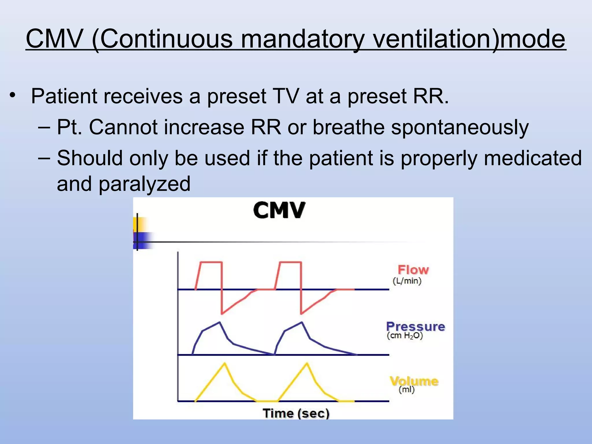
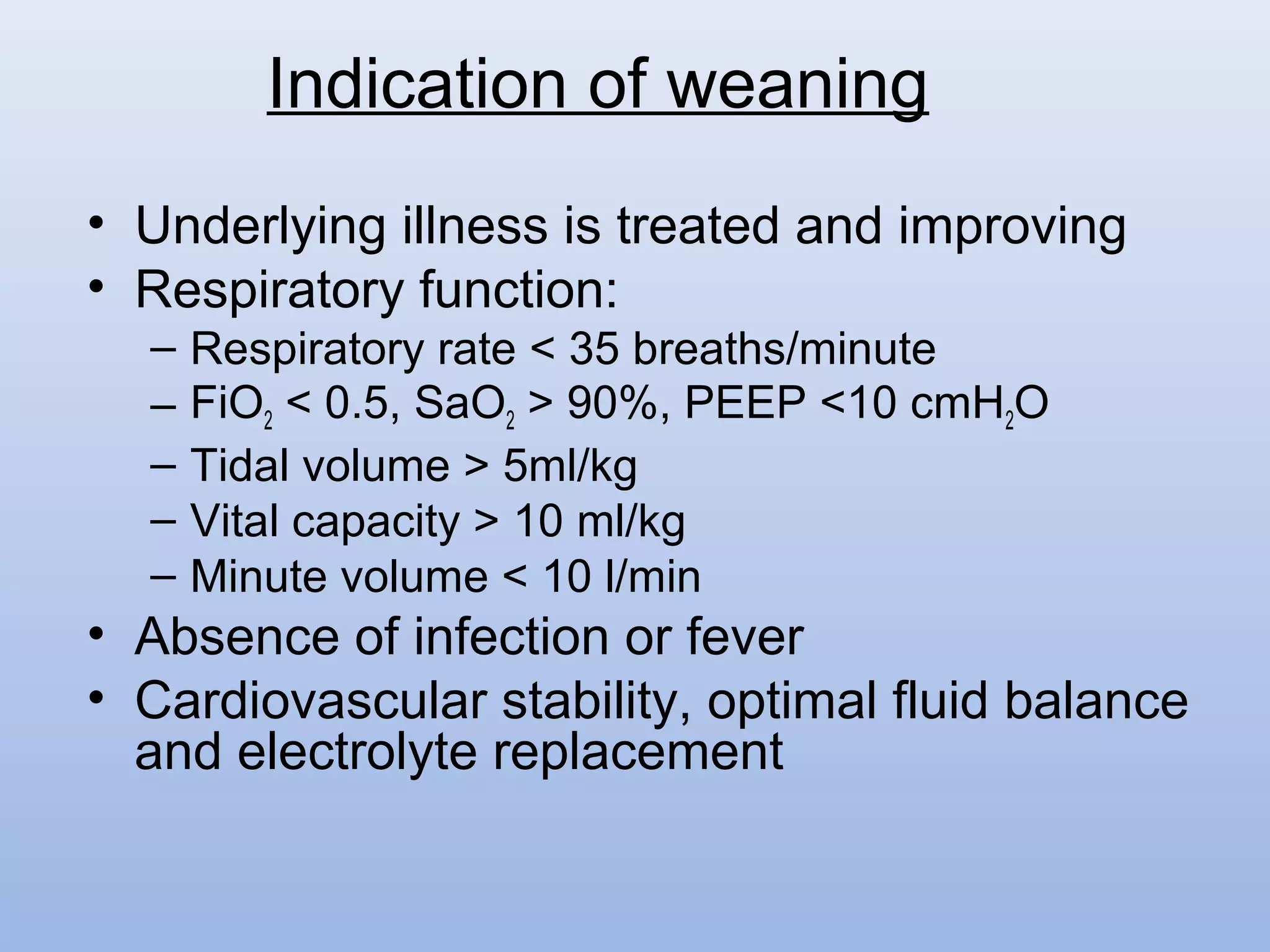
welcome
LATEST VENTILATOR MODES
INTRODUCTION
Patient from an initial event such as an accident
location all the way until he/she is released from
hospital, mechanical ventilation is necessary and
used in many areas of patient care. Even during
transportation, ventilation is provided using an
emergency ventilator. During the operation in the
hospital an anesthesia machine provides
ventilation. Intensive care ventilators are available
during the critical stay in intensive care. Even
during the subsequent treatment on intermediate
care wards, some patients require mechanical
breathing support.
HISTORY
Andreas Vesalius (1555) is credited with the first
description of positive-pressure ventilation, but it
took 400 years to apply his concept to patient care.
The occasion was the polio epidemic of 1955, when
the demand for assisted ventilation outgrew the
supply of negative-pressure tank ventilators (known
as iron lungs).
Invasive ventilation first used at Massachusetts
General Hospital in 1955.
• Criteria for starting
mechanical ventilation are
difficult to define and the
decision is often a clinical
one.
Indication for mechanical ventilation
• Respiratory rate >35 or <5 breaths/ minute
• Exhaustion, with laboured pattern of breathing
• Hypoxia - central cyanosis, SaO2 <90% on oxygen
or PaO2 < 60 mmHg (8kPa)
• Hypercarbia - PaCO2 > 60 mmHg (8kPa)
• Decreasing conscious level
• Significant chest trauma
• Tidal volume < 5ml/kg or Vital capacity <15ml/kg
Indication (Physical parameters)
• Control of intracranial pressure in head injury
• Airway protection following drug overdose
• Following cardiac arrest
• For recovery after prolonged major surgery or
trauma
Indication (non- respiratory condition)
• Non-invasive ventilation - eg. Iron lung(negative
pressure), Musk & adapter (positive pressure)
• Invasive ventilation- eg. IPPV
Type of ventilation
A. Trigger
B. Limit
C. Cycling
How a ventilator ventilates ?
A
B C
• Patient effort- pressure triggering
• After a predetermined time-time triggering
• Manually - manual/flow triggering
Start Inflation (Trigger)
Pressure Triggering
• Breath is delivered when ventilator senses patients
spontaneous inspiratory effort.
• sensitivity refers to the amount of negative pressure the
patient must generate to receive a breath/gas flow.
• If the sensitivity is set at 1 cm then the patient must
generate 1 cm H2O of negative pressure for the
machine to sense the patient's effort and deliver a
breath.
• Acceptable range -1 to -5 cm H2O below patient s
baseline pressure
Time triggering
Flow Triggering
• The flow triggered system has two preset variables for
triggering, the base flow and flow sensitivity.
• The base flow consists of fresh gas that flows
continuously through the circuit. The patient’s earliest
demand for flow is satisfied by the base flow.
• The flow sensitivity is computed as the difference
between the base flow and the exhaled flow
• Here delivered flow= base flow- returned flow
• Hence the flow sensitivity is the magnitude of the flow
diverted from the exhalation circuit into the patient’s
lungs. As the subject inhales and the set flow sensitivity
is reached the flow pressure control algorithm is
activated, the proportional valve opens, and fresh gas is
delivered.
• Pressure
• Volume
• Flow
• Time
Stop Inflation (Cycling)
• Terminates inspiratory phase at a preset
PIP. TV varies directly with lung
compliance and inversely with airway
resistance.
• Advantages: reduced barotrauma which
has been implicated secondary to high
PIP.
• Disadvantages: if lung compliance is
less, will lead to respiratory acidosis.
• Important to monitor patient’s expired
TV.
Pressure Cycled Ventilation
• Terminates inspiratory phase at a preset TV.
• Advantages: patient is guaranteed to receive a preset
TV under normal operating conditions.
• Disadvantages: PIP may rise high enough to cause
barotrauma.
Volume Cycled Ventilation
• Terminates the inspiratory phase when inspiratory
flow reaches a predetermined minimal level.
• Measured during spontaneous ventilation
• Mostly seen in pressure support modes of
ventilation.
Flow cycled ventilation
• Terminates the inspiratory phase when a preset
inspiratory time has been reached.
• Advantages: ease to regulate I:E ratio especially
when inverse ratio ventilation is desired.
• Disadvantages: delivered TV is dependent on
airway resistance and compliance characteristics.
Time Cycled Ventilation
Basic definitions
– Peak Inspiratory Pressure (PIP)
– Plateau pressures
– Positive End Expiratory Pressure (PEEP)
– Continuous Positive Airway Pressure (CPAP)
• Peak Inspiratory Pressure (PIP)-
The peak pressure is the maximum pressure
obtainable during active gas delivery. This pressure
a function of the compliance of the lung and thorax
and the airway resistance including the contribution
made by the tracheal tube and the ventilator circuit.
– Maintained at <45cm H2O to minimize barotrauma
• Plateau Pressure-
The plateau pressure is defined as the end inspiratory
pressure during a period of no gas flow. The plateau
pressure reflects lung and chest wall compliance.
• Mean Airway Pressure-
The mean airway pressure is an average of the
system pressure over the entire ventilatory period.
• End Expiratory Pressure-
End expiratory pressure is the airway pressure at the
termination of the expiratory phase and is normally
equal to atmospheric or the applied PEEP level.
PEEP
• Positive end expiratory pressure (PEEP) refers to the
application of a fixed amount of positive pressure
applied during mechanical ventilation cycle
• Continuous positive airway pressure (CPAP) refers to
the addition of a fixed amount of positive airway
pressure to spontaneous respirations, in the presence
or absence of an endotracheal tube.
• PEEP and CPAP are not separate modes of ventilation
as they do not provide ventilation. Rather they are
used together with other modes of ventilation or during
spontaneous breathing to improve oxygenation, recruit
alveoli, and / or decrease the work of breathing
Advantages
• Ability to increase functional residual capacity (FRC)
and keep FRC above Closing Capacity.
• The increase in FRC is accomplished by increasing
alveolar volume and through the recruitment of
alveoli that would not otherwise contribute to gas
exchange. Thus increasing oxygenation and lung
compliance
• The potential ability of PEEP and CPAP to open
closed lung units increases lung compliance and
tends to make regional impedances to ventilation
more homogenous.
Physiology of PEEP
• Reinflates collapsed alveoli and maintains alveolar
inflation during exhalation
PEEP
Decreases alveolar distending pressure
Increases FRC by alveolar recruitment
Improves ventilation
Increases V/Q, improves oxygenation, decreases work of
breathing
Dangers of PEEP
• High intra-thoracic pressures can cause decreased
venous return and decreased cardiac output
• May produce pulmonary barotrauma
• May worsen air-trapping in obstructive pulmonary
disease
• Increases intracranial pressure
• Alterations of renal functions and water metabolism
AutoPEEP
• During expiration alveolar pressure is greater than
circuit pressure until expiratory flow ceases. If
expiratory flow does not cease prior to the initiation
of the next breath gas trapping may occur. Gas
trapping increases the pressure in the alveoli at the
end of expiration and has been termed:
– dynamic hyperinflation;
– autoPEEP;
– inadvertent PEEP;
– intrinsic PEEP; and
– occult PEEP
Effects of autoPEEP can predispose the patient to:
• Increased risk of barotrauma
• Fall in cardiac output
• Hypotension
• Fluid retention
• Increased work of breathing
Modes of Ventilation
• Controlled
– Pressure Control (PC)
– Volume Control (VC)
• Supported
– Continuous Positive Airway Pressure (CPAP)
– Pressure Support (PS)
• Combined
– SIMV (PC) + PS
– SIMV (VC) + PS
• CMV
• SIMV
• Spont
• APRV
• ASV
• Duopap, BiPAP
• NIV
Modes of latest ventilation
• Patient receives a preset TV at a preset RR.
– Pt. Cannot increase RR or breathe spontaneously
– Should only be used if the patient is properly medicated
and paralyzed
CMV (Continuous mandatory ventilation)mode
• Indications:
- bucking during initial stages of vent support
- flail chests
- who otherwise need complete respiratory rest
• Complications: a disconnect will lead to apnea and
hypoxia.
• Disadvantages:
- Muscles of respiration weaken making weaning
more difficult.
CMV (Continuous mandatory ventilation)mode
- May lead to a rapid type of disuse atrophy involving
the diaphragmatic muscle fibers, which can develop
within the first day of mechanical ventilation.
-
May cause atrophy in all respiratory related muscles
during CMV
CMV (Continuous mandatory ventilation)mode
• Ventilator delivers a pre-set tidal volume at a pre-set
respiratory rate when there is not respiratory effort from
the patient.
• But if the patient triggers a spontaneous respiratory
effort earlier than the time interval created by the set
respiratory rate, the ventilator will still deliver the breath
at the set tidal volume and then resets the time interval
for the next breath.
• All breaths are delivered at the set tidal volume whether
it was ventilator triggered or patient triggered.
AC ( Assist control ventilation) mode
• Indications: Myasthenia gravis, GBS, post cardiac /
resp arrest, ARDS, pulmonary oedema.
• Advantages: minimal work of breathing and patient
controls RR which helps normalize PaCO2.
AC ( Assist control ventilation) mode
• Ventilator delivers either assisted breaths to the patient
at the beginning of a spontaneous breath or time
triggered mandatory breaths.
• Synchronization window- time interval just prior to time
triggering.
• Breath stacking is avoided as mandatory breaths are
synchronized with spontaneous breaths.
• In between mandatory breaths patient is allowed to
take spontaneous breath at any TV.
• It provides partial ventilatory support
SIMV(Synchronized Intermittent Mandatory
Ventilation )
SIMV(Synchronized Intermittent Mandatory
Ventilation )
• Advantages:
– Maintain respiratory muscle strength.
– Reduce ventilation to perfusion mismatch.
– Decreases mean airway pressure.
– Facilitates weaning.
SIMV(Synchronized Intermittent Mandatory
Ventilation )
• Supports spontaneous breathing of the patients.
• Each inspiratory effort is augmented by ventilator at
a preset level of inspiratory pressure.
• Patient triggered, flow cycled and pressure
controlled mode.
• Applies pressure plateau to patient airway during
spontaneous breathing.
• Commonly applied to SIMV mode during
spontaneous ventilation to facilitate weaning
PSV (Pressure Support Ventilation) mode
• Indications:
As an adjunct to SIMV. Not used during machine
breaths. This will increase pts. Spontaneous TV,
decrease spontaneous RR and decrease work of
breathing
• Disadvantages-
Not suitable for patients with central apnea.
(hypoventilation)
Development of high airway pressure. (hemodynamic
distubances)
Hypoventilation, if inspiratory time is short.
PSV (Pressure Support Ventilation) mode
• Similar to CPAP as patient breathes spontaneously.
• Airway pressure is maintained at moderately high level
(15-20 cmH2O) throughout most of respiratory cycle with
brief periods of lower pressure to allow deflation of
lungs.
• Increased pressure ensures alveolar recruitment &
oxygenation & brief deflation allows CO2 elimination
without alveolar collapse.
• Indicated as an alternative to conventional volume
cycled ventilation for patients with decreased lung
compliance (ARDS), as chances of barotrauma is less
due to less PAW.
APRV (Airway Pressure Release Ventilation) Mode
• a ventilator targeting scheme in which one variable is
automatically adjusted to achieve a predetermined value
of another variable.
• Patient body weight (deadspace) & percent minute
volume are feed in ventilator.
• Inspiratory pressure is automatically adjusted by the
ventilator to achieve a minute volume target.
• Automatically adjust the inspiratory flow to maintain a
constant I:E ratio.
• Using artificial intelligence application to conduct
ventilation
ASV (Adaptive Support Ventilation ) Mode
• Used in conjunction with other
modes Prevents closing of alveoli
by increasing baseline airway
pressure.
• Indications: intrapulmonary shunt
and refractory hypoxemia,
decrease FRC and lung
compliance.
• Complications: decrease venous
return, barotrauma, increased ICP
and alterations in renal functions
and water metabolism.
PEEP (Positive end expiratory Pressure)
CPAP and BiPAP
CPAP is essentially constant PEEP; BiPAP is CPAP plus PS
• Parameters
•CPAP – PEEP set at 5-10 cm H2O
•BiPAP – CPAP with Pressure Support (5-20 cm H2O)
•Shown to reduce need for intubation and mortality in COPD
pts
•Indications
•When medical therapy fails (tachypnea, hypoxemia,
respiratory acidosis)
•Use in conjunction with bronchodilators, steroids,
oral/parenteral steroids, antibiotics to prevent/delay intubation
•Weaning protocols
•Obstructive Sleep Apnea
- Decrease cardiac output
- Barotrauma
- Nosocomial pneumonia
- Positive water balance
- Decrease renal perfusion
- Increase ICP
- Hepatic congestion
- Worsening of intracardiac shunt
Complications
• Unsupported spontaneous breathing trials.
- The machine support is withdrawn
- T-Piece (or CPAP) circuit can be attached .
• Intermittent mandatory ventilation (IMV) weaning.
- The ventilator delivers a preset minimum minute volume
- Synchronized (SIMV) to the patient's own resp efforts.
• Pressure support weaning.
- Patient initiates all breaths and these are 'boosted' by the
ventilator.
- Gradually reducing the level of pressure support,
- Once the level of pressure support is low (5-10 cmH2O
above PEEP), a trial of T-Piece or CPAP weaning should
Modes of Weaning
• Underlying illness is treated and improving
• Respiratory function:
– Respiratory rate < 35 breaths/minute
– FiO2 < 0.5, SaO2 > 90%, PEEP <10 cmH2O
– Tidal volume > 5ml/kg
– Vital capacity > 10 ml/kg
– Minute volume < 10 l/min
• Absence of infection or fever
• Cardiovascular stability, optimal fluid balance
and electrolyte replacement
Indication of weaning
Conclusion
Ventilator mode
Ventilator mode

Ventilator mode

  • 1.
  • 2.
  • 3.
    INTRODUCTION Patient from aninitial event such as an accident location all the way until he/she is released from hospital, mechanical ventilation is necessary and used in many areas of patient care. Even during transportation, ventilation is provided using an emergency ventilator. During the operation in the hospital an anesthesia machine provides ventilation. Intensive care ventilators are available during the critical stay in intensive care. Even during the subsequent treatment on intermediate care wards, some patients require mechanical breathing support.
  • 4.
    HISTORY Andreas Vesalius (1555)is credited with the first description of positive-pressure ventilation, but it took 400 years to apply his concept to patient care. The occasion was the polio epidemic of 1955, when the demand for assisted ventilation outgrew the supply of negative-pressure tank ventilators (known as iron lungs). Invasive ventilation first used at Massachusetts General Hospital in 1955.
  • 5.
    • Criteria forstarting mechanical ventilation are difficult to define and the decision is often a clinical one. Indication for mechanical ventilation
  • 6.
    • Respiratory rate>35 or <5 breaths/ minute • Exhaustion, with laboured pattern of breathing • Hypoxia - central cyanosis, SaO2 <90% on oxygen or PaO2 < 60 mmHg (8kPa) • Hypercarbia - PaCO2 > 60 mmHg (8kPa) • Decreasing conscious level • Significant chest trauma • Tidal volume < 5ml/kg or Vital capacity <15ml/kg Indication (Physical parameters)
  • 7.
    • Control ofintracranial pressure in head injury • Airway protection following drug overdose • Following cardiac arrest • For recovery after prolonged major surgery or trauma Indication (non- respiratory condition)
  • 8.
    • Non-invasive ventilation- eg. Iron lung(negative pressure), Musk & adapter (positive pressure) • Invasive ventilation- eg. IPPV Type of ventilation
  • 9.
    A. Trigger B. Limit C.Cycling How a ventilator ventilates ? A B C
  • 10.
    • Patient effort-pressure triggering • After a predetermined time-time triggering • Manually - manual/flow triggering Start Inflation (Trigger)
  • 11.
    Pressure Triggering • Breathis delivered when ventilator senses patients spontaneous inspiratory effort. • sensitivity refers to the amount of negative pressure the patient must generate to receive a breath/gas flow. • If the sensitivity is set at 1 cm then the patient must generate 1 cm H2O of negative pressure for the machine to sense the patient's effort and deliver a breath. • Acceptable range -1 to -5 cm H2O below patient s baseline pressure
  • 12.
  • 13.
    Flow Triggering • Theflow triggered system has two preset variables for triggering, the base flow and flow sensitivity. • The base flow consists of fresh gas that flows continuously through the circuit. The patient’s earliest demand for flow is satisfied by the base flow. • The flow sensitivity is computed as the difference between the base flow and the exhaled flow • Here delivered flow= base flow- returned flow • Hence the flow sensitivity is the magnitude of the flow diverted from the exhalation circuit into the patient’s lungs. As the subject inhales and the set flow sensitivity is reached the flow pressure control algorithm is activated, the proportional valve opens, and fresh gas is delivered.
  • 14.
    • Pressure • Volume •Flow • Time Stop Inflation (Cycling)
  • 15.
    • Terminates inspiratoryphase at a preset PIP. TV varies directly with lung compliance and inversely with airway resistance. • Advantages: reduced barotrauma which has been implicated secondary to high PIP. • Disadvantages: if lung compliance is less, will lead to respiratory acidosis. • Important to monitor patient’s expired TV. Pressure Cycled Ventilation
  • 16.
    • Terminates inspiratoryphase at a preset TV. • Advantages: patient is guaranteed to receive a preset TV under normal operating conditions. • Disadvantages: PIP may rise high enough to cause barotrauma. Volume Cycled Ventilation
  • 17.
    • Terminates theinspiratory phase when inspiratory flow reaches a predetermined minimal level. • Measured during spontaneous ventilation • Mostly seen in pressure support modes of ventilation. Flow cycled ventilation
  • 18.
    • Terminates theinspiratory phase when a preset inspiratory time has been reached. • Advantages: ease to regulate I:E ratio especially when inverse ratio ventilation is desired. • Disadvantages: delivered TV is dependent on airway resistance and compliance characteristics. Time Cycled Ventilation
  • 19.
    Basic definitions – PeakInspiratory Pressure (PIP) – Plateau pressures – Positive End Expiratory Pressure (PEEP) – Continuous Positive Airway Pressure (CPAP)
  • 20.
    • Peak InspiratoryPressure (PIP)- The peak pressure is the maximum pressure obtainable during active gas delivery. This pressure a function of the compliance of the lung and thorax and the airway resistance including the contribution made by the tracheal tube and the ventilator circuit. – Maintained at <45cm H2O to minimize barotrauma • Plateau Pressure- The plateau pressure is defined as the end inspiratory pressure during a period of no gas flow. The plateau pressure reflects lung and chest wall compliance.
  • 22.
    • Mean AirwayPressure- The mean airway pressure is an average of the system pressure over the entire ventilatory period. • End Expiratory Pressure- End expiratory pressure is the airway pressure at the termination of the expiratory phase and is normally equal to atmospheric or the applied PEEP level.
  • 23.
    PEEP • Positive endexpiratory pressure (PEEP) refers to the application of a fixed amount of positive pressure applied during mechanical ventilation cycle • Continuous positive airway pressure (CPAP) refers to the addition of a fixed amount of positive airway pressure to spontaneous respirations, in the presence or absence of an endotracheal tube. • PEEP and CPAP are not separate modes of ventilation as they do not provide ventilation. Rather they are used together with other modes of ventilation or during spontaneous breathing to improve oxygenation, recruit alveoli, and / or decrease the work of breathing
  • 24.
    Advantages • Ability toincrease functional residual capacity (FRC) and keep FRC above Closing Capacity. • The increase in FRC is accomplished by increasing alveolar volume and through the recruitment of alveoli that would not otherwise contribute to gas exchange. Thus increasing oxygenation and lung compliance • The potential ability of PEEP and CPAP to open closed lung units increases lung compliance and tends to make regional impedances to ventilation more homogenous.
  • 25.
    Physiology of PEEP •Reinflates collapsed alveoli and maintains alveolar inflation during exhalation PEEP Decreases alveolar distending pressure Increases FRC by alveolar recruitment Improves ventilation Increases V/Q, improves oxygenation, decreases work of breathing
  • 26.
    Dangers of PEEP •High intra-thoracic pressures can cause decreased venous return and decreased cardiac output • May produce pulmonary barotrauma • May worsen air-trapping in obstructive pulmonary disease • Increases intracranial pressure • Alterations of renal functions and water metabolism
  • 27.
    AutoPEEP • During expirationalveolar pressure is greater than circuit pressure until expiratory flow ceases. If expiratory flow does not cease prior to the initiation of the next breath gas trapping may occur. Gas trapping increases the pressure in the alveoli at the end of expiration and has been termed: – dynamic hyperinflation; – autoPEEP; – inadvertent PEEP; – intrinsic PEEP; and – occult PEEP
  • 28.
    Effects of autoPEEPcan predispose the patient to: • Increased risk of barotrauma • Fall in cardiac output • Hypotension • Fluid retention • Increased work of breathing
  • 29.
    Modes of Ventilation •Controlled – Pressure Control (PC) – Volume Control (VC) • Supported – Continuous Positive Airway Pressure (CPAP) – Pressure Support (PS) • Combined – SIMV (PC) + PS – SIMV (VC) + PS
  • 30.
    • CMV • SIMV •Spont • APRV • ASV • Duopap, BiPAP • NIV Modes of latest ventilation
  • 31.
    • Patient receivesa preset TV at a preset RR. – Pt. Cannot increase RR or breathe spontaneously – Should only be used if the patient is properly medicated and paralyzed CMV (Continuous mandatory ventilation)mode
  • 32.
    • Indications: - buckingduring initial stages of vent support - flail chests - who otherwise need complete respiratory rest • Complications: a disconnect will lead to apnea and hypoxia. • Disadvantages: - Muscles of respiration weaken making weaning more difficult. CMV (Continuous mandatory ventilation)mode
  • 33.
    - May leadto a rapid type of disuse atrophy involving the diaphragmatic muscle fibers, which can develop within the first day of mechanical ventilation. - May cause atrophy in all respiratory related muscles during CMV CMV (Continuous mandatory ventilation)mode
  • 34.
    • Ventilator deliversa pre-set tidal volume at a pre-set respiratory rate when there is not respiratory effort from the patient. • But if the patient triggers a spontaneous respiratory effort earlier than the time interval created by the set respiratory rate, the ventilator will still deliver the breath at the set tidal volume and then resets the time interval for the next breath. • All breaths are delivered at the set tidal volume whether it was ventilator triggered or patient triggered. AC ( Assist control ventilation) mode
  • 35.
    • Indications: Myastheniagravis, GBS, post cardiac / resp arrest, ARDS, pulmonary oedema. • Advantages: minimal work of breathing and patient controls RR which helps normalize PaCO2. AC ( Assist control ventilation) mode
  • 36.
    • Ventilator deliverseither assisted breaths to the patient at the beginning of a spontaneous breath or time triggered mandatory breaths. • Synchronization window- time interval just prior to time triggering. • Breath stacking is avoided as mandatory breaths are synchronized with spontaneous breaths. • In between mandatory breaths patient is allowed to take spontaneous breath at any TV. • It provides partial ventilatory support SIMV(Synchronized Intermittent Mandatory Ventilation )
  • 37.
  • 38.
    • Advantages: – Maintainrespiratory muscle strength. – Reduce ventilation to perfusion mismatch. – Decreases mean airway pressure. – Facilitates weaning. SIMV(Synchronized Intermittent Mandatory Ventilation )
  • 39.
    • Supports spontaneousbreathing of the patients. • Each inspiratory effort is augmented by ventilator at a preset level of inspiratory pressure. • Patient triggered, flow cycled and pressure controlled mode. • Applies pressure plateau to patient airway during spontaneous breathing. • Commonly applied to SIMV mode during spontaneous ventilation to facilitate weaning PSV (Pressure Support Ventilation) mode
  • 40.
    PSV (Pressure SupportVentilation) mode
  • 41.
    • Indications: As anadjunct to SIMV. Not used during machine breaths. This will increase pts. Spontaneous TV, decrease spontaneous RR and decrease work of breathing • Disadvantages- Not suitable for patients with central apnea. (hypoventilation) Development of high airway pressure. (hemodynamic distubances) Hypoventilation, if inspiratory time is short. PSV (Pressure Support Ventilation) mode
  • 42.
    • Similar toCPAP as patient breathes spontaneously. • Airway pressure is maintained at moderately high level (15-20 cmH2O) throughout most of respiratory cycle with brief periods of lower pressure to allow deflation of lungs. • Increased pressure ensures alveolar recruitment & oxygenation & brief deflation allows CO2 elimination without alveolar collapse. • Indicated as an alternative to conventional volume cycled ventilation for patients with decreased lung compliance (ARDS), as chances of barotrauma is less due to less PAW. APRV (Airway Pressure Release Ventilation) Mode
  • 43.
    APRV (Airway PressureRelease Ventilation) Mode
  • 44.
    • a ventilatortargeting scheme in which one variable is automatically adjusted to achieve a predetermined value of another variable. • Patient body weight (deadspace) & percent minute volume are feed in ventilator. • Inspiratory pressure is automatically adjusted by the ventilator to achieve a minute volume target. • Automatically adjust the inspiratory flow to maintain a constant I:E ratio. • Using artificial intelligence application to conduct ventilation ASV (Adaptive Support Ventilation ) Mode
  • 45.
    • Used inconjunction with other modes Prevents closing of alveoli by increasing baseline airway pressure. • Indications: intrapulmonary shunt and refractory hypoxemia, decrease FRC and lung compliance. • Complications: decrease venous return, barotrauma, increased ICP and alterations in renal functions and water metabolism. PEEP (Positive end expiratory Pressure)
  • 46.
    CPAP and BiPAP CPAPis essentially constant PEEP; BiPAP is CPAP plus PS • Parameters •CPAP – PEEP set at 5-10 cm H2O •BiPAP – CPAP with Pressure Support (5-20 cm H2O) •Shown to reduce need for intubation and mortality in COPD pts •Indications •When medical therapy fails (tachypnea, hypoxemia, respiratory acidosis) •Use in conjunction with bronchodilators, steroids, oral/parenteral steroids, antibiotics to prevent/delay intubation •Weaning protocols •Obstructive Sleep Apnea
  • 47.
    - Decrease cardiacoutput - Barotrauma - Nosocomial pneumonia - Positive water balance - Decrease renal perfusion - Increase ICP - Hepatic congestion - Worsening of intracardiac shunt Complications
  • 48.
    • Unsupported spontaneousbreathing trials. - The machine support is withdrawn - T-Piece (or CPAP) circuit can be attached . • Intermittent mandatory ventilation (IMV) weaning. - The ventilator delivers a preset minimum minute volume - Synchronized (SIMV) to the patient's own resp efforts. • Pressure support weaning. - Patient initiates all breaths and these are 'boosted' by the ventilator. - Gradually reducing the level of pressure support, - Once the level of pressure support is low (5-10 cmH2O above PEEP), a trial of T-Piece or CPAP weaning should Modes of Weaning
  • 49.
    • Underlying illnessis treated and improving • Respiratory function: – Respiratory rate < 35 breaths/minute – FiO2 < 0.5, SaO2 > 90%, PEEP <10 cmH2O – Tidal volume > 5ml/kg – Vital capacity > 10 ml/kg – Minute volume < 10 l/min • Absence of infection or fever • Cardiovascular stability, optimal fluid balance and electrolyte replacement Indication of weaning
  • 50.