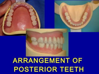
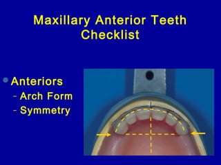
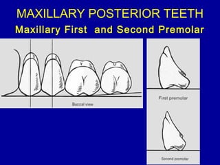
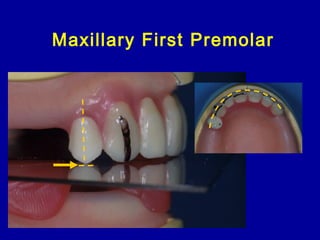
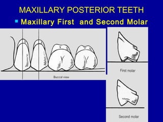
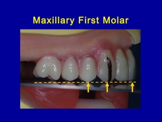
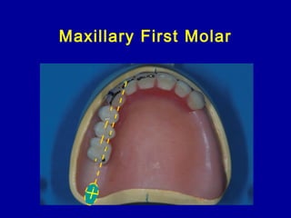
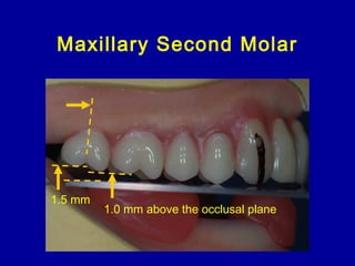
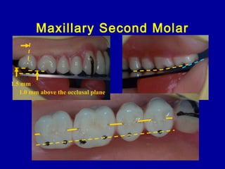
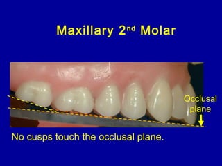
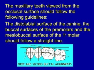
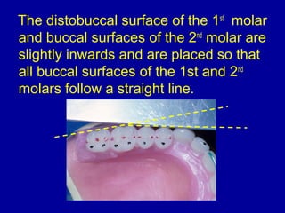
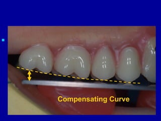
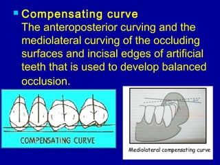
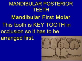
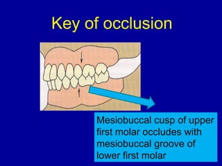
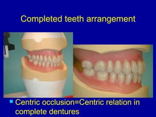
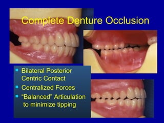
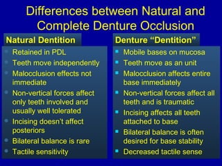
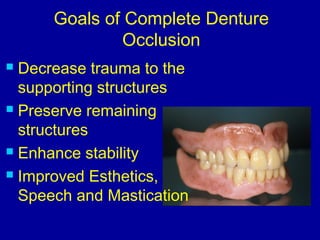
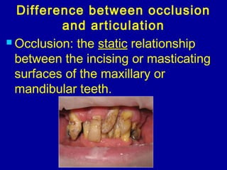
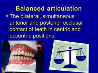
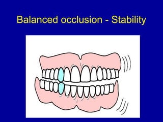
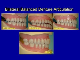
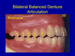
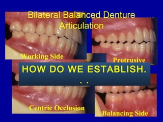
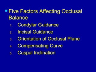
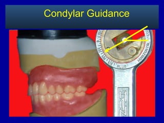
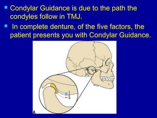
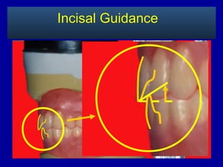
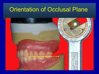
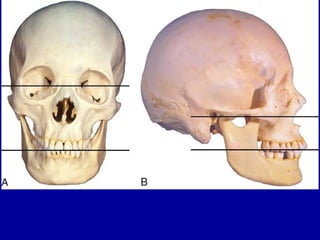
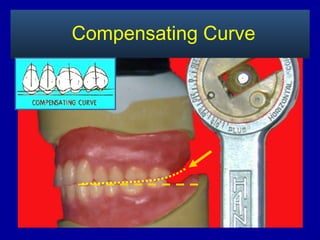
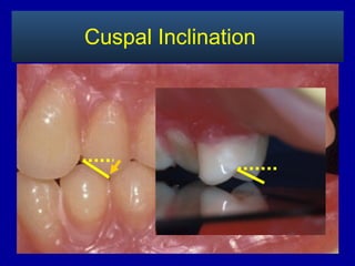
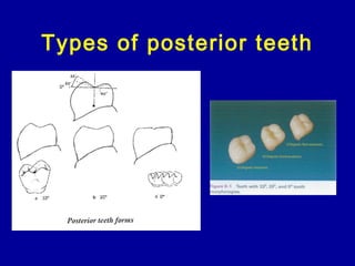
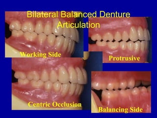
8. The long axis of the premolars should be
perpendicular to the occlusal plane and the
buccal and lingual cusp tips should touch the
occlusal plane.
Maxillary Premolar Teeth
Occlusal
plane
9. Both the buccal and lingual cusp tips
of the maxillary premolars should
contact the plane of occlusion.
The Maxillary Premolars
13. Maxillary 1st
Molar
The mesio-palatal cusp tip of the 1st
molar
contacts the occlusal plane but the buccal
cusp tips and the distal lingual cusp are
elevated about 0.5mm off the occlusal plane.
17. The maxillary teeth viewed from the
occlusal surface should follow the
following guidelines:
The distolabial surface of the canine, the
buccal surfaces of the premolars and the
mesiobuccal surface of the 1st
molar
should follow a straight line.
18. The distobuccal surface of the 1st
molar
and buccal surfaces of the 2nd
molar are
slightly inwards and are placed so that
all buccal surfaces of the 1st and 2nd
molars follow a straight line.
20. Compensating curve
The anteroposterior curving and the
mediolateral curving of the occluding
surfaces and incisal edges of artificial
teeth that is used to develop balanced
occlusion.
24. Mesiobuccal cusp of upper
first molar occludes with
mesiobuccal groove of
lower first molar
Key of occlusion
25. Setting the Mandibular Posterior Teeth
The Mandibular 1st Molar
Begin by positioning the mandibular 1st
molar. The
mesial buccal cusp tip should engage the embrasure
between the 1st
molar and 2nd
premolar.
26. Mandibular 2nd
Premolar
Position the mandibular 2nd
premolar. The
buccal cusp of the 2nd
premolar should
occlude with the adjacent marginal ridges
of the maxillary 1st
and 2nd
premolars.
27. The Mandibular 1st
Premolar
The buccal cusp of the mandibular 1st
premolar should engage the mesial
marginal ridge of the opposing 1st
premolar.
28. The Mandibular 2nd
Molar
The mandibular 2nd
molar is placed with
the mesiobuccal cusp should engage
between the maxillary 1st
and 2nd
molars
and contact with their marginal ridges.
29. Mandibular Posterior Teeth
The mandibular cusps tips are
designed to engage the embrasures of
the opposing maxillary teeth.
Completed set up
Centric
30. Additional Guidelines for Arranging the
Mandibular Posterior Denture Teeth
2/3 up the retromolar pad
31. Verify that the posterior
mandibular teeth are centered
over the crest of the ridge.
34. Mobile bases on mucosa
Teeth move as an unit
Malocclusion affects entire
base immediately
Non-vertical forces affect all
teeth and is traumatic
Incising affects all teeth
attached to base
Bilateral balance is often
desired for base stability
Decreased tactile sense
Retained in PDL
Teeth move independently
Malocclusion effects not
immediate
Non-vertical forces affect
only teeth involved and
usually well tolerated
Incising doesn’t affect
posteriors
Bilateral balance is rare
Tactile sensitivity
Natural Dentition Denture “Dentition”
Differences between Natural and
Complete Denture Occlusion
35. Goals of Complete Denture
Occlusion
Decrease trauma to the
supporting structures
Preserve remaining
structures
Enhance stability
Improved Esthetics,
Speech and Mastication
36. Difference between occlusion
and articulation
Occlusion: the static relationship
between the incising or masticating
surfaces of the maxillary or
mandibular teeth.
37. Articulation: the
static and
dynamic contact
relationship
between the
occlusal
surfaces of the
teeth during
function.
38. Balanced articulation
The bilateral, simultaneous
anterior and posterior occlusal
contact of teeth in centric and
eccentric positions.
46. Condylar Guidance is due to the path the
condyles follow in TMJ.
In complete denture, of the five factors, the
patient presents you with Condylar Guidance.