Downloaded 1,047 times
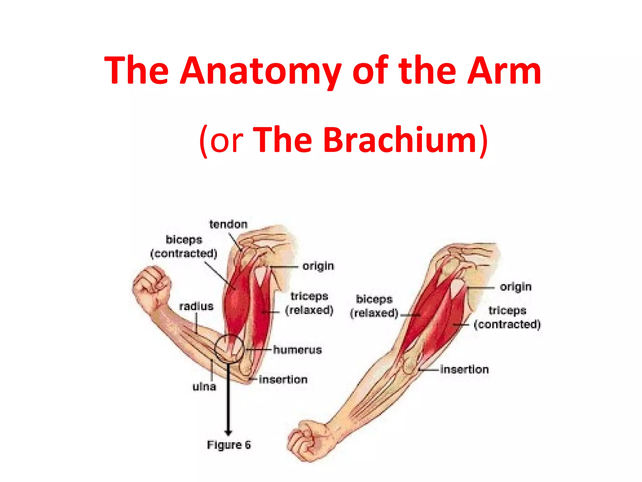


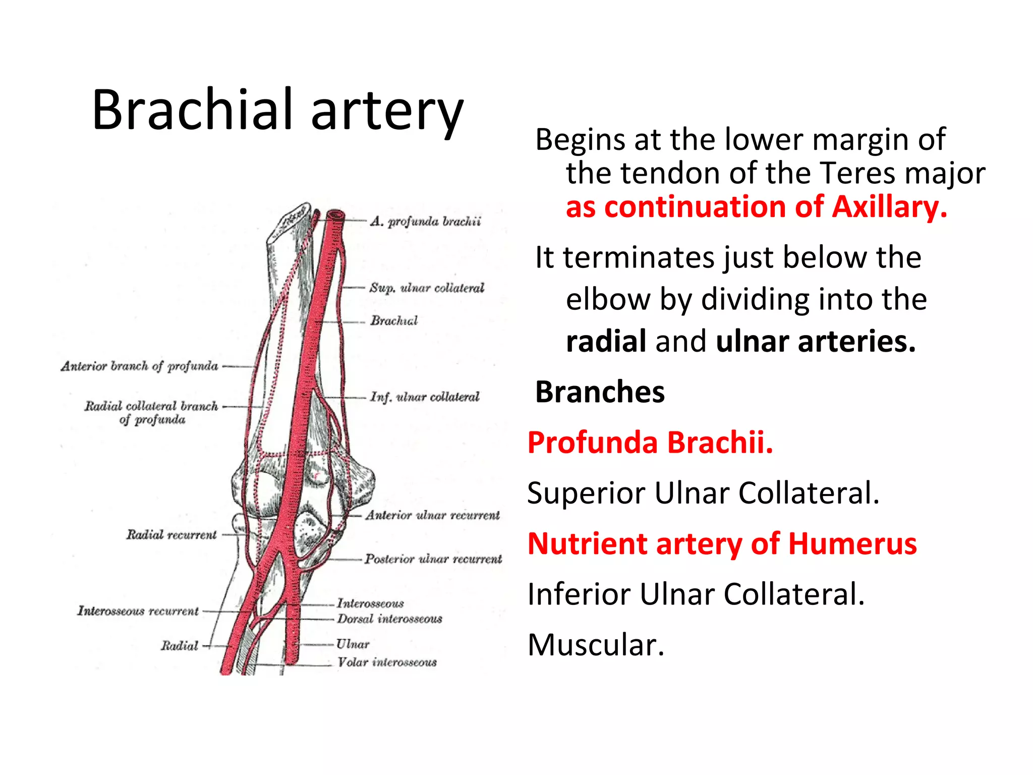
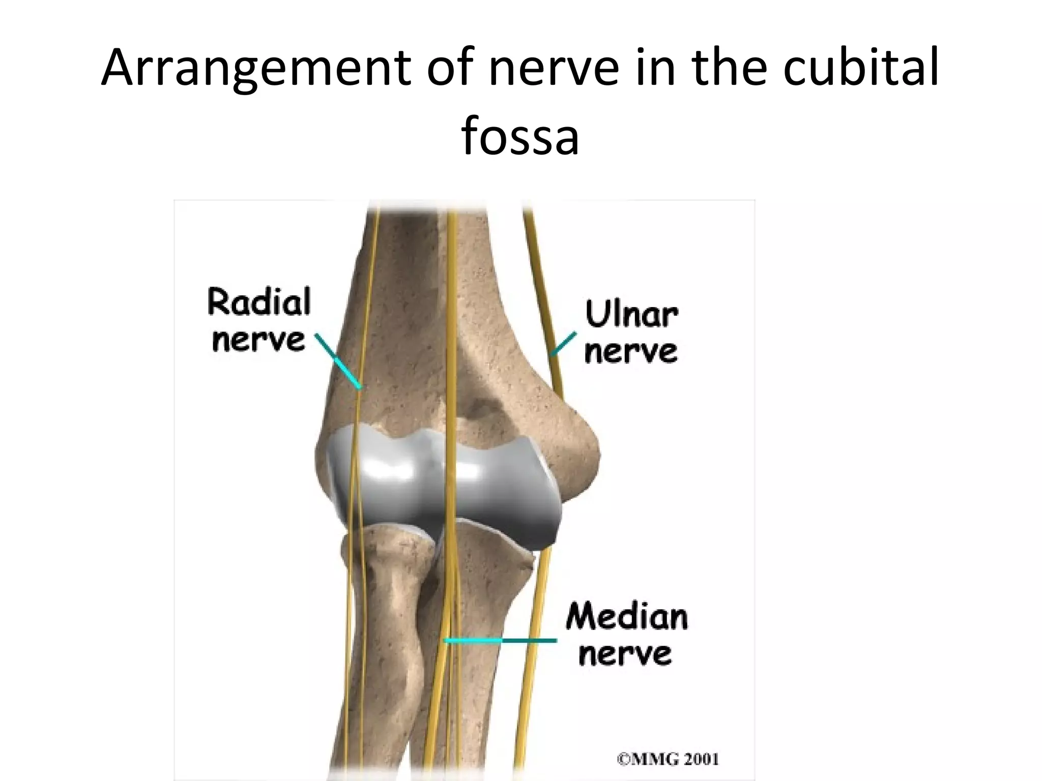


The document describes the anatomy of the arm, including: - The biceps brachii muscle, which flexes the forearm. - The brachial artery, which supplies blood to the arm and divides into the radial and ulnar arteries near the elbow. - The radial, median, and musculocutaneous nerves which innervate muscles of the arm. - Joints of the elbow and proximal radioulnar joint which allow flexion/extension and supination/pronation.


