Downloaded 366 times


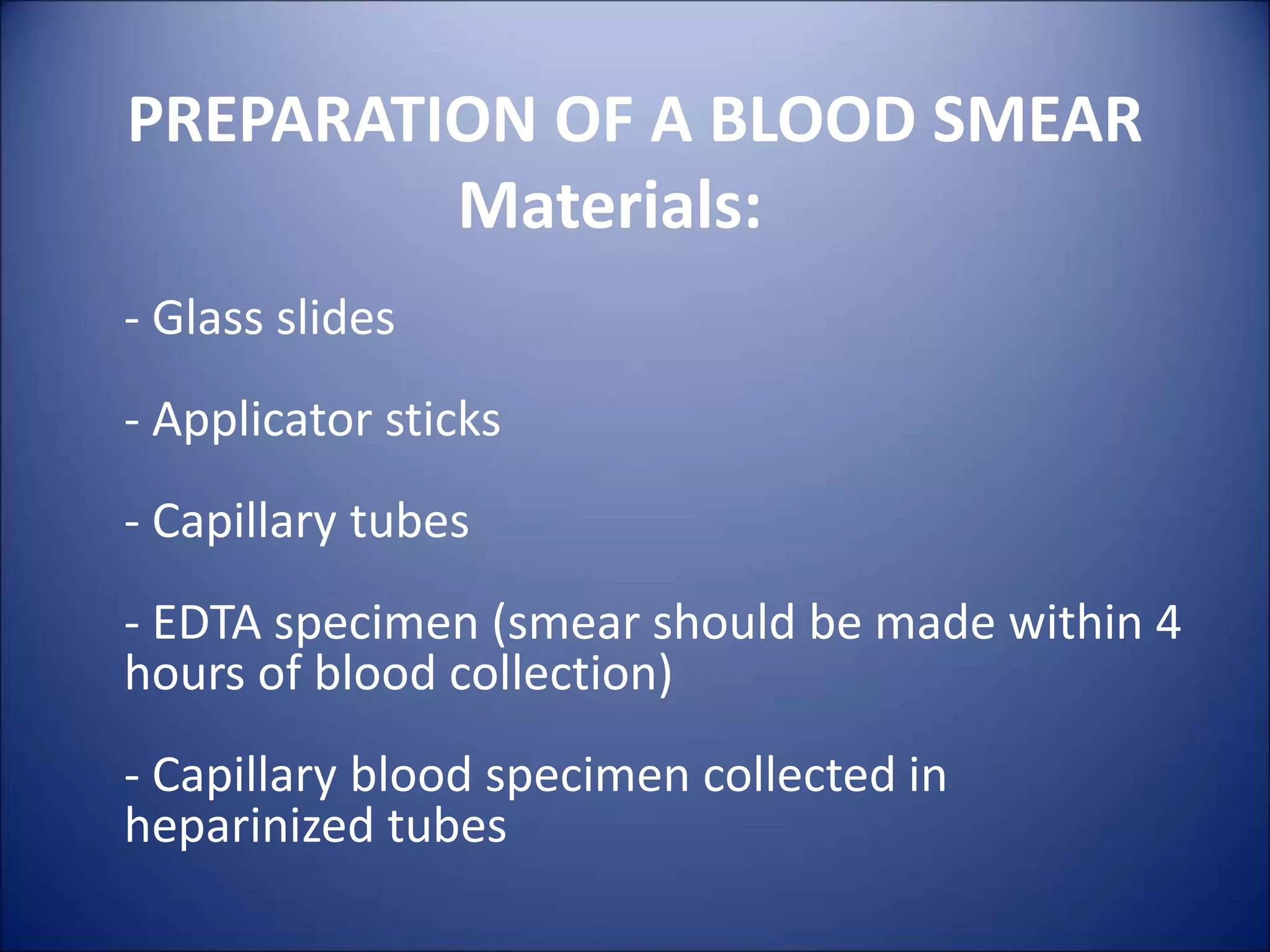


The document discusses the importance of blood smears in diagnosing various hematological disorders. It states that blood smears provide a rapid and inexpensive way to examine blood cell morphology and detect abnormalities. While automated analyzers are used more often, blood smears remain an important diagnostic tool, especially when clinical findings require further investigation or there are discrepancies with previous test results. The document also provides guidance on properly preparing blood smears to obtain diagnostic quality samples.


![Peripheral blood smear [autosaved]](https://cdn.slidesharecdn.com/ss_thumbnails/peripheralbloodsmearautosaved-201029200454-thumbnail.jpg?width=640&height=640&fit=bounds)