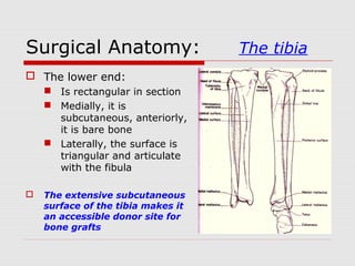
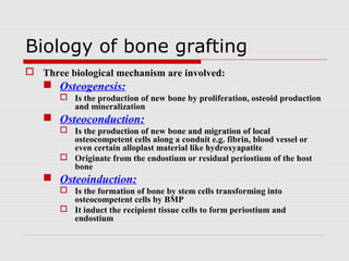
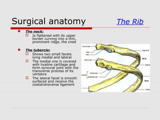
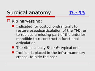
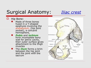
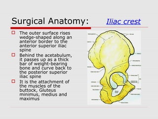
Bone Grafting and
Reconstruction
A.SELVA AROCKIAM
CRI
CSICDSR
Introduction
 Historical background:
 Surgeons have gained their experience
in reconstruction from the numerous
wars
 Civilian injuries produces the largest
number and the most extensive tissue
loss almost indistinguishable from ware
injuries
Introduction
 It started in WW I and concentrated around
reconstruction of the mandible but without
antibiotic support
 In WW II distant bone blocks were transplanted
from the ilium, rib and tibia with routine use of
antibiotic
 No cancellous cellular marrow
Introduction
 Mowlem in 1944, introduced the concept of
“Iliac cancellous bone chips” beginning the
evolution of predictable bony reconstruction of
the jaw bone
 Boyne brought about the “use of particulate
bone and cancellous marrow” with metallic
trays splinted to large acellular cortical bone
Biology of bone grafting
 Three biological mechanism are involved:
 Osteogenesis:
 Is the production of new bone by proliferation, osteoid production
and mineralization
 Osteoconduction:
 Is the production of new bone and migration of local
osteocompetent cells along a conduit e.g. fibrin, blood vessel or
even certain alloplast material like hydroxyapatite
 Originate from the endostium or residual periostium of the host
bone
 Osteoinduction:
 Is the formation of bone by stem cells transforming into
osteocompetent cells by BMP
 It induct the recipient tissue cells to form periostium and
endostium
The Rib
Surgical anatomy The Rib
 The first, eleventh and
twelfth ribs are atypical
 A typical rib has a head,
a neck and a shaft.
 The shaft slopes down
and laterally to an angle
and then curve forward
 The upper border is
blunt and lateral to the
angle the lower border
form a sharp ridge
sheltering a costal
groove
 This feature identify
right from left ribs
Surgical anatomy The Rib
 Typical rib:
 The head:
 Bevelled by two articular
facets that slope away
from a dividing ridge.
 The lower one is vertical
and articulate with the
upper border of its own
vertebra
 The upper facets faces
up and articulate with
the lower border of the
vertebra above
 Each form a synovial
joint separated by a
ligament attached to the
ridge
Surgical anatomy The Rib
 The neck:
 Is flattened with its upper
border curving into a thin,
prominent ridge, the crest
 The tubercle:
 Shows two small facets
lying medial and lateral
 The medial one is covered
with hyaline cartilage and
form synovial joint with the
transverse process of its
vertebra
 The lateral facet is smooth
surfaced and receive the
costotransverse ligament
Surgical anatomy The Rib
 Costal cartilages:
 They form a primary
cartilaginous joints at the
extremities of all twelve
ribs
 The first is short and
articulate with the
manubrium and the
clavicle
 They increase in length
below and the seventh has
the longest.
 They are bend from a
downward slope with the
rib to upward slope toward
the sternum
Surgical anatomy The Rib
 Rib harvesting:
 Indicated for costochondral graft to
restore pseudoarticulation of the TMJ, or
to replace a missing part of the anterior
mandible to reconstruct a functional
articulation
 The rib is usually 5th
or 6th
typical one
 Incision is placed in the infra-mammary
crease, to hide the scar
Surgical anatomy The Rib
 Right rib is always preferred because:
 It could be contoured to fit either side of
the mandible or facial bones
 Postoperative pain is less likely to be
confused with cardiogenic pain
 The 6th
rib is where the distal origin of the
pectoralis major muscle, dissection transect the
muscle minimally
 Sharp dissection is carried through full thickness
of skin, subcutaneous tissues and the muscle, to
expose the rib periostium, the chest wall cortex
Surgical anatomy The Rib
 The periostium is incised from 1 cm onto
the rib cartilage to the full desired length,
the anterior border of the latissimus dorsi
muscle, about 12 cm.
 Reflected carefully from the chest wall
cortex around the inferior and superior rib
edges to the pleural cortex periostium,
using a maxillofacial surgery periosteal
elevator rather than Doyen rib stripper
Surgical anatomy The Rib
 This is to avoid creating pleural tear,
because of the irregularities and bony
projection to which periostium and lung
pleura are firmly attached, leading to
pneumothorax
 A releasing incision made at right angle
to the rib incision carried to the rib edges
help in reflecting the perichondrium and
gaining access to the cartilage
Surgical anatomy The Rib
 The cartilage is separated first by scalpel
blade and the proximal part is cut with a
saw or rib cutter after lifting the rib and
carefully separating any adherent
periosteal membrane from the pleural
cortex
 The closure is layered, periostium,
subcutaneous tissue, dermis and lastly
skin
 Drain is not necessary
Surgical anatomy The Rib
 The length of the cartilage is related to the growth of
the graft not to the prevention of bony ankylosis
 Disadvantages:
 Longer length create a longer lever arm, promoting
separation (2-3 mm)
 Associated with overgrowth
 Incorporation of the perichondrium or periostium
sleeve, in the graft does not enhance survival or
stability of the graft
 In children the cartilage is easily separated from
bone, sleeve reduce the chance of separation
 In adult the cartilage is firmly incorporated to bone
 Increases the probability of pneumothorax
Surgical anatomy The Rib
 It is recommended, a 2 – 3 mm of
cartilage length without adherent
periostium of perichondrium for both
costochondral growth grafts in
children and articulation graft in adult
The Iliac crest
Surgical Anatomy: Iliac crest
 Hip Bone:
 Made of three bones
fused in a Y-shaped
epiphysis involving the
acetabulum, (hip joint
socket), a concave
hemisphere,
 Pubis and ischium
form incomplete bony
wall for pelvic cavity,
their outer surface gives
attachment to the thigh
muscles
 The ilium forms a brim
between the hip joint
and the joint with the
sacrum
Surgical Anatomy: Iliac crest
 The anterior 2/3 is thin
bone forming the iliac
fossa, posterior
abdominal wall
 The posterior 1/3 is
thick bone and carries
the articular surface
for the sacrum
 The ilium is nearly at
right angle to the
other two bones
Surgical Anatomy: Iliac crest
 The outer surface rises
wedge-shaped along an
anterior border to the
anterior superior iliac
spine
 Behind the acetabulum,
it passes up as a thick
bar of weight-bearing
bone and curve back to
the posterior superior
iliac spine
 It is the attachment of
the muscles of the
buttock, Gluteus
minimus, medius and
maximus
Surgical Anatomy: Iliac crest
 The upper border between
the anterior and posterior
superior iliac spines , the
iliac crest, has a bold
upward convexity and
curve from front backward
in a sinuous bend
 The anterior part is curved
outwards and it’s external
rim has a more prominent
convexity behind the
anterior superior iliac
crest spine, the iliac
tubercle
Surgical Anatomy: Iliac crest
 The gluteal surface:
 Convex in front,
concave behind,
conforming to the
curvature of the iliac
crest
 The anterior border:
 Shows a gentle S-
shaped bend
 Sartorius muscle is
attached a finger
breadth below the
anterior spine
 The posterior part of the
crest is thicker than the
rest
Surgical Anatomy: Iliac crest
 The inner surface:
 The iliac fossa, shows
a gentle concavity
and is paper thin in
it’s deepest part
 The lower 2/3 is bare
bone
 The iliacus muscle
and fascia are
attached to the inner
lip of the crest over
the whole area
Surgical Anatomy: Iliac crest
 Bone harvesting:
 The lateral approach to the anterior ilium
affect the gait the most
 The medial anterior approach involve the
large iliacus muscle which is not
necessary for normal gait but large
medial haematoma might produce gait
disturbances
Surgical Anatomy: Iliac crest
 Surgical access:
 Incision should be placed 1 cm posterior to the
anterior superior spine and extend to the iliac
tubercle
 It should be placed lateral to the bony
prominence to prevent irritation by tight cloths
or belt
 Proceed down to bone medial to the muscles,
tensor fascia lata and gluteus medius and lateral
to the iliacus and the external abdominal
muscles
Surgical Anatomy: Iliac crest
 Cancellous bone is available in the anterior ilium
within the upper 2 – 3 cm and between the
tubercle and the anterior superior spine, IliacIliac
crest graftcrest graft.
 “Trap door” is one of the most common
osteotomy used for anterior ilium harvest
 During closure, strict attention should be
followed in order to reorient and reposition the
muscles in their original positions
 A drain is required to because of the dead space
and should be placed within the bony cavity
The tibia
Surgical Anatomy: The tibia
 Is the largest and
medial bone of the
lower leg, has a large
upper end and a
smaller lower one
 The shaft is vertical
and triangular in
cross-section
 Its anterior and
posterior borders with
the medial surface
between them are
subcutaneous
Surgical Anatomy: The tibia
 The anterior border is
sharp above and blunt
below where it
continue with medial
malleolus
 The posterior border is
blunt and run down
into the posterior
border of the medial
malleolus
 On the fibular side it
has a sharp
interosseous border
Surgical Anatomy: The tibia
 The upper end:
 Expand widely with
prominent
tuberosity
projecting
anteriorly from its
lower part
 The surface bone is
a very thin
compact-type which
is fragile around the
margins
Surgical Anatomy: The tibia
 The superior articular
surface or plateau
shows a pair of
condylar concavity to
articulate with
meniscus and the
condyle of the femur
 Between the condylar
surfaces, the plateau
is elevated into
intercondylar
eminence and
grooved by the
medial and lateral
tubercles
Surgical Anatomy: The tibia
 The lower end:
 Is rectangular in section
 Medially, it is
subcutaneous, anteriorly,
it is bare bone
 Laterally, the surface is
triangular and articulate
with the fibula
 The extensive subcutaneous
surface of the tibia makes it
an accessible donor site for
bone grafts
Surgical Anatomy: The tibia
 Bone harvesting:
 The tibial plateau is an excellent reservoir for
cancellous bone
 It can provide up to 40 cc of bone without
affecting the structural support of the tibia
 Indication:
 Small bony defects
 Non-union,
 Osteotomy defects
 Dentoalveolar defects
 Sinus lift procedure
Surgical Anatomy: The tibia
 Surgical access:
 Could be done under local anaesthesia and
conscious sedation
 Incision over the lateral tubercle best
accomplished by flexing the leg at the knee joint
 It is 6 – 10 mm from the skin and dissection is
made through the thin subcutaneous tissue
 Sharp dissection to reflect the tensor fascia lata
band and make 1 cm opening into the cortex
and the cancellous bone could be harvested
lateral and inferior to the midline to avoid
damage to the knee
THANK YOU

Bone graft

  • 1.
  • 2.
    Introduction  Historical background: Surgeons have gained their experience in reconstruction from the numerous wars  Civilian injuries produces the largest number and the most extensive tissue loss almost indistinguishable from ware injuries
  • 3.
    Introduction  It startedin WW I and concentrated around reconstruction of the mandible but without antibiotic support  In WW II distant bone blocks were transplanted from the ilium, rib and tibia with routine use of antibiotic  No cancellous cellular marrow
  • 4.
    Introduction  Mowlem in1944, introduced the concept of “Iliac cancellous bone chips” beginning the evolution of predictable bony reconstruction of the jaw bone  Boyne brought about the “use of particulate bone and cancellous marrow” with metallic trays splinted to large acellular cortical bone
  • 5.
    Biology of bonegrafting  Three biological mechanism are involved:  Osteogenesis:  Is the production of new bone by proliferation, osteoid production and mineralization  Osteoconduction:  Is the production of new bone and migration of local osteocompetent cells along a conduit e.g. fibrin, blood vessel or even certain alloplast material like hydroxyapatite  Originate from the endostium or residual periostium of the host bone  Osteoinduction:  Is the formation of bone by stem cells transforming into osteocompetent cells by BMP  It induct the recipient tissue cells to form periostium and endostium
  • 6.
  • 7.
    Surgical anatomy TheRib  The first, eleventh and twelfth ribs are atypical  A typical rib has a head, a neck and a shaft.  The shaft slopes down and laterally to an angle and then curve forward  The upper border is blunt and lateral to the angle the lower border form a sharp ridge sheltering a costal groove  This feature identify right from left ribs
  • 8.
    Surgical anatomy TheRib  Typical rib:  The head:  Bevelled by two articular facets that slope away from a dividing ridge.  The lower one is vertical and articulate with the upper border of its own vertebra  The upper facets faces up and articulate with the lower border of the vertebra above  Each form a synovial joint separated by a ligament attached to the ridge
  • 9.
    Surgical anatomy TheRib  The neck:  Is flattened with its upper border curving into a thin, prominent ridge, the crest  The tubercle:  Shows two small facets lying medial and lateral  The medial one is covered with hyaline cartilage and form synovial joint with the transverse process of its vertebra  The lateral facet is smooth surfaced and receive the costotransverse ligament
  • 10.
    Surgical anatomy TheRib  Costal cartilages:  They form a primary cartilaginous joints at the extremities of all twelve ribs  The first is short and articulate with the manubrium and the clavicle  They increase in length below and the seventh has the longest.  They are bend from a downward slope with the rib to upward slope toward the sternum
  • 11.
    Surgical anatomy TheRib  Rib harvesting:  Indicated for costochondral graft to restore pseudoarticulation of the TMJ, or to replace a missing part of the anterior mandible to reconstruct a functional articulation  The rib is usually 5th or 6th typical one  Incision is placed in the infra-mammary crease, to hide the scar
  • 12.
    Surgical anatomy TheRib  Right rib is always preferred because:  It could be contoured to fit either side of the mandible or facial bones  Postoperative pain is less likely to be confused with cardiogenic pain  The 6th rib is where the distal origin of the pectoralis major muscle, dissection transect the muscle minimally  Sharp dissection is carried through full thickness of skin, subcutaneous tissues and the muscle, to expose the rib periostium, the chest wall cortex
  • 13.
    Surgical anatomy TheRib  The periostium is incised from 1 cm onto the rib cartilage to the full desired length, the anterior border of the latissimus dorsi muscle, about 12 cm.  Reflected carefully from the chest wall cortex around the inferior and superior rib edges to the pleural cortex periostium, using a maxillofacial surgery periosteal elevator rather than Doyen rib stripper
  • 14.
    Surgical anatomy TheRib  This is to avoid creating pleural tear, because of the irregularities and bony projection to which periostium and lung pleura are firmly attached, leading to pneumothorax  A releasing incision made at right angle to the rib incision carried to the rib edges help in reflecting the perichondrium and gaining access to the cartilage
  • 15.
    Surgical anatomy TheRib  The cartilage is separated first by scalpel blade and the proximal part is cut with a saw or rib cutter after lifting the rib and carefully separating any adherent periosteal membrane from the pleural cortex  The closure is layered, periostium, subcutaneous tissue, dermis and lastly skin  Drain is not necessary
  • 16.
    Surgical anatomy TheRib  The length of the cartilage is related to the growth of the graft not to the prevention of bony ankylosis  Disadvantages:  Longer length create a longer lever arm, promoting separation (2-3 mm)  Associated with overgrowth  Incorporation of the perichondrium or periostium sleeve, in the graft does not enhance survival or stability of the graft  In children the cartilage is easily separated from bone, sleeve reduce the chance of separation  In adult the cartilage is firmly incorporated to bone  Increases the probability of pneumothorax
  • 17.
    Surgical anatomy TheRib  It is recommended, a 2 – 3 mm of cartilage length without adherent periostium of perichondrium for both costochondral growth grafts in children and articulation graft in adult
  • 18.
  • 19.
    Surgical Anatomy: Iliaccrest  Hip Bone:  Made of three bones fused in a Y-shaped epiphysis involving the acetabulum, (hip joint socket), a concave hemisphere,  Pubis and ischium form incomplete bony wall for pelvic cavity, their outer surface gives attachment to the thigh muscles  The ilium forms a brim between the hip joint and the joint with the sacrum
  • 20.
    Surgical Anatomy: Iliaccrest  The anterior 2/3 is thin bone forming the iliac fossa, posterior abdominal wall  The posterior 1/3 is thick bone and carries the articular surface for the sacrum  The ilium is nearly at right angle to the other two bones
  • 21.
    Surgical Anatomy: Iliaccrest  The outer surface rises wedge-shaped along an anterior border to the anterior superior iliac spine  Behind the acetabulum, it passes up as a thick bar of weight-bearing bone and curve back to the posterior superior iliac spine  It is the attachment of the muscles of the buttock, Gluteus minimus, medius and maximus
  • 22.
    Surgical Anatomy: Iliaccrest  The upper border between the anterior and posterior superior iliac spines , the iliac crest, has a bold upward convexity and curve from front backward in a sinuous bend  The anterior part is curved outwards and it’s external rim has a more prominent convexity behind the anterior superior iliac crest spine, the iliac tubercle
  • 23.
    Surgical Anatomy: Iliaccrest  The gluteal surface:  Convex in front, concave behind, conforming to the curvature of the iliac crest  The anterior border:  Shows a gentle S- shaped bend  Sartorius muscle is attached a finger breadth below the anterior spine  The posterior part of the crest is thicker than the rest
  • 24.
    Surgical Anatomy: Iliaccrest  The inner surface:  The iliac fossa, shows a gentle concavity and is paper thin in it’s deepest part  The lower 2/3 is bare bone  The iliacus muscle and fascia are attached to the inner lip of the crest over the whole area
  • 25.
    Surgical Anatomy: Iliaccrest  Bone harvesting:  The lateral approach to the anterior ilium affect the gait the most  The medial anterior approach involve the large iliacus muscle which is not necessary for normal gait but large medial haematoma might produce gait disturbances
  • 26.
    Surgical Anatomy: Iliaccrest  Surgical access:  Incision should be placed 1 cm posterior to the anterior superior spine and extend to the iliac tubercle  It should be placed lateral to the bony prominence to prevent irritation by tight cloths or belt  Proceed down to bone medial to the muscles, tensor fascia lata and gluteus medius and lateral to the iliacus and the external abdominal muscles
  • 27.
    Surgical Anatomy: Iliaccrest  Cancellous bone is available in the anterior ilium within the upper 2 – 3 cm and between the tubercle and the anterior superior spine, IliacIliac crest graftcrest graft.  “Trap door” is one of the most common osteotomy used for anterior ilium harvest  During closure, strict attention should be followed in order to reorient and reposition the muscles in their original positions  A drain is required to because of the dead space and should be placed within the bony cavity
  • 28.
  • 29.
    Surgical Anatomy: Thetibia  Is the largest and medial bone of the lower leg, has a large upper end and a smaller lower one  The shaft is vertical and triangular in cross-section  Its anterior and posterior borders with the medial surface between them are subcutaneous
  • 30.
    Surgical Anatomy: Thetibia  The anterior border is sharp above and blunt below where it continue with medial malleolus  The posterior border is blunt and run down into the posterior border of the medial malleolus  On the fibular side it has a sharp interosseous border
  • 31.
    Surgical Anatomy: Thetibia  The upper end:  Expand widely with prominent tuberosity projecting anteriorly from its lower part  The surface bone is a very thin compact-type which is fragile around the margins
  • 32.
    Surgical Anatomy: Thetibia  The superior articular surface or plateau shows a pair of condylar concavity to articulate with meniscus and the condyle of the femur  Between the condylar surfaces, the plateau is elevated into intercondylar eminence and grooved by the medial and lateral tubercles
  • 33.
    Surgical Anatomy: Thetibia  The lower end:  Is rectangular in section  Medially, it is subcutaneous, anteriorly, it is bare bone  Laterally, the surface is triangular and articulate with the fibula  The extensive subcutaneous surface of the tibia makes it an accessible donor site for bone grafts
  • 34.
    Surgical Anatomy: Thetibia  Bone harvesting:  The tibial plateau is an excellent reservoir for cancellous bone  It can provide up to 40 cc of bone without affecting the structural support of the tibia  Indication:  Small bony defects  Non-union,  Osteotomy defects  Dentoalveolar defects  Sinus lift procedure
  • 35.
    Surgical Anatomy: Thetibia  Surgical access:  Could be done under local anaesthesia and conscious sedation  Incision over the lateral tubercle best accomplished by flexing the leg at the knee joint  It is 6 – 10 mm from the skin and dissection is made through the thin subcutaneous tissue  Sharp dissection to reflect the tensor fascia lata band and make 1 cm opening into the cortex and the cancellous bone could be harvested lateral and inferior to the midline to avoid damage to the knee
  • 36.