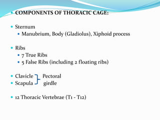
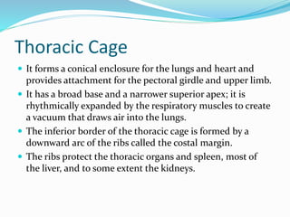
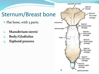
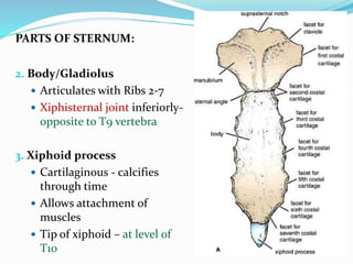
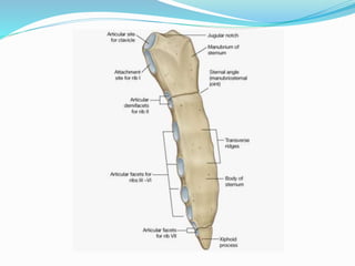
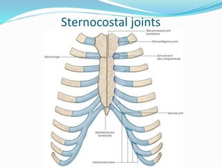
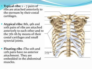
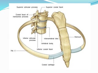
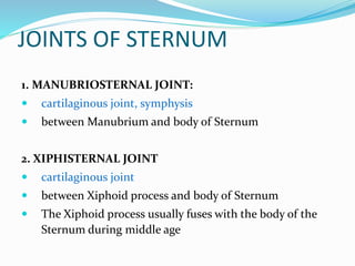
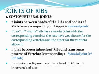
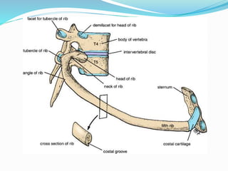
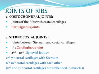
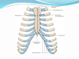
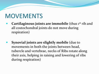
Dr. Aunum Iqbal
 COMPONENTS OF THORACIC CAGE:
 Sternum
 Manubrium, Body (Gladiolus), Xiphoid process
 Ribs
 7 True Ribs
 5 False Ribs (including 2 floating ribs)
 Clavicle Pectoral
 Scapula girdle
 12 Thoracic Vertebrae (T1 - T12)
Thoracic Cage
 It forms a conical enclosure for the lungs and heart and
provides attachment for the pectoral girdle and upper limb.
 It has a broad base and a narrower superior apex; it is
rhythmically expanded by the respiratory muscles to create
a vacuum that draws air into the lungs.
 The inferior border of the thoracic cage is formed by a
downward arc of the ribs called the costal margin.
 The ribs protect the thoracic organs and spleen, most of
the liver, and to some extent the kidneys.
Sternum/Breast bone
 Flat bone, with 3 parts:
1. Manubrium sterni
2. Body/Gladiolus
3. Xiphoid process
PARTS OF STERNUM:
1. Manubrium sterni
 Jugular/suprasternal notch
 Articulates with Clavicles
and Ribs 1 and 2
 Lies opposite to T3 and T4
vertebrae
 Manubriosternal joint
inferiorly – called Sternal
Angle/Angle of Louis –
opposite articulation with
2nd rib – at the level of
intervertbral disc between
T4 and T5 vertebrae
(imp. for counting the ribs)
PARTS OF STERNUM:
2. Body/Gladiolus
 Articulates with Ribs 2-7
 Xiphisternal joint inferiorly-
opposite to T9 vertebra
3. Xiphoid process
 Cartilaginous - calcifies
through time
 Allows attachment of
muscles
 Tip of xiphoid – at level of
T10
Clinical Anatomy
1. Bone marrow biopsy (to take sample of bone
marrow)
1. Median sternotomy (Sternum is split in half,
longitudinally to gain access to thoracic organs for
surgery)
Sternocostal joints
Ribs
 Typical Ribs 2-7
 Head
 Neck
 Tubercle
 Angle
 Shaft
 Subcostal groove
 Atypical Ribs 1, 8 -10
•Rib 1 - short, flat and
supports Subclavian
vessels
• Ribs 1, 10-12 - articulate
with only 1 vertebra
• Ribs 11 and 12 –
“floating ribs” – do not
articulate with Transverse
processes of Vertebrae or
Sternum
 Typical ribs: 1 – 7 pairs of
ribs are attached anteriorly to
the sternum by their costal
cartilages.
 Atypical ribs: 8th, 9th and
10th pairs of ribs are attached
anteriorly to each other and to
the 7th rib by means of their
costal cartilages and small
synovial joints.
 Floating ribs :The 11th and
12th pairs have no anterior
attachment. They are
embedded in the abdominal
muscles.
Typical Ribs (2 -7)
• Long, twisted, flat bone
• The anterior end of each rib is attached to the corresponding costal
cartilage
• A rib has a head, neck, tubercle, shaft, and angle
• Head – located posteriorly - has 2 facets for articulation – one for the
numerically corresponding vertebral body and the other for the vertebral
body immediately above it.
• Neck is a constricted portion - between the head and the tubercle.
• The Tubercle is a prominence on outer surface of the rib - at the
junction of the neck with the shaft. It has a facet for articulation with the
transverse process of the numerically corresponding vertebra.
• The Shaft is thin, flat and twisted on its long axis.
• It has a rounded, smooth superior border and a sharp, thin inferior
border which has costal groove (it accommodates the intercostal
vessels and nerve (VAN ) The angle is where the shaft of the rib bends
sharply forward.
Atypical Rib (1st Rib)
• The first rib has a close relationship to the lower nerves of the
Brachial plexus, Subclavian artery and vein
• This rib is small and flattened from above downward
• Scalenus anterior muscle is attached to its upper surface and inner
border
• Anterior to the attachment of Scalenus anterior, the Subclavian
vein crosses the rib
• Posterior to the attachment of Scalenus anterior, the Subclavian
artery and the lower trunk of the Brachial plexus cross the rib
and lie in contact with the bone
Clinical Anatomy
• Fracture of 1st rib may cause:
• Injury to lower trunk of Brachial plexus: Klumpke’s
paralysis
• Injury to Subclavian vessels:
Hemorrhage/Ischemia
• Thoracic outlet syndrome: Compression of
Subclavian vessels/Brachial plexus between 1st Rib
and Clavicle – Klumpke’s paralysis and ischemia.
JOINTS OF STERNUM
1. MANUBRIOSTERNAL JOINT:
 cartilaginous joint, symphysis
 between Manubrium and body of Sternum
2. XIPHISTERNAL JOINT
 cartilaginous joint
 between Xiphoid process and body of Sternum
 The Xiphoid process usually fuses with the body of the
Sternum during middle age
JOINTS OF RIBS
1. COSTOVERTEBRAL JOINTS:
 2 joints between heads of the Ribs and bodies of
Vertebrae (corresponding and upper)- Synovial joints
 1st, 10th, 11th and 12th rib has 1 synovial joint with the
corresponding vertebra, the rest have 2 each; one for the
corresponding vertebra and the other for the vertebra
above it
 1 joint between tubercle of Ribs and transverse
process of Vertebra (corresponding) - Synovial joint (1st-
10th Rib)
 Intra articular ligament connects head of Rib to the
intervertebral disc
JOINTS OF RIBS
2. COSTOCHONDRAL JOINTS:
 Joints of the Ribs with costal cartilages
 Cartilaginous joints
3. STERNOCOSTAL JOINTS:
 Joints between Sternum and costal cartilages
 1st : Cartilaginous joint
 2nd – 10th : Synovial joints=
2nd-7th costal cartilages with Sternum
8th-10th costal cartilages with each other
(11th and 12th costal cartilages are embedded in muscles)
MOVEMENTS
 Cartilaginous joints are immobile (thus 1st rib and
all costochondral joints do not move during
respiration)
 Synovial joints are slightly mobile (due to
movements in both the joints between head,
tubercle and vertebrae, necks of Ribs rotate along
their axis, helping in raising and lowering of ribs
during respiration)
Cervical Rib (Accessory Rib)
 Occurs in 0.5% population
 There is an extra pair of ribs just above the 1st rib
 They arise from the transverse process of C7 vertebrae
 Anteriorly, they may be attached to 1st Rib or may be
free
 Clinical Anatomy: Cervical Rib may compress Brachial
plexus/Subclavian artery; causing Klumpke’s
paralysis/Ischemia

thoraciccage-160601033405.pdf

  • 1.
  • 2.
     COMPONENTS OFTHORACIC CAGE:  Sternum  Manubrium, Body (Gladiolus), Xiphoid process  Ribs  7 True Ribs  5 False Ribs (including 2 floating ribs)  Clavicle Pectoral  Scapula girdle  12 Thoracic Vertebrae (T1 - T12)
  • 4.
    Thoracic Cage  Itforms a conical enclosure for the lungs and heart and provides attachment for the pectoral girdle and upper limb.  It has a broad base and a narrower superior apex; it is rhythmically expanded by the respiratory muscles to create a vacuum that draws air into the lungs.  The inferior border of the thoracic cage is formed by a downward arc of the ribs called the costal margin.  The ribs protect the thoracic organs and spleen, most of the liver, and to some extent the kidneys.
  • 5.
    Sternum/Breast bone  Flatbone, with 3 parts: 1. Manubrium sterni 2. Body/Gladiolus 3. Xiphoid process
  • 6.
    PARTS OF STERNUM: 1.Manubrium sterni  Jugular/suprasternal notch  Articulates with Clavicles and Ribs 1 and 2  Lies opposite to T3 and T4 vertebrae  Manubriosternal joint inferiorly – called Sternal Angle/Angle of Louis – opposite articulation with 2nd rib – at the level of intervertbral disc between T4 and T5 vertebrae (imp. for counting the ribs)
  • 7.
    PARTS OF STERNUM: 2.Body/Gladiolus  Articulates with Ribs 2-7  Xiphisternal joint inferiorly- opposite to T9 vertebra 3. Xiphoid process  Cartilaginous - calcifies through time  Allows attachment of muscles  Tip of xiphoid – at level of T10
  • 9.
    Clinical Anatomy 1. Bonemarrow biopsy (to take sample of bone marrow) 1. Median sternotomy (Sternum is split in half, longitudinally to gain access to thoracic organs for surgery)
  • 10.
  • 11.
    Ribs  Typical Ribs2-7  Head  Neck  Tubercle  Angle  Shaft  Subcostal groove  Atypical Ribs 1, 8 -10 •Rib 1 - short, flat and supports Subclavian vessels • Ribs 1, 10-12 - articulate with only 1 vertebra • Ribs 11 and 12 – “floating ribs” – do not articulate with Transverse processes of Vertebrae or Sternum
  • 12.
     Typical ribs:1 – 7 pairs of ribs are attached anteriorly to the sternum by their costal cartilages.  Atypical ribs: 8th, 9th and 10th pairs of ribs are attached anteriorly to each other and to the 7th rib by means of their costal cartilages and small synovial joints.  Floating ribs :The 11th and 12th pairs have no anterior attachment. They are embedded in the abdominal muscles.
  • 13.
    Typical Ribs (2-7) • Long, twisted, flat bone • The anterior end of each rib is attached to the corresponding costal cartilage • A rib has a head, neck, tubercle, shaft, and angle • Head – located posteriorly - has 2 facets for articulation – one for the numerically corresponding vertebral body and the other for the vertebral body immediately above it. • Neck is a constricted portion - between the head and the tubercle. • The Tubercle is a prominence on outer surface of the rib - at the junction of the neck with the shaft. It has a facet for articulation with the transverse process of the numerically corresponding vertebra. • The Shaft is thin, flat and twisted on its long axis. • It has a rounded, smooth superior border and a sharp, thin inferior border which has costal groove (it accommodates the intercostal vessels and nerve (VAN ) The angle is where the shaft of the rib bends sharply forward.
  • 19.
    Atypical Rib (1stRib) • The first rib has a close relationship to the lower nerves of the Brachial plexus, Subclavian artery and vein • This rib is small and flattened from above downward • Scalenus anterior muscle is attached to its upper surface and inner border • Anterior to the attachment of Scalenus anterior, the Subclavian vein crosses the rib • Posterior to the attachment of Scalenus anterior, the Subclavian artery and the lower trunk of the Brachial plexus cross the rib and lie in contact with the bone
  • 20.
    Clinical Anatomy • Fractureof 1st rib may cause: • Injury to lower trunk of Brachial plexus: Klumpke’s paralysis • Injury to Subclavian vessels: Hemorrhage/Ischemia • Thoracic outlet syndrome: Compression of Subclavian vessels/Brachial plexus between 1st Rib and Clavicle – Klumpke’s paralysis and ischemia.
  • 23.
    JOINTS OF STERNUM 1.MANUBRIOSTERNAL JOINT:  cartilaginous joint, symphysis  between Manubrium and body of Sternum 2. XIPHISTERNAL JOINT  cartilaginous joint  between Xiphoid process and body of Sternum  The Xiphoid process usually fuses with the body of the Sternum during middle age
  • 24.
    JOINTS OF RIBS 1.COSTOVERTEBRAL JOINTS:  2 joints between heads of the Ribs and bodies of Vertebrae (corresponding and upper)- Synovial joints  1st, 10th, 11th and 12th rib has 1 synovial joint with the corresponding vertebra, the rest have 2 each; one for the corresponding vertebra and the other for the vertebra above it  1 joint between tubercle of Ribs and transverse process of Vertebra (corresponding) - Synovial joint (1st- 10th Rib)  Intra articular ligament connects head of Rib to the intervertebral disc
  • 27.
    JOINTS OF RIBS 2.COSTOCHONDRAL JOINTS:  Joints of the Ribs with costal cartilages  Cartilaginous joints 3. STERNOCOSTAL JOINTS:  Joints between Sternum and costal cartilages  1st : Cartilaginous joint  2nd – 10th : Synovial joints= 2nd-7th costal cartilages with Sternum 8th-10th costal cartilages with each other (11th and 12th costal cartilages are embedded in muscles)
  • 29.
    MOVEMENTS  Cartilaginous jointsare immobile (thus 1st rib and all costochondral joints do not move during respiration)  Synovial joints are slightly mobile (due to movements in both the joints between head, tubercle and vertebrae, necks of Ribs rotate along their axis, helping in raising and lowering of ribs during respiration)
  • 30.
    Cervical Rib (AccessoryRib)  Occurs in 0.5% population  There is an extra pair of ribs just above the 1st rib  They arise from the transverse process of C7 vertebrae  Anteriorly, they may be attached to 1st Rib or may be free  Clinical Anatomy: Cervical Rib may compress Brachial plexus/Subclavian artery; causing Klumpke’s paralysis/Ischemia