dusjagr & nano talk on open tools for agriculture research and learning
45 hormones and the endocrine system
1. 993
45
Hormones and
the Endocrine System
The Body’s Long-Distance Regulators
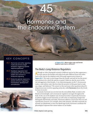
Although we often distinguish animals of different species by their appearance,
in many species the females and males look quite different from each other.
Such is the case for the elephant seals (Mirounga angustirostris) shown in
Figure 45.1. The male is much larger than the female, and only he has the promi-
nent proboscis for which the species is named. Males are also far more territo-
rial and aggressive than females. A sex-determining gene on the Y chromosome
makes a seal embryo male. But how does the presence of this gene lead to male
size, shape, and behavior? The answer to this and many other questions about bi-
ological processes involves signaling molecules called hormones (from the Greek
horman, to excite).
In animals, hormones are secreted into the extracellular fluid, circulate in the
blood (or hemolymph), and communicate regulatory messages throughout the
body. In the case of the elephant seal, increased secretion of particular hormones
at puberty triggers sexual maturation, as well as the accompanying changes that
distinguish adult females and males. Hormones influence much more than sex and
reproduction, however. For example, when seals, humans, and other mammals are
stressed, are dehydrated, or have low blood sugar levels, hormones coordinate the
physiological responses that restore balance in our bodies.
▲ Figure 45.1 What makes male and female
elephant seals look so different?
993
K E Y C O N C E P T S
45.1 Hormones and other signaling
molecules bind to target
receptors, triggering specific
response pathways
45.2 Feedback regulation and
coordination with the nervous
system are common in
endocrine signaling
45.3 Endocrine glands respond to
diverse stimuli in regulating
homeostasis, development,
and behavior
◀ Male elephant seals sparring
2. 994 UNIT SEVEN Animal Form and Function
Paracrine and Autocrine Signaling
Many types of cells produce and secrete local regulators,
molecules that act over short distances and reach their target
cells solely by diffusion. Once secreted, local regulators act on
their target cells within seconds or even milliseconds. Local
Each hormone binds to specific receptors in the body. Al-
though a given hormone can reach all cells of the body, only
some cells have receptors for that hormone. A hormone elic-
its a response—such as a change in metabolism—in specific
target cells, those that have the matching receptor. Cells lack-
ing a receptor for that hormone are unaffected.
Chemical signaling by hormones is the function of the
endocrine system, one of the two basic systems for commu-
nication and regulation in the animal body. The other major
communication and control system is the nervous system,
a network of specialized cells—neurons—that transmit sig-
nals along dedicated pathways. These signals in turn regulate
neurons, muscle cells, and endocrine cells. Because signaling
by neurons can regulate the release of hormones, the nervous
and endocrine systems often overlap in function.
In this chapter, we’ll begin with an overview of the differ-
ent types of chemical signaling in animals. We’ll then explore
how hormones regulate target cells, how hormone secretion
is regulated, and how hormones help maintain homeostasis.
We’ll also consider the ways in which endocrine and nervous
system activities are coordinated and examine the role of hor-
mones in regulating growth and development.
C O N C E P T 45.1
Hormones and other signaling
molecules bind to target receptors,
triggering specific response pathways
We’ll begin our consideration of the endocrine system by
examining the diverse ways that animal cells use chemical
signals to communicate.
Intercellular Communication
Communication between animal cells via secreted signals is
often classified by two criteria: the type of secreting cell and
the route taken by the signal in reaching its target. Figure 45.2
illustrates five forms of signaling distinguished in this manner.
We’ll explore these forms in turn.
Endocrine Signaling
In endocrine signaling (see Figure 45.2a), hormones secreted
into extracellular fluid by endocrine cells reach target cells
via the bloodstream (or hemolymph). One function of en-
docrine signaling is to maintain homeostasis. Hormones
regulate properties that include blood pressure and volume,
energy metabolism and allocation, and solute concentra-
tions in body fluids. Endocrine signaling also mediates
responses to environmental stimuli, regulates growth and
development, and, as discussed above, triggers physical
and behavioral changes underlying sexual maturity and
reproduction.
Synapse
Blood
vessel
Neurosecretory
cell
Neuron
Blood
vessel
(a) In endocrine signaling, secreted molecules diffuse into the blood-
stream and trigger responses in target cells anywhere in the body.
(b) In paracrine signaling, secreted molecules diffuse locally and
trigger a response in neighboring cells.
(c) In autocrine signaling, secreted molecules diffuse locally and trigger
a response in the cells that secrete them.
(d) In synaptic signaling, neurotransmitters diffuse across synapses
and trigger responses in cells of target tissues (neurons, muscles,
or glands).
(e) In neuroendocrine signaling, neurohormones diffuse into the
bloodstream and trigger responses in target cells anywhere in
the body.
RESPONSE
RESPONSE
RESPONSE
RESPONSE
RESPONSE
▲ Figure 45.2 Intercellular communication by secreted mol-
ecules. In each type of signaling, secreted molecules ( ) bind to a
specific receptor protein ( ) expressed by target cells. Some receptors
are located inside cells, but for simplicity, here all are drawn on the
cell surface.
3. CHAPTER 45 Hormones and the Endocrine System 995
communicate with each other via pheromones, chemicals
that are released into the external environment. For ex-
ample, when a foraging ant discovers a new food source, it
marks its path back to the nest with a pheromone. Ants also
use pheromones for guidance when a colony migrates to a
new location (Figure 45.3).
Pheromones serve a wide range of functions that include
defining territories, warning of predators, and attracting
potential mates. The polyphemus moth (Antheraea polyphe-
mus) provides a noteworthy example: The sex pheromone
released into the air by a female enables her to attract a male
of the species from up to 4.5 km away. You’ll read more
about pheromone function when we take up the topic of
animal behavior in Chapter 51.
Chemical Classes of Local Regulators
and Hormones
Molecules used in intercellular signaling vary substantially
in size and chemical properties. Let’s take a look at some of
the most common classes of local regulators and hormones.
Classes of Local Regulators
One group of local regulators, the prostaglandins, are modified
fatty acids. Many other local regulators, including cytokines
and growth factors, are polypeptides, and some are gases.
Nitric oxide (NO), a gas, functions in the body as both
a local regulator and a neurotransmitter. When the level of
oxygen in the blood falls, endothelial cells in blood vessel
walls synthesize and release NO. After diffusing into the sur-
rounding smooth muscle cells, NO activates an enzyme that
relaxes the cells. The result is vasodilation, which increases
blood flow to tissues.
In human males, NO’s ability to promote vasodilation en-
ables sexual function by increasing blood flow into the penis,
producing an erection. The drug Viagra (sildenafil citrate), a
regulators include cytokines, which enable communication
between immune cells (see Figure 43.16 and Figure 43.18),
and growth factors, which promote the growth, division, and
development of many types of cells.
Depending on the target cell, signaling by local regulators
can be either paracrine or autocrine. In paracrine signaling
(from the Greek para, to one side of), target cells lie near
the secreting cell (see Figure 45.2b). In autocrine signaling
(from the Greek auto, self), the secreting cells themselves
are the target cells (see Figure 45.2c).
Paracrine and autocrine signaling play roles in many
physiological processes, including blood pressure regulation,
nervous system function, and reproduction. Local regula-
tors that mediate such signaling include the prostaglandins,
so named because they were first discovered in prostate
gland secretions that contribute to semen. In the reproduc-
tive tract of a female, prostaglandins introduced in semen
stimulate the smooth muscles of the uterine wall to con-
tract, helping sperm reach an egg. At the onset of childbirth,
prostaglandins produced by the placenta cause the uterine
muscles to become more excitable, helping to induce labor
(see Figure 46.18).
Prostaglandins also act in the immune system, promoting
inflammation and the sensation of pain in response to injury.
That is why drugs that block prostaglandin synthesis, such
as aspirin and ibuprofen, have both anti-inflammatory and
pain-relieving effects. Prostaglandins also help regulate the
aggregation of platelets, one step in the formation of blood
clots. Because blood clots in vessels that supply the heart can
block blood flow, causing a heart attack (see Concept 42.4),
some physicians recommend that people at risk for a heart
attack take aspirin on a regular basis.
Synaptic and Neuroendocrine Signaling
Secreted molecules are crucial for two types of signaling by
neurons. In synaptic signaling, neurons form specialized junc-
tions called synapses with target cells, such as other neurons
and muscle cells. At most synapses, neurons secrete molecules
called neurotransmitters that diffuse a very short distance
and bind to receptors on the target cells (see Figure 45.2d).
Neurotransmitters are central to sensation, memory, cogni-
tion, and movement (as we’ll explore in Chapters 48–50).
In neuroendocrine signaling, specialized neurons called neu-
rosecretory cells secrete neurohormones, which diffuse from
nerve cell endings into the bloodstream (see Figure 45.2e).
One example of a neurohormone is antidiuretic hormone,
which is essential to kidney function and water balance (see
Concept 44.5). Many neurohormones function in the regula-
tion of endocrine signaling, as we’ll discuss later in this chapter.
Signaling by Pheromones
Not all secreted signaling molecules act within the body.
Members of a particular animal species sometimes
▲ Figure 45.3 Signaling by pheromones. Using their lowered
antennae, these Asian army ants (Leptogenys distinguenda) follow
a pheromone-marked trail as they carry pupae and larvae to a new
nest site.
4. 996 UNIT SEVEN Animal Form and Function
To focus this and subsequent discussions, we’ll restrict our
presentation to endocrine signaling.
One difference in cellular hormone response path-
ways is the location of the target cells’ receptor proteins.
Water-soluble hormones are secreted by exocytosis and
travel freely in the bloodstream. Being insoluble in lipids,
they cannot diffuse through the plasma membranes of
target cells. Instead, these hormones bind to cell-surface
receptors, inducing changes in cytoplasmic molecules and
sometimes altering gene transcription (Figure 45.5a). In
contrast, lipid-soluble hormones diffuse out across the
membranes of endocrine cells. Outside the cell, they bind
to transport proteins that keep them soluble in the aqueous
environment of the blood. Upon leaving the blood, they dif-
fuse into target cells and typically bind to receptors in the
cytoplasm or nucleus (Figure 45.5b). The hormone-bound
receptor then triggers changes in gene transcription.
treatment for male erectile dysfunction, sustains an erection
by prolonging activity of the NO response pathway.
Classes of Hormones
Hormones fall into three major chemical classes: polypep-
tides, steroids, and amines (Figure 45.4). The hormone in-
sulin, for example, is a polypeptide that contains two chains
in its active form. Steroid hormones, such as cortisol, are
lipids that contain four fused carbon rings. All are derived
from the steroid cholesterol (see Figure 5.12). Epinephrine
and thyroxine are amine hormones, each synthesized from a
single amino acid, either tyrosine or tryptophan.
As Figure 45.4 indicates, hormones vary in their solubility
in aqueous and lipid-rich environments. Polypeptides and
most amine hormones are water-soluble, whereas steroid
hormones and other largely nonpolar (hydrophobic) hor-
mones, such as thyroxine, are lipid-soluble.
Cellular Response Pathways
There are several differences between the response path-
ways for water-soluble and lipid-soluble signaling molecules.
0.8 nm
O
HO OH
O
OH
CortisolInsulin
ThyroxineEpinephrine
HO
O
I
I
I
I
HOOC NH2
CH3
CH3
H3C
HO
OH
N
H
HO
Water-soluble (hydrophilic) Lipid-soluble (hydrophobic)
Polypeptides
Amines
Steroids
▲ Figure 45.4 Differences in hormone solubility and structure.
M A K E C O N N E C T I O N S Cells synthesize epinephrine from the
amino acid tyrosine (see Figure 5.14). On the structure of epinephrine
shown above, draw an arrow pointing to the position corresponding to
the α carbon of tyrosine.
SECRETORY
CELL
Water-
soluble
hormone
Blood
vessel
OR
NUCLEUS
Receptor protein
Gene
regulation
TARGET
CELL
(a) Water-soluble hormone;
receptor in plasma
membrane
Cytoplasmic
response
SECRETORY
CELL
Lipid-
soluble
hormone
Receptor
protein
Blood
vessel
Transport
protein
NUCLEUS
Gene
regulation
TARGET
CELL
(b) Lipid-soluble hormone;
receptor in nucleus or
cytoplasm
Cytoplasmic
response
▼ Figure 45.5 Variation in hormone receptor location.
W H AT I F ? Suppose you are studying a cell’s response to a particular
hormone. You observe that the cell produces the same response to the
hormone whether or not the cell is treated with a chemical that blocks
transcription. What can you surmise about the hormone and its receptor?
5. CHAPTER 45 Hormones and the Endocrine System 997
that the liver releases glucose into the bloodstream, provid-
ing the fuel you need to chase the departing bus.
Pathway for Lipid-Soluble Hormones
Intracellular receptors for lipid-soluble hormones perform
the entire task of transducing a signal within a target cell.
The hormone activates the receptor, which then directly
triggers the cell’s response. In most cases, the response to a
lipid-soluble hormone is a change in gene expression.
Most steroid hormone receptors are predominantly lo-
cated in the cytosol prior to binding to a hormone. When a
steroid hormone binds to its cytosolic receptor, a hormone-
receptor complex forms, which moves into the nucleus (see
Figure 18.9). There, the receptor portion of the complex
alters transcription of particular genes by interacting with
a specific DNA-binding protein or response element in the
DNA. (In some cell types, steroid hormones trigger addi-
tional responses by interacting with other kinds of receptor
proteins located at the cell surface).
Among the best-characterized steroid hormone receptors
are those that bind to estrogens, steroid hormones necessary
for female reproductive function in vertebrates. For exam-
ple, in female birds and frogs, estradiol, a form of estrogen,
binds to a specific cytoplasmic receptor in liver cells. Bind-
ing of estradiol to this receptor activates transcription of the
gene for the protein vitellogenin (Figure 45.7). Following
translation of the messenger RNA, vitellogenin is secreted
To follow the distinct cellular responses to water-soluble
and lipid-soluble hormones, we’ll examine the two response
pathways in turn.
Pathway for Water-Soluble Hormones
The binding of a water-soluble hormone to a receptor pro-
tein triggers events at the plasma membrane that result in a
cellular response. The response may be the activation of an
enzyme, a change in the uptake or secretion of specific mol-
ecules, or a rearrangement of the cytoskeleton. In addition,
some cell-surface receptors cause proteins in the cytoplasm
to move into the nucleus and alter the transcription of spe-
cific genes.
The series of changes in cellular proteins that converts
the extracellular chemical signal to a specific intracellular
response is called signal transduction. A signal transduc-
tion pathway typically involves multiple steps, each involv-
ing specific molecular interactions (see Chapter 11).
To explore the role of signal transduction in hormone
signaling, consider one response to short-term stress. When
you are in a stressful situation, perhaps running to catch a
bus, the adrenal glands that lie atop your kidneys secrete
the hormone epinephrine, also known as adrenaline.
Upon reaching the liver, epinephrine binds to a G protein-
coupled receptor in the plasma membrane of target cells
(Figure 45.6). The binding of hormone to receptor triggers
a cascade of events in liver cells involving synthesis of cyclic
AMP (cAMP) as a short-lived second messenger. Activation
of protein kinase A by cAMP leads to activation of an en-
zyme required for glycogen breakdown and inactivation of
an enzyme needed for glycogen synthesis. The net result is
Second
messenger
G protein-coupled
receptor
G protein
Adenylyl
cyclase
cAMP
Inhibition of
glycogen synthesis
Promotion of
glycogen breakdown
Hormone (epinephrine)
EXTRACELLULAR
FLUID
CYTOPLASM
ATP
GTP
Protein
kinase A
▲ Figure 45.6 Signal transduction triggered by a cell-surface
hormone receptor.
Hormone
(estradiol)
EXTRACELLULAR
FLUID
Plasma
membrane
Estradiol
receptor
Hormone-receptor
complex
mRNA
for vitellogenin
DNA
NUCLEUS
CYTOPLASM
Vitellogenin
▲ Figure 45.7 Direct regulation of gene expression by a
steroid hormone receptor.
6. 998 UNIT SEVEN Animal Form and Function
Lipid-soluble hormones also exert different effects on dif-
ferent target cells. For example, estradiol, which stimulates a
bird’s liver to synthesize the yolk protein vitellogenin, stimu-
lates the reproductive system to synthesize proteins that
form the egg white.
Endocrine Tissues
and Organs
Some endocrine cells are found in organs that are part of
other organ systems. For example, the stomach contains
isolated endocrine cells that help regulate digestive pro-
cesses by secreting the hormone gastrin. More often, endo-
crine cells are grouped in ductless organs called endocrine
glands, such as the thyroid and parathyroid glands and the
gonads, either testes in males or ovaries in females. The en-
docrine glands of humans are illustrated in Figure 45.9. This
overview of endocrine glands and the hormones that they
produce will serve as a useful point of reference as you move
through the chapter.
Note that endocrine glands secrete hormones directly
into the surrounding fluid. In contrast, exocrine glands,
and transported in the blood to the reproductive system,
where it is used to produce egg yolk.
Thyroxine, vitamin D, and other lipid-soluble hormones
that are not steroids have receptors that are typically located
in the nucleus. These receptors bind to hormone molecules
that diffuse from the bloodstream across both the plasma
membrane and nuclear envelope. Once bound to a hor-
mone, the receptor binds to specific sites in the cell’s DNA
and stimulates the transcription of specific genes.
Multiple Effects of Hormones
Many hormones elicit more than one type of response in the
body. Consider, for example, epinephrine. As we noted ear-
lier, this hormone triggers glycogen breakdown in the liver.
However, epinephrine also increases blood flow to major
skeletal muscles and decreases blood flow to the digestive
tract. These varied responses enhance the rapid reactions of
the body in emergencies.
How can a hormone such as epinephrine have such
widely varying effects? A single hormone can elicit multiple
responses if its target cells differ in their receptor type or in
the molecules that produce the response.
Consider the target cells for epinephrine:
In liver cells, epinephrine binds to a
β-type receptor in the plasma mem-
brane. This receptor activates the en-
zyme protein kinase A, which in turn
regulates enzymes of glycogen metab-
olism, causing release of glucose into
the blood (Figure 45.8a). (Note that
this is the signal transduction pathway
illustrated in Figure 45.6.)
In the smooth muscle cells lining
blood vessels that supply skeletal mus-
cle, the same kinase activated by the
same epinephrine receptor inactivates
a muscle-specific enzyme. The result
is smooth muscle relaxation, leading
to vasodilation and hence increased
blood flow to skeletal muscles
(Figure 45.8b).
In the smooth muscle cells lining
blood vessels of the intestines, epi-
nephrine binds to an α-type receptor
(Figure 45.8c). Rather than activating
protein kinase A, this receptor triggers
a signaling pathway involving a differ-
ent G protein and different enzymes.
The result is smooth muscle contrac-
tion that brings about vasoconstric-
tion, restricting blood flow to the
intestines.
Different receptors
Same receptors but different
intracellular proteins (not shown)
Epinephrine
Glycogen
deposits
Glucose
Glycogen breaks down and
glucose is released from cell.
Cell relaxes.
(c) Smooth muscle cell in
wall of blood vessel
that supplies intestines
(b) Smooth muscle cell in
wall of blood vessel that
supplies skeletal muscle
(a) Liver cell
β receptor
Epinephrine
β receptor
Blood glucose level
increases.
Blood vessel dilates,
increasing flow to
skeletal muscle.
Cell contracts.
Epinephrine
α receptor
Blood vessel constricts,
decreasing flow to
intestines.
▲ Figure 45.8 One hormone, different effects. Epinephrine, the primary “fight-or-flight”
hormone, produces different responses in different target cells. Target cells with the same receptor
exhibit different responses if they have different signal transduction pathways or effector proteins;
compare (a) with (b). Target cells with different receptors for the hormone may also exhibit differ-
ent responses; compare (b) with (c).
7. CHAPTER 45 Hormones and the Endocrine System 999
Endocrine gland Hormones
Pineal gland Participates in regulation of biological rhythms.
Raises blood calcium level
Relea Regulate anterior pituitary
3 and 4 Stimulates and maintains
metabolic processes
Lowers blood calcium level
Pancreas
*Found in both males and females, but with a major role in one sex
Lowers blood glucose level
Raises blood glucose level
Stimulate uterine lining growth; promote
development and maintenance of female secondary sex
characteristics
Promote uterine lining growth
Support sperm formation; promote
development and maintenance of male secondary sex
characteristics
and
Stimulate ovaries and testes
Stimulates thyroid gland
Stimulates
adrenal cortex
Stimulates mammary gland cells
Stimulates growth and
metabolic functions
Stimulates contraction of smooth muscle cells
in uterus and mammary glands
(also called ):
Promotes retention of water by kidneys; influences social
behavior and bonding
and Raise blood glucose
level; increase metabolic activities; constrict certain blood vessels.
Raise blood glucose level
Promote reabsorption of Na+ and
excretion of K+ in kidneys
Adrenal cortex
Adrenal medulla
Anterior pituitary
Posterior pituitary
C O N C E P T C H E C K 4 5 . 1
1. How do response mechanisms in target cells differ for
water-soluble and lipid-soluble hormones?
2. In what way does one activity described for prostaglan-
dins resemble that of a pheromone?
3. M A K E C O N N E C T I O N S How is the action of epi-
nephrine on blood flow to muscles in the fight-or-flight
response similar to the action of the plant hormone auxin
in apical dominance (see Concept 39.2)?
For suggested answers, see Appendix A.
▶ Figure 45.9 Human endocrine
glands and their hormones. This
figure highlights the location and pri-
mary functions of the major human
endocrine glands. Endocrine tissues
and cells are also located in the thy-
mus, heart, liver, stomach, kidneys,
and small intestine.
such as salivary glands, have ducts that carry secreted
substances onto body surfaces or into body cavities. This
distinction is reflected in the glands’ names: The Greek
endo (within) and exo (out of) refer to secretion into or out
of body fluids, while crine (from the Greek word meaning
“separate”) refers to movement away from the secreting
cell. In the case of the pancreas, endocrine and exocrine tis-
sues are found in the same gland: Ductless tissues secrete
hormones, whereas tissues with ducts secrete enzymes and
bicarbonate.
8. 1000 UNIT SEVEN Animal Form and Function
As an example of a simple endocrine pathway, we’ll con-
sider the control of pH in the duodenum, the first part of
the small intestine. The digestive juices of the stomach are
extremely acidic and must be neutralized before further
digestion can occur. As the stomach contents enter the duo-
denum, their low pH stimulates endocrine cells in the lining
of the duodenum to secrete the hormone secretin into the
extracellular fluid (see Figure 45.10). From there, secretin
diffuses into the blood. Circulating secretin reaches target
cells in the pancreas, which respond by releasing bicarbon-
ate into ducts leading to the duodenum. This response—the
release of bicarbonate—raises the pH in the duodenum,
neutralizing the stomach acid.
Neuroendocrine pathways include additional steps and
involve more than one cell type. In a simple neuroendocrine
pathway, the stimulus is received by a sensory neuron,
which stimulates a neurosecretory cell (Figure 45.11). The
neurosecretory cell then secretes a neurohormone, which
diffuses into the bloodstream and travels to target cells.
As an example of a simple neuroendocrine pathway,
consider the regulation of milk release during nursing
in mammals. Suckling by an infant stimulates sensory
Low pH in
duodenum
S cells of duodenum
Secretin ( )
Pancreatic cells
Bicarbonate release
Hormone
Circulation
throughout
body via
blood
Target
cells
Example: secretin signalingSimple endocrine pathway
Negativefeedback
Endocrine
cell
STIMULUS
–
RESPONSE
▲ Figure 45.10 A simple endocrine pathway. Endocrine cells re-
spond to a change in some internal or external variable—the stimulus—
by secreting hormone molecules that trigger a specific response by tar-
get cells. In the case of secretin signaling, the simple endocrine pathway
is self-limiting because the response to secretin (bicarbonate release)
reduces the stimulus (low pH) through negative feedback.
Oxytocin ( )
Smooth muscle in
mammary glands
Milk release
Suckling
Sensory
neuron
Hypothalamus/
posterior pituitary
+
Neurohormone
Neurosecretory
cell
Circulation
throughout
body via
blood
Target
cells
Example: oxytocin signalingSimple neuroendocrine pathway
Positivefeedback
STIMULUS
RESPONSE
▲ Figure 45.11 A simple neuroendocrine pathway. Sensory
neurons respond to a stimulus by sending nerve impulses to a neuro-
secretory cell, triggering secretion of a neurohormone. Upon reaching
its target cells, the neurohormone binds to its receptor, triggering a
specific response. In oxytocin signaling, the response increases the
stimulus, forming a positive-feedback loop that amplifies signaling.
C O N C E P T 45.2
Feedback regulation and coordination
with the nervous system are common in
endocrine signaling
So far, we have explored forms of intercellular signaling as
well as hormone structure, recognition, and response. We
turn now to considering how regulatory pathways that con-
trol hormone secretion are organized.
Simple Hormone Pathways
In examining the regulation of hormone secretion, we begin
with two basic types of organization—simple endocrine
and simple neuroendocrine pathways. In a simple endocrine
pathway, endocrine cells respond directly to an internal or
environmental stimulus by secreting a particular hormone
(Figure 45.10). The hormone travels in the bloodstream
to target cells, where it interacts with its specific receptors.
Signal transduction within target cells brings about a physi-
ological response.
9. CHAPTER 45 Hormones and the Endocrine System 1001
neurons in the nipples, generating signals in the nervous
system that reach the hypothalamus. Nerve impulses from
the hypothalamus then trigger the release of the neuro-
hormone oxytocin from the posterior pituitary gland (see
Figure 45.11). In response to circulating oxytocin, the
mammary glands secrete milk.
Feedback Regulation
A feedback loop linking the response back to the initial stim-
ulus is characteristic of control pathways. Often, regulation
involves negative feedback, in which the response reduces
the initial stimulus. For instance, bicarbonate release in re-
sponse to secretin increases pH in the intestine, eliminating
the stimulus and thereby shutting off secretin release (see
Figure 45.10). By decreasing hormone signaling, negative-
feedback regulation prevents excessive pathway activity.
Whereas negative feedback dampens a stimulus, positive
feedback reinforces a stimulus, leading to an even greater
response. For example, in the oxytocin pathway outlined in
Figure 45.11, the mammary glands secrete milk in response
to circulating oxytocin. Milk released in response to the
oxytocin leads to more suckling and therefore more stimula-
tion. Activation of the pathway is sustained until the baby
stops suckling. When mammals give birth, oxytocin induces
target cells in the uterine muscles to contract. This pathway
is also characterized by positive-feedback regulation, such
that it drives the birth process to completion.
While positive feedback amplifies both stimulus and re-
sponse, negative feedback helps restore a preexisting state. It
is not surprising, therefore, that hormone pathways involved
in homeostasis typically involve negative feedback. Often
such pathways are paired, providing even more balanced
control. For example, the regulation of blood glucose levels
relies on the antagonistic effects of insulin and glucagon (see
Figure 41.21).
Coordination of Endocrine and Nervous
Systems
In a wide range of animals, endocrine organs in the brain
integrate function of the endocrine system with that of the
nervous system. We’ll explore the basic principles of such
integration in invertebrates and vertebrates.
Invertebrates
The control of development in a moth
illustrates neuroendocrine coordina-
tion in invertebrates. A moth larva,
such as this colorful caterpillar of the
giant silk moth (Hyalophora cecropia), grows in stages. Be-
cause its exoskeleton cannot stretch, the larva must periodi-
cally molt, shedding the old exoskeleton and secreting a new
one. The endocrine pathway that controls molting originates
in the larval brain (Figure 45.12). There, neurosecretory cells
Prothoracic
gland
ADULTPUPA
LATER
LARVA
EARLY
LARVA
Ecdysteroid
Brain
Neurosecretory cells
Corpora cardiaca
High JH
Low
JH
PTTH
Corpora allata
Neurosecretory cells in the brain
produce prothoracicotropic
hormone (PTTH), which is stored in
the corpora cardiaca until release.
1
2 PTTH signals its main target
organ, the prothoracic gland,
to produce the hormone
ecdysteroid.
Ecdysteroid secretion from
the prothoracic gland is
episodic, with each release
stimulating a molt.
3
Juvenile hormone (JH), secreted by
the corpora allata, determines the
result of the molt. At relatively high
concentrations, JH suppresses
metamorphosis. Under these
conditions, molting stimulated by
ecdysteroid produces another larval
stage. When JH drops below a certain
concentration, a pupa forms at the
next ecdysteroid-induced molt. The
adult insect emerges from the pupa.
4
▶ Figure 45.12 Regulation of insect
development and metamorphosis. As
shown here for a giant silk moth, most insects
go through a series of larval stages, with each
molt (shedding of the old exoskeleton) leading
to a larger larva. Molting of the final larval stage
gives rise to a pupa, in which metamorphosis
produces the adult form of the insect. Neurohormones
and hormones control the progression of stages.
10. 1002 UNIT SEVEN Animal Form and Function
from nerves throughout the body and, in response, initiates
endocrine signaling appropriate to environmental condi-
tions. In many vertebrates, for example, nerve signals from
the brain pass sensory information to the hypothalamus
about seasonal changes. The hypothalamus, in turn, regu-
lates the release of reproductive hormones required during
the breeding season.
Signals from the hypothalamus travel to the pituitary
gland, a gland located at the base of the hypothalamus (see
Figure 45.13). Roughly the size and shape of a lima bean, the
pituitary has discrete posterior and anterior parts, or lobes,
which are actually two fused glands that perform very dif-
ferent functions. The posterior pituitary is an extension of
the hypothalamus. Hypothalamic axons that reach into the
posterior pituitary secrete neurohormones synthesized in
the hypothalamus. In contrast, the anterior pituitary is an
endocrine gland that synthesizes and secretes hormones in
response to hormones from the hypothalamus.
Posterior Pituitary Hormones
Neurosecretory cells of the hypothalamus synthesize the
two posterior pituitary hormones: antidiuretic hormone and
oxytocin. After traveling to the posterior pituitary within
the long axons of the neurosecretory cells, these neurohor-
mones are stored, to be released in response to nerve im-
pulses transmitted by the hypothalamus (Figure 45.14).
produce PTTH, a polypeptide neurohormone. When PTTH
reaches an endocrine organ called the prothoracic gland, it
directs release of a second hormone, ecdysteroid. Bursts of
ecdysteroid trigger each successive molt.
Ecdysteroid also controls a remarkable change in form
called metamorphosis. Within the larva lie islands of tissues
that will become the eyes, wings, brain, and other structures
of the adult. Once the plump, crawling larva becomes a sta-
tionary pupa, these islands of cells take over. They complete
their program of development, while many larval tissues
undergo programmed cell death. The end result is the trans-
formation of the crawling caterpillar into a free-flying moth.
Given that ecdysteroid can cause molting or metamorpho-
sis, what determines which process takes place? The answer
is another signal, juvenile hormone (JH), secreted by a pair of
endocrine glands behind the brain. JH modulates the activity
of ecdysteroid. As long as the level of JH is high, ecdysteroid
stimulates molting (and thus maintains the “juvenile” larval
state). When the JH level drops, ecdysteroid induces forma-
tion of a pupa, within which metamorphosis occurs.
Understanding the coordination between the nervous
system and endocrine system in insects has led to advance-
ments in agricultural pest control. For example, one type of
chemical developed to control insect pests is a compound
that binds to the ecdysteroid receptor, causing insect larvae
to molt prematurely and die.
Vertebrates
In vertebrates, coordination of endocrine signaling relies
heavily on a region of the brain called the hypothalamus
(Figure 45.13). The hypothalamus receives information
Posterior
pituitary
Neurosecretory
cells of the
hypothalamus
Neurohormone
Anterior
pituitary
Hypothalamus
Axons
HORMONE ADH Oxytocin
TARGET Kidney tubules Mammary glands,
uterine muscles
▲ Figure 45.14 Production and release of posterior pituitary
hormones. The posterior pituitary gland is an extension of the hy-
pothalamus. Certain neurosecretory cells in the hypothalamus make
antidiuretic hormone (ADH) and oxytocin, which are transported to the
posterior pituitary, where they are stored. Nerve signals from the brain
trigger release of these neurohormones.
Pineal
gland
Cerebellum
Spinal cord
Cerebrum
Thalamus
Hypothalamus
Pituitary
gland
Posterior
pituitary
Anterior
pituitary
Hypothalamus
▲ Figure 45.13 En-
docrine glands in the
human brain. This side
view of the brain indicates
the position of the hypo-
thalamus, the pituitary
gland, and the pineal
gland. (The pineal gland
plays a role in regulating
biological rhythms.)
11. CHAPTER 45 Hormones and the Endocrine System 1003
controlled by at least one releasing hormone. Some, such
as prolactin, have both a releasing hormone and an inhibit-
ing hormone.
The hypothalamic releasing and inhibiting hormones
are secreted near capillaries at the base of the hypothala-
mus. The capillaries drain into short blood vessels, called
portal vessels, which subdivide into a second capillary bed
within the anterior pituitary. Releasing and inhibiting hor-
mones thus have direct access to the gland they control.
Sets of hormones from the hypothalamus, the anterior
pituitary, and a target endocrine gland are often organized
into a hormone cascade pathway. Signals to the brain stimu-
late the hypothalamus to secrete a hormone that stimulates
or inhibits release of an anterior pituitary hormone. The an-
terior pituitary hormone in turn acts on another endocrine
organ, stimulating secretion of yet another hormone, which
exerts effects on specific target tissues.
In a sense, hormone cascade pathways redirect signals
from the hypothalamus to other endocrine glands. For this
reason, the anterior pituitary hormones in these pathways
are called tropic hormones or tropins, from the Greek word
for bending or turning. For example, the hormones FSH and
LH secreted by the anterior pituitary are called gonadotro-
pins because they convey signals from the hypothalamus to
the gonads (testes or ovaries). To learn more about tropic
hormones and hormone cascade pathways, we’ll turn next
to thyroid gland function and regulation.
Anterior pituitary hormones
Endocrine cells of
the anterior pituitary
Neurosecretory cells
of the hypothalamus
Portal vessels
Posterior pituitary
Hypothalamic
releasing and
inhibiting
hormones
Tropic effects only Nontropic effects only Tropic and nontropic
effects
FSH and LH TSH
Thyroid
MSH
Melanocytes
ACTH
Adrenal
cortex
Testes or
ovaries
GH
Liver, bones,
other tissues
Prolactin
Mammary
glands
HORMONE
TARGET
▶ Figure 45.15 Production and release of anterior
pituitary hormones. The release of hormones synthesized
in the anterior pituitary gland is controlled by hypotha-
lamic releasing and inhibiting hormones. The hypothalamic
hormones are secreted by neurosecretory cells and enter
a capillary network within the hypothalamus. These capil-
laries drain into portal vessels that connect with a second
capillary network in the anterior pituitary.
Antidiuretic hormone (ADH), or vasopressin, regulates
kidney function. Secretion of ADH increases water retention
in the kidneys, helping maintain normal blood osmolarity
(see Concept 44.5). ADH also has an important role in social
behavior (see Concept 51.4).
Oxytocin has multiple functions related to reproduction.
As we have seen, in female mammals oxytocin controls milk
secretion by the mammary glands and regulates uterine con-
tractions during birthing. In addition, oxytocin has targets in
the brain, where it influences behaviors related to maternal
care, pair bonding, and sexual activity.
Anterior Pituitary Hormones
Hormones secreted by the anterior pituitary control a di-
verse set of processes in the human body, including metabo-
lism, osmoregulation, and reproduction. As illustrated in
Figure 45.15, many anterior pituitary hormones, but not all,
regulate the activity of other endocrine glands or tissues.
Hormones secreted by the hypothalamus control the re-
lease of all anterior pituitary hormones. Each hypothalamic
hormone is either a releasing hormone or an inhibiting hor-
mone, reflecting its role in promoting or inhibiting release
of one or more specific hormones by the anterior pituitary.
Prolactin-releasing hormone, for example, is a hypotha-
lamic hormone that stimulates the anterior pituitary to
secrete prolactin, which has activities that include stimu-
lating milk production. Every anterior pituitary hormone is
12. 1004 UNIT SEVEN Animal Form and Function
Thyroid Regulation: A Hormone Cascade
Pathway
In humans and other mammals, thyroid hormone regulates
bioenergetics; helps maintain normal blood pressure, heart
rate, and muscle tone; and regulates digestive and reproduc-
tive functions. If the level of thyroid hormone in the blood
drops, the hypothalamus responds by initiating a hormone
cascade pathway (Figure 45.16). The hypothalamus secretes
thyrotropin-releasing hormone (TRH), causing the ante-
rior pituitary to secrete a tropic hormone known as either
thyroid-stimulating hormone (TSH) or thyrotropin. TSH
stimulates release of thyroid hormone by the thyroid gland,
an organ in the neck consisting of two lobes on the ventral
surface of the trachea. As thyroid hormone accumulates, it
increases metabolic rate, while also initiating negative feed-
back that prevents its overproduction.
Disorders of Thyroid Function and Regulation
Disruption of thyroid hormone production and regula-
tion can result in serious disorders. Hypothyroidism, the
secretion of too little thyroid hormone, can cause weight
gain, lethargy, and intolerance to cold in adults. In contrast,
excessive secretion of thyroid hormone, known as hyper-
thyroidism, can lead to high body temperature, profuse
sweating, weight loss, irritability, and high blood pressure.
The most common form of hyperthyroidism in humans is
Graves’ disease. Protruding eyes, caused by fluid accumula-
tion behind the eyes, are a typical symptom. In this autoim-
mune disorder, the body produces antibodies that bind to
and activate the receptor for TSH, causing sustained thyroid
hormone production.
Some thyroid disorders reflect the unusual chemical
makeup of thyroid hormone. The term thyroid hormone
actually refers to a pair of very similar hormones derived
from the amino acid tyrosine. Triiodothyronine (T3) con-
tains three iodine atoms, whereas tetraiodothyronine, or
thyroxine (T4), contains four (see Figure 45.4). Because thy-
roid hormone production requires iodine, it is an essential
mineral.
Although iodine is readily obtained from seafood or io-
dized salt, people in many parts of the world suffer from
inadequate iodine in their diet and therefore cannot synthe-
size adequate amounts of thyroid hormone. The low blood
levels of thyroid hormone are insufficient to provide the
usual negative feedback on the hypothalamus and anterior
pituitary (see Figure 45.16). As a consequence, the pituitary
continues to secrete TSH. Elevated TSH levels cause an en-
largement of the thyroid gland, resulting in a swelling of the
neck known as goiter.
Proper thyroid function is also required for normal de-
velopment. Humans and other vertebrates require thyroid
hormone for the normal functioning of bone-forming cells,
TSH
Thyroid
hormone
Thyroid
gland
TRH causes the
anterior pituitary to
secrete thyroid-
stimulating hormone
(TSH, also known as
thyrotropin ) into
the circulatory
system.
TSH stimulates
endocrine cells in the
thyroid gland to
secrete thyroid
hormone (T3 and T4 )
into the circulatory
system.
Thyroid hormone
levels increase in the
blood and body
tissues, returning to
the normal range.
Thyroid hormone
acts on target cells
throughout the
body to control
bioenergetics; help
maintain normal
blood pressure,
heart rate, and
muscle tone; and
regulate digestive
and reproductive
functions.
Sensory
neuron
Neurosecretory
cell
TRH
Anterior
pituitary
Hypothalamus
2
3
4
5
Thyroid hormone blocks TRH
release from the hypothalamus and
TSH release from the anterior pituitary,
forming a negative-feedback loop that
prevents overproduction of thyroid
hormone.
6
The hypothalamus
secretes thyrotropin-
releasing hormone
(TRH ) into the
blood. Portal vessels
carry TRH to the
anterior pituitary.
1 Thyroid hormone
levels drop below the
normal range.
Circulation
throughout
body
via blood
Circulation
throughout
body
via blood
Negativefeedback
STIMULUS
–
–
RESPONSE
▲ Figure 45.16 Regulation of thyroid hormone secretion:
a hormone cascade pathway.
13. CHAPTER 45 Hormones and the Endocrine System 1005
is an overgrowth of the extremities called acromegaly (from
the Greek acros, extreme, and mega, large).
Hyposecretion of GH in childhood retards long-bone
growth and can lead to pituitary dwarfism. Individuals with
this disorder are for the most part properly proportioned
but generally reach a height of only about 1.2 m (4 feet). If
diagnosed before puberty, pituitary dwarfism can be treated
successfully with human GH (also called HGH). Since the
mid-1980s, recombinant DNA technology has been used to
produce HGH in bacteria (see Concept 20.4). Treatment of af-
fected children with recombinant HGH is now fairly routine.
as well as for the branching of nerve cells during embryonic
development of the brain. In humans, congenital hypothy-
roidism, an inherited condition of thyroid deficiency, re-
sults in markedly retarded skeletal growth and poor mental
development. These defects can often be avoided, at least
partially, if treatment with thyroid hormone begins early
in life. Iodine deficiency in childhood causes the same de-
fects, but it is fully preventable if iodized salt is used in food
preparation.
The fact that iodine in the body is dedicated to the pro-
duction of thyroid hormone provides a novel diagnostic tool
for disorders of thyroid function: Radioactive forms of iodine
enable specific imaging of the thyroid gland (Figure 45.17).
Hormonal Regulation of Growth
Growth hormone (GH), which is secreted by the anterior
pituitary, stimulates growth through both tropic and non-
tropic effects. A major target, the liver, responds to GH by
releasing insulin-like growth factors (IGFs), which circu-
late in the blood and directly stimulate bone and cartilage
growth. (IGFs also appear to play a key role in aging in many
animal species.) In the absence of GH, the skeleton of an
immature animal stops growing. GH also exerts diverse
metabolic effects that tend to raise blood glucose levels, thus
opposing the effects of insulin.
Abnormal production of GH in humans can result in sev-
eral disorders, depending on when the problem occurs and
whether it involves hypersecretion (too much) or hyposecre-
tion (too little). Hypersecretion of GH during childhood can
lead to gigantism, in which the person grows unusually tall but
retains relatively normal body proportions (Figure 45.18).
Excessive GH production in adulthood stimulates bony
growth in the few body parts that are still responsive to the
hormone—predominantly the face, hands, and feet. The result
High level of
iodine uptake
Low level of
iodine uptake
▲ Figure 45.17 Thyroid scan. Physicians can use a radioactive iso-
tope of iodine to detect abnormal patterns of iodine uptake that could
indicate a thyroid disorder.
▲ Figure 45.18 Effect of growth hormone overproduction.
Shown here surrounded by his family, Robert Wadlow grew to a height
of 2.7 m (8 feet 11 inches) by age 22, making him the tallest man in
history. His height was due to excess secretion of growth hormone by
his pituitary gland.
C O N C E P T C H E C K 4 5 . 2
1. What are the roles of oxytocin and prolactin in regulating
the mammary glands?
2. How do the two fused glands of the pituitary gland differ
in function?
3. W H AT I F ? Propose an explanation for why people with
defects in specific endocrine pathways typically have de-
fects in the final gland in the pathway rather than in the
hypothalamus or pituitary.
4. W H AT I F ? Lab tests of two patients, each diagnosed
with excessive thyroid hormone production, revealed
elevated levels of TSH in one but not the other. Was the
diagnosis of one patient necessarily incorrect? Explain.
For suggested answers, see Appendix A.
14. 1006 UNIT SEVEN Animal Form and Function
and enhances Ca2+
excretion by the kidneys. In fishes, ro-
dents, and some other animals, calcitonin is required for
Ca2+
homeostasis. In humans, however, calcitonin is ap-
parently needed only during the extensive bone growth of
childhood.
Adrenal Hormones: Response to Stress
The adrenal glands of vertebrates are located atop the kid-
neys (the renal organs). In mammals, each adrenal gland
is actually made up of two glands with different cell types,
functions, and embryonic origins: the adrenal cortex, the
outer portion, and the adrenal medulla, the central portion.
The adrenal cortex consists of true endocrine cells, whereas
the secretory cells of the adrenal medulla develop from neu-
ral tissue. Thus, like the pituitary gland, each adrenal gland
is a fused endocrine and neuroendocrine gland.
Catecholamines from the Adrenal Medulla
Imagine that while walking in the woods at night you hear
a growling noise nearby. “A bear?” you wonder. Your heart
beats faster, your breathing quickens, your muscles tense,
and your thoughts speed up. These and other rapid re-
sponses to perceived danger comprise the “fight-or-flight”
response. This coordinated set of physiological changes
is triggered by two hormones of the adrenal medulla, epi-
nephrine (adrenaline) and norepinephrine (also known as
noradrenaline). Both are catecholamines, a class of amine
hormones synthesized from the amino acid tyrosine.
The adrenal medulla secretes epinephrine and norepi-
nephrine in response to short-term stress—whether extreme
C O N C E P T 45.3
Endocrine glands respond to diverse
stimuli in regulating homeostasis,
development, and behavior
In the remainder of this chapter, we’ll focus on endocrine
function in homeostasis, development, and behavior. We’ll
begin with another example of a simple hormone pathway,
the regulation of calcium ion concentration in the circula-
tory system.
Parathyroid Hormone and Vitamin D:
Control of Blood Calcium
Because calcium ions (Ca2+
) are essential to the normal
functioning of all cells, homeostatic control of blood cal-
cium level is vital. If the blood Ca2+
level falls substantially,
skeletal muscles begin to contract convulsively, a potentially
fatal condition. If the blood Ca2+
level rises substantially,
precipitates of calcium phosphate can form in body tissues,
leading to widespread organ damage.
In mammals, the parathyroid glands, a set of four small
structures embedded in the posterior surface of the thyroid
(see Figure 45.9), play a major role in blood Ca2+
regulation.
When the blood Ca2+
level falls below a set point of about
10 mg/100 mL, these glands release parathyroid hormone
(PTH).
PTH raises the level of blood Ca2+
through direct effects
in bones and the kidneys and an indirect effect on the intes-
tines (Figure 45.19). In bones, PTH causes the mineralized
matrix to break down, releasing Ca2+
into the blood. In the
kidneys, PTH directly stimulates reabsorption of Ca2+
through the renal tubules. In addition, PTH indirectly
raises blood Ca2+
levels by promoting production
of vitamin D. A precursor form of vitamin D is ob-
tained from food or synthesized by skin exposed
to sunlight. Conversion of this precursor to ac-
tive vitamin D begins in the liver. PTH acts
in the kidney to stimulate completion of the
Blood Ca2+
level rises.
PTH
Active vitamin D
increases Ca2+
uptake in
intestines.
PTH stimulates
Ca2+ release
from bones.
PTH stimulates Ca2+
uptake in kidneys and
promotes activation of
vitamin D by kidney.
Parathyroid
glands
release PTH.
Blood Ca2+ level falls
(such as when diet
provides less calcium
than is excreted in urine).
NORMAL BLOOD
Ca2+ LEVEL
(about 10 mg/100 mL)
▲ Figure 45.19 The roles of parathyroid hormone (PTH) in regulating blood calcium
levels in mammals.
conversion process. Vitamin D in turn
acts on the intestines, stimulating the
uptake of Ca2+
from food. As the blood
Ca2+
level rises, a negative-feedback
loop inhibits further release of PTH
from the parathyroid glands (not shown
in Figure 45.19).
The thyroid gland can also contribute
to calcium homeostasis. If the blood
Ca2+
level rises above the set point, the
thyroid gland releases calcitonin, a
hormone that inhibits bone breakdown
15. CHAPTER 45 Hormones and the Endocrine System 1007
stronger effect on heart and metabolic rates, while norepi-
nephrine primarily modulates blood pressure.
Note that epinephrine and norepinephrine are produced
not only in the adrenal medulla, but also in the nervous sys-
tem. In the latter location they function as neurotransmit-
ters, as you’ll read in Chapter 48.
Steroid Hormones from the Adrenal Cortex
Whereas the adrenal medulla responds to short-term
stress, the adrenal cortex functions in the body’s response
to long-term stress (Figure 45.20b). In contrast to the ad-
renal medulla, which reacts to nervous input, the adrenal
cortex responds to endocrine signals. Stressful stimuli
cause the hypothalamus to secrete a releasing hormone
that stimulates the anterior pituitary to release adreno-
corticotropic hormone (ACTH), a tropic hormone. When
ACTH reaches the adrenal cortex via the bloodstream, it
stimulates the endocrine cells to synthesize and secrete
a family of steroids called corticosteroids. The two main
types of corticosteroids in humans are glucocorticoids and
mineralocorticoids.
pleasure or life-threatening danger. A major activity of these
hormones is to increase the amount of chemical energy
available for immediate use (Figure 45.20a). Both epineph-
rine and norepinephrine increase the rate of glycogen break-
down in the liver and skeletal muscles, and both promote
the release of glucose by liver cells and of fatty acids from
fat cells. The released glucose and fatty acids circulate in the
blood and can be used by body cells as fuel.
In addition to increasing the availability of energy
sources, epinephrine and norepinephrine exert profound
effects on the cardiovascular and respiratory systems. For
example, they increase heart rate and stroke volume and
dilate the bronchioles in the lungs, actions that raise the rate
of oxygen delivery to body cells. For this reason, doctors
may prescribe epinephrine as a heart stimulant or to open
the airways during an asthma attack. These catecholamines
also alter blood flow, causing constriction of some blood
vessels and dilation of others (see Figure 45.8). The overall
effect is to shunt blood away from the skin, digestive organs,
and kidneys, while increasing the blood supply to the heart,
brain, and skeletal muscles. Epinephrine generally has a
Nerve
impulses
Neuron
Neuron
Spinal cord
(cross section)
Adrenal
medulla
The adrenal medulla
secretes epinephrine
and norepinephrine.
Anterior pituitary
Hypothalamus
Releasing
hormone
Blood vessel
Adrenal
cortex
Adrenal
gland
ACTH
Stress
Kidney
Glycogen broken down to glucose; increased blood glucose
Increased blood pressure
Increased breathing rate
Increased metabolic rate
Change in blood flow patterns, leading to increased
alertness and decreased digestive, excretory, and
reproductive system activity
Effects of epinephrine and norepinephrine:
Proteins and fats broken down
and converted to glucose, leading
to increased blood glucose
Partial suppression of
immune system
Effects of
glucocorticoids:
Retention of sodium
ions and water by
kidneys
Increased blood
volume and blood
pressure
Effects of
mineralocorticoids:
(a) Short-term stress response and the adrenal medulla (b) Long-term stress response and the adrenal cortex
1
2
Stressful stimuli
cause the
hypothalamus to
activate the adrenal
medulla via nerve
impulses.
1 Stressful stimuli
cause the hypothalamus
to activate the adrenal
cortex via hormonal
signals.
2 The adrenal cortex
secretes mineralo-
corticoids and
glucocorticoids.
▼ Figure 45.20 Stress and the adrenal gland.
16. 1008 UNIT SEVEN Animal Form and Function
As reflected in their name, glucocorticoids, such as
cortisol (see Figure 45.4), have a primary effect on glucose
metabolism. Augmenting the fuel-mobilizing effects of glu-
cagon from the pancreas, glucocorticoids promote glucose
synthesis from noncarbohydrate sources, such as proteins,
making more glucose available as fuel. Glucocorticoids also
act on skeletal muscle, causing the breakdown of muscle
proteins. The resulting amino acids are transported to the
liver and kidneys, where they are converted to glucose and
released into the blood. The synthesis of glucose upon the
breakdown of muscle proteins provides circulating fuel
when the body requires more glucose than the liver can mo-
bilize from its glycogen stores.
When glucocorticoids are introduced into the body at
levels above those normally present, they suppress certain
components of the body’s immune system. Because of this
anti-inflammatory effect, glucocorticoids are sometimes
used to treat inflammatory diseases such as arthritis. How-
ever, long-term use can have serious side effects, reflect-
ing the potent activity of glucocorticoids on metabolism.
For these reasons, nonsteroidal anti-inflammatory drugs
(NSAIDs), including aspirin and ibuprofen, are generally
preferred for treating chronic inflammatory conditions.
Mineralocorticoids, named for their effects on mineral
metabolism, act principally in maintaining salt and water
balance. For example, the mineralocorticoid aldosterone
functions in ion and water homeostasis of the blood (see
Figure 44.21). Aldosterone also functions in the body’s re-
sponse to severe stress.
Glucocorticoids and mineralocorticoids not only mediate
stress responses, but also participate in homeostatic regula-
tion of metabolism. In the Scientific Skills Exercise, you
can explore an experiment investigating changes in ACTH
secretion as humans awaken from sleep.
Sex Hormones
Sex hormones affect growth, development, reproductive
cycles, and sexual behavior. Whereas the adrenal glands se-
crete small quantities of these hormones, the gonads (testes
of males and ovaries of females) are their principal sources.
The gonads produce and secrete three major types of steroid
sex hormones: androgens, estrogens, and progestins. All
three types are found in both males and females but in dif-
ferent proportions.
The testes primarily synthesize androgens, the main one
being testosterone. In humans, testosterone first functions
before birth, promoting development of male reproductive
structures (Figure 45.21). Androgens play a major role again
at puberty, when they are responsible for the development
of male secondary sex characteristics. High concentrations
of androgen lead to a low voice and male patterns of hair
growth, as well as increases in muscle and bone mass. The
Designing a Controlled Experiment
How is Nighttime ACTH Secretion Related to Expected Sleep
Duration? Humans secrete increasing amounts of adrenocorticotropic
hormone (ACTH) during the late stages of normal sleep, with the peak
secretion occurring at the time of spontaneous waking. Because ACTH
is released in response to stressful stimuli, scientists hypothesized that
ACTH secretion prior to waking might be an anticipatory response to
the stress associated with transitioning from sleep to a more active
state. If so, an individual’s expectation of waking at a particular time
might influence the timing of ACTH secretion. How can such a hy-
pothesis be tested? In this exercise, you will examine how researchers
designed a controlled experiment to study the role of expectation.
How the Experiment Was Done Researchers studied 15 healthy
volunteers in their mid-twenties over three nights. Each night each
subject was told when he or she would be awakened: 6:00 or 9:00
a.m. The subjects went to sleep at midnight. Subjects in the “short” or
“long” protocol group were awakened at the expected time (6:00 or
9:00 a.m., respectively). Subjects in the “surprise” protocol group were
told they would be awakened at 9:00 a.m., but were actually awak-
ened three hours early, at 6:00 a.m. At set times, blood samples were
drawn to determine plasma levels of ACTH. To determine the change
(∆) in ACTH concentration post-waking, the researchers compared
samples drawn at waking and 30 minutes later.
Data from the Experiment
Mean Plasma ACTH Level (pg/mL)
Sleep
Protocol
Expected
Wake
Time
Actual
Wake
Time 1:00 a.m. 6:00 a.m.
∆ in the
30 Minutes
Post-waking
Short
Long
Surprise
Interpret the Data
1. Describe the role of the “surprise” protocol in the experimental
design.
2. Each subject was given a different protocol on each of the three
nights, and the order of the protocols was varied among the subjects
that so that one third had each protocol each night. What factors
were the researchers attempting to control for with this approach?
3. For subjects in the short protocol, what was the mean ACTH level
at waking? Using the data in the last two columns, calculate the
mean level 30 minutes later. Was the rate of change faster or
slower in that 30-minute period than during the interval from 1:00
to 6:00 a.m.?
4. How does the change in ACTH levels between 1:00 and 6:00 a.m.
for the surprise protocol compare to that for the short and long
protocols? Does this result support the hypothesis being tested?
Explain.
5. Using the data in the last two columns, calculate the mean ACTH
concentration 30 minutes post-waking for the surprise protocol
and compare to your answer for Question 3. What do your results
suggest about a person’s physiological response immediately after
waking?
6. What are some variables that weren’t controlled for in this experi-
ment that could be explored in a follow-up study?
A version of this Scientific Skills Exercise can be assigned in
MasteringBiology.
Data from J. Born, et al., Timing the end of nocturnal sleep, Nature 397:29-30 (1999).
S C I E N T I F I C S K I L L S E X E R C I S E
17. CHAPTER 45 Hormones and the Endocrine System 1009
relationships that regulate gonadal hormone secretion in de-
tail when we discuss animal reproduction in Chapter 46.
Endocrine Disruptors
Between 1938 and 1971, some pregnant women at risk for
pregnancy complications were prescribed a synthetic es-
trogen called diethylstilbestrol (DES). What was not known
until 1971 was that exposure to DES can alter reproductive
system development in the fetus. Daughters of women who
took DES are more frequently afflicted with certain repro-
ductive abnormalities, including vaginal and cervical cancer,
structural changes in the reproductive organs, and increased
risk of miscarriage (spontaneous abortion). DES is now rec-
ognized as an endocrine disruptor, a foreign molecule that
interrupts the normal function of a hormone pathway.
In recent years, some scientists have hypothesized that
molecules in the environment also act as endocrine disrup-
tors. For example, bisphenol A, a chemical used in making
some plastics, has been studied for potential interference
with normal reproduction and development. In addition,
it has been suggested that some estrogen-like molecules,
such as those present in soybeans and other edible plant
products, have the beneficial effect of lowering breast can-
cer risk. Sorting out such effects, whether harmful or ben-
eficial, has proven quite difficult, in part because enzymes
in the liver change the properties of any such molecules
entering the body through the digestive system.
Hormones and Biological Rhythms
There is still much to be learned about the hormone
melatonin, a modified amino acid that regulates functions
related to light and the seasons. Melatonin is produced by
the pineal gland, a small mass of tissue near the center of
the mammalian brain (see Figure 45.13).
Although melatonin affects skin pigmentation in many
vertebrates, its primary effects relate to biological rhythms
associated with reproduction and with daily activity levels
(see Figure 40.9). Melatonin is secreted at night, and the
amount released depends on the length of the night. In win-
ter, for example, when days are short and nights are long,
more melatonin is secreted. There is also good evidence that
nightly increases in the levels of melatonin play a significant
role in promoting sleep.
The release of melatonin by the pineal gland is controlled
by a group of neurons in the hypothalamus called the supra-
chiasmatic nucleus (SCN). The SCN functions as a biologi-
cal clock and receives input from specialized light-sensitive
neurons in the retina of the eye. Although the SCN regulates
melatonin production during the 24-hour light/dark cycle,
melatonin also influences SCN activity. We’ll consider bio-
logical rhythms further in Chapter 49, where we analyze
experiments on SCN function.
muscle-building, or anabolic, action of testosterone and
related steroids has enticed some athletes to take them as
supplements, despite prohibitions against their use in nearly
all sports. Use of anabolic steroids, while effective in increas-
ing muscle mass, can cause severe acne outbreaks and liver
damage, as well as significant decreases in sperm count and
testicular size.
Estrogens, of which the most important is estradiol, are
responsible for the maintenance of the female reproduc-
tive system and for the development of female secondary
sex characteristics. In mammals, progestins, which include
progesterone, are primarily involved in preparing and
maintaining tissues of the uterus required to support the
growth and development of an embryo.
Estrogens and other gonadal sex hormones are compo-
nents of hormone cascade pathways. Synthesis of these hor-
mones is controlled by two gonadotropins from the anterior
pituitary gland, follicle-stimulating hormone and luteinizing
hormone (see Figure 45.15). Gonadotropin secretion is in
turn controlled by GnRH (gonadotropin-releasing hor-
mone), from the hypothalamus. We’ll examine the feedback
Embryo (XY or XX)
Male (XY) fetus Female (XX) fetus
Female duct
(Müllerian)
Male duct
(Wolffian)
Testis
Vas
deferens
Bladder
Uterus
Ovary
Oviduct
Bladder
Absence of male
hormones
Seminal
vesicle
Bipotential gonad
Testosterone
AMH
▲ Figure 45.21 Sex hormones regulate formation of internal
reproductive structures in human development. In a male (XY)
embryo, the bipotential gonads become the testes, which secrete
testosterone and anti-Müllerian hormone (AMH). Testosterone directs
formation of sperm-carrying ducts (vas deferens and seminal vesicles),
while AMH causes the female ducts to degenerate. In the absence of
these testis hormones, the male ducts degenerate and female struc-
tures form, including the oviduct, uterus, and vagina.
18. Evolution of Hormone Function
E VO L U T I O N Many of the hormones de-
scribed in this chapter are found in a broad
range of animal species. Over the course of
evolution, however, the functions of par-
ticular hormones have diverged. Thyroid
hormone, which regulates metabolism in a
number of animals, provides one example. In
frogs, thyroid hormone (thyroxine) has taken
on an apparently unique function: stimulat-
ing tail resorption during metamorphosis,
the change in form from tadpole to adult frog
(Figure 45.22).
Diverse functions have also evolved for
many other vertebrate hormones. Prolactin,
a product of the anterior pituitary, has an especially broad
range of activities: It stimulates mammary gland growth and
milk synthesis in mammals, regulates fat metabolism and
reproduction in birds, delays metamorphosis in amphibians,
and regulates salt and water balance in freshwater fishes.
These varied activities suggest that prolactin is an ancient
hormone with functions that have diversified during the
evolution of vertebrate groups.
Melanocyte-stimulating hormone (MSH), secreted
by the anterior pituitary, provides another example of a
hormone with distinct functions in different evolutionary
lineages. In amphibians, fishes, and reptiles, MSH regulates
skin color by controlling pigment distribution in skin cells
called melanocytes. In mammals, MSH functions in hunger
and metabolism in addition to skin coloration.
The specialized action of MSH that has evolved in the
mammalian brain may prove to be of particular medical
importance. Many patients with late-stage cancer, AIDS,
tuberculosis, and certain aging disorders suffer from a dev-
astating wasting condition called cachexia. Characterized by
weight loss, muscle atrophy, and loss of appetite, cachexia
responds poorly to existing therapies. However, it turns
C O N C E P T C H E C K 4 5 . 3
1. If a hormone pathway produces a transient response to
a stimulus, how would shortening the stimulus duration
affect the need for negative feedback?
2. How would a decrease in the number of corticosteroid
receptors in the hypothalamus affect levels of corticoste-
roids in the blood?
3. W H AT I F ? Suppose you receive an injection of corti-
sone, a glucocorticoid, in an inflamed joint. What aspect
of glucocorticoid activity would you be exploiting? If a
glucocorticoid pill were also effective at treating the in-
flammation, why would it still be preferable to introduce
the drug locally?
For suggested answers, see Appendix A.
▲ Tadpole
▲ Adult frog
◀ Figure 45.22 Specialized role of a
hormone in frog metamorphosis. The
hormone thyroxine is responsible for the
resorption of the tadpole’s tail as the frog
develops into its adult form.
out that activation of a brain receptor for MSH produces
some of the same changes seen in cachexia. Moreover, in
experiments on mice with mutations that cause cancer and
consequently cachexia, treatment with drugs that blocked
the brain MSH receptor prevented cachexia. Whether such
drugs can be used to treat cachexia in humans is an area of
active study.
45 Chapter Review
Endocrine signals, or hormones, are secreted into the extracel-
lular fluid by endocrine cells or ductless glands and reach target
cells via circulatory fluids. Paracrine signals act on neighboring
cells, whereas autocrine signals act on the secreting cell itself.
Neurotransmitters also act locally, but neurohormones can
act throughout the body. Pheromones are released into the
environment for communication between animals of the same
species.
Local regulators, which carry out paracrine and autocrine
signaling, include cytokines and growth factors (polypeptides),
prostaglandins (modified fatty acids), and nitric oxide (a gas).
SUMMARY OF KEY CONCEPTS
C O N C E P T 45.1
Hormones and other signaling molecules bind to target
receptors, triggering specific response pathways
(pp. 994–999)
The forms of signaling between animal cells differ in the type
of secreting cell and the route taken by the signal to its target.
1010 UNIT SEVEN Animal Form and Function
19. CHAPTER 45 Hormones and the Endocrine System 1011
C O N C E P T 45.3
Endocrine glands respond to diverse stimuli in regulating
homeostasis, development, and behavior (pp. 1006–1010)
Parathyroid hormone (PTH), secreted by the parathyroid
glands, causes bone to release Ca2+
into the blood and stimu-
lates reabsorption of Ca2+
in the kidneys. PTH also stimulates
the kidneys to activate vitamin D, which promotes intestinal
uptake of Ca2+
from food. Calcitonin, secreted by the thyroid,
has the opposite effects in bones and kidneys as PTH. Calcitonin
is important for calcium homeostasis in adults of some verte-
brates, but not humans.
In response to stress, neurosecretory cells in the adrenal me-
dulla release epinephrine and norepinephrine, which mediate
various fight-or-flight responses. The adrenal cortex releases
glucocorticoids, such as cortisol, which influence glucose
metabolism and the immune system. It also releases mineralo-
corticoids, primarily aldosterone, which help regulate salt and
water balance.
Sex hormones regulate growth, development, reproduction, and
sexual behavior. Although the adrenal cortex produces small
amounts of these hormones, the gonads (testes and ovaries)
serve as the major source. All three types—androgens,
estrogens, and progestins—are produced in males and females,
but in different proportions.
The pineal gland, located within the brain, secretes melatonin,
which functions in biological rhythms related to reproduction
and sleep. Release of melatonin is controlled by the SCN, the
region of the brain that functions as a biological clock.
Hormones have acquired distinct roles in different species over the
course of evolution. Prolactin stimulates milk production in mam-
mals but has diverse effects in other vertebrates. Melanocyte-
stimulating hormone (MSH) influences fat metabolism in mam-
mals and skin pigmentation in other vertebrates.
? ADH and epinephrine act as hormones when released into the
bloodstream and as neurotransmitters when released in synapses be-
tween neurons. What is similar about the endocrine glands that pro-
duce these two molecules?
TEST YOUR UNDERSTANDING
LEVEL 1: KNOWLEDGE/COMPREHENSION
1. Which of the following is not an accurate statement?
a. Hormones are chemical messengers that travel to target
cells through the circulatory system.
b. Hormones often regulate homeostasis through antagonistic
functions.
c. Hormones of the same chemical class usually have the same
function.
d. Hormones are often regulated through feedback loops.
2. The hypothalamus
a. synthesizes all of the hormones produced by the pituitary
gland.
b. influences the function of only one lobe of the pituitary
gland.
c. produces only inhibitory hormones.
d. regulates both reproduction and body temperature.
3. Growth factors are local regulators that
a. are produced by the anterior pituitary.
b. are modified fatty acids that stimulate bone and cartilage
growth.
c. are found on the surface of cancer cells and stimulate ab-
normal cell division.
d. bind to cell-surface receptors and stimulate growth and de-
velopment of target cells.
Polypeptides, steroids, and amines comprise the major classes
of animal hormones. Depending on whether they are water-
soluble or lipid-soluble, hormones activate different response
pathways.
OR
NUCLEUS
Receptor protein
Receptor protein
Gene
regulation
TARGET
CELL
Water-soluble
hormone
Lipid-soluble
hormone
Cytoplasmic
response
NUCLEUS
Gene
regulation
TARGET
CELL
Cytoplasmic
response
? Predict what would happen if you injected a water-soluble hor-
mone directly into the cytosol of a target cell.
C O N C E P T 45.2
Feedback regulation and coordination with the nervous
system are common in endocrine signaling
(pp. 1000–1005)
Hormone pathways may be regulated by negative feedback,
which dampens the stimulus, or positive feedback, which am-
plifies the stimulus and drives the response to completion.
In insects, molting and development are controlled by three hor-
mones: PTTH; ecdysteroid, whose release is triggered by PTTH;
and juvenile hormone. Coordination of signals from the nervous
and endocrine systems and modulation of one hormone activity
by another bring about the sequence of developmental stages
that lead to an adult form.
In vertebrates, neurosecretory cells in the hypothalamus pro-
duce two hormones that are secreted by the posterior pituitary
and that act directly on nonendocrine tissues: oxytocin, which
induces uterine contractions and release of milk from mammary
glands, and antidiuretic hormone (ADH), which enhances
water reabsorption in the kidneys.
Other hypothalamic cells produce hormones that are trans-
ported to the anterior pituitary, where they stimulate or inhibit
the release of particular hormones.
Often, anterior pituitary hormones act in a cascade. For ex-
ample, the secretion of thyroid-stimulating hormone (TSH) is
regulated by thyrotropin-releasing hormone (TRH). TSH in turn
induces the thyroid gland to secrete thyroid hormone, a com-
bination of the iodine-containing hormones T3 and T4. Thyroid
hormone stimulates metabolism and influences development
and maturation.
Most anterior pituitary hormones are tropic hormones, acting
on endocrine tissues or glands to regulate hormone secretion.
Tropic hormones of the anterior pituitary include TSH, follicle-
stimulating hormone (FSH), luteinizing hormone (LH), and
adrenocorticotropic hormone (ACTH). Growth hormone
(GH) has both tropic and nontropic effects. It promotes growth
directly, affects metabolism, and stimulates the production of
growth factors by other tissues.
? Which major endocrine organs described in Figure 45.9 are regu-
lated independently of the hypothalamus and pituitary?
20. 1012 UNIT SEVEN Animal Form and Function
11. WRITE ABOUT A THEME: INTERACTIONS
In a short essay (100–150 words), use specific examples to dis-
cuss the role of hormones in an animal’s responses to changes
in its environment.
12. SYNTHESIZE YOUR KNOWLEDGE
The frog on the left
was injected with
MSH, causing a
change in skin color
within minutes due to
a rapid redistribution
of pigment granules in
specialized skin cells.
Using what you know
about neuroendocrine
signaling, explain how
a frog could use MSH
to match its skin col-
oration to that of its
surroundings.
For selected answers, see Appendix A.
4. Which hormone is incorrectly paired with its action?
a. oxytocin—stimulates uterine contractions during
childbirth
b. thyroxine—inhibits metabolic processes
c. ACTH—stimulates the release of glucocorticoids by the
adrenal cortex
d. melatonin—affects biological rhythms and seasonal
reproduction
LEVEL 2: APPLICATION/ANALYSIS
5. Steroid and peptide hormones typically have in common
a. their solubility in cell membranes.
b. their requirement for travel through the bloodstream.
c. the location of their receptors.
d. their reliance on signal transduction in the cell.
6. Which of the following is the most likely explanation for hypo-
thyroidism in a patient whose iodine level is normal?
a. greater production of T3 than of T4
b. hyposecretion of TSH
c. hypersecretion of MSH
d. a decrease in the thyroid secretion of calcitonin
7. The relationship between the insect hormones ecdysteroid and
PTTH is an example of
a. an interaction of the endocrine and nervous systems.
b. homeostasis achieved by positive feedback.
c. homeostasis maintained by antagonistic hormones.
d. competitive inhibition of a hormone receptor.
8. D R AW I T In mammals, milk production by mammary glands
is controlled by prolactin and prolactin-releasing hormone.
Draw a simple sketch of this pathway, including glands, tissues,
hormones, routes for hormone movement, and effects.
LEVEL 3: SYNTHESIS/EVALUATION
9. EVOLUTION CONNECTION
The intracellular receptors used by all the steroid and thyroid
hormones are similar enough in structure that they are all
considered members of one “superfamily” of proteins. Propose
a hypothesis for how the genes encoding these receptors may
have evolved. (Hint: See Figure 21.13.) How could you test your
hypothesis using DNA sequence data?
10. SCIENTIFIC INQUIRY
I N T E R P R E T T H E DATA Chronically high levels of glucocor-
ticoids can result in obesity, muscle weakness, and depression,
a combination of symptoms called Cushing’s syndrome. Exces-
sive activity of either the pituitary or the adrenal gland can be
the cause. To determine which gland has abnormal activity in a
particular patient, doctors use the drug dexamethasone, a syn-
thetic glucocorticoid that blocks ACTH release. Based on the
graph, which gland is affected in patient X?
Normal
Cortisollevel
inblood
Patient X
None
Drug administered
Dexamethasone
Students Go to MasteringBiology for assignments, the eText, and the
Study Area with practice tests, animations, and activities.
Instructors Go to MasteringBiology for automatically graded tutorials and
questions that you can assign to your students, plus Instructor Resources.