13 meiosis and sexual life cycles

The document discusses the mechanisms of inheritance and genetic variation through sexual reproduction. It begins by explaining that offspring inherit genes from their parents in the form of DNA contained within chromosomes. These chromosomes are passed from parents to offspring during fertilization. In humans, somatic cells contain 46 chromosomes in two sets, while gametes like eggs and sperm contain one set of 23 chromosomes. The number of chromosome sets is reduced from two to one during meiosis in gamete formation, and is restored to two upon fertilization, maintaining the chromosome number between generations. This alternation between diploid and haploid number of chromosome sets, along with the mixing of parental chromosomes during fertilization, contributes to genetic variation in offspring.

Recommended
More Related Content
What's hot
What's hot (20)
Similar to 13 meiosis and sexual life cycles
Similar to 13 meiosis and sexual life cycles (20)
More from Renee Ariesen
More from Renee Ariesen (10)
Recently uploaded
Recently uploaded (20)
13 meiosis and sexual life cycles
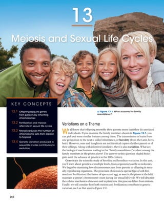
- 1. ▲ Figure 13.1 What accounts for family resemblance? Variations on a Theme We all know that offspring resemble their parents more than they do unrelated individuals. If you examine the family members shown in Figure 13.1, you can pick out some similar features among them. The transmission of traits from one generation to the next is called inheritance, or heredity (from the Latin heres, heir). However, sons and daughters are not identical copies of either parent or of their siblings. Along with inherited similarity, there is also variation. What are the biological mechanisms leading to the “family resemblance” evident among the family members in the photo above? The answer to this question eluded biolo- gists until the advance of genetics in the 20th century. Genetics is the scientific study of heredity and hereditary variation. In this unit, you’ll learn about genetics at multiple levels, from organisms to cells to molecules. We begin by examining how chromosomes pass from parents to offspring in sexu- ally reproducing organisms. The processes of meiosis (a special type of cell divi- sion) and fertilization (the fusion of sperm and egg, as seen in the photo at the left) maintain a species’ chromosome count during the sexual life cycle. We will describe the cellular mechanics of meiosis and explain how this process differs from mitosis. Finally, we will consider how both meiosis and fertilization contribute to genetic variation, such as that seen in Figure 13.1. K E Y C O N C E P T S 13.1 Offspring acquire genes from parents by inheriting chromosomes 13.2 Fertilization and meiosis alternate in sexual life cycles 13.3 Meiosis reduces the number of chromosome sets from diploid to haploid 13.4 Genetic variation produced in sexual life cycles contributes to evolution 252 13 Meiosis and Sexual Life Cycles
- 2. CHAPTER 13 Meiosis and Sexual Life Cycles 253 Comparison of Asexual and Sexual Reproduction Only organisms that reproduce asexually have offspring that are exact genetic copies of themselves. In asexual re- production, a single individual is the sole parent and passes copies of all its genes to its offspring without the fusion of gametes. For example, single-celled eukaryotic organisms can reproduce asexually by mitotic cell division, in which DNA is copied and allocated equally to two daughter cells. The genomes of the offspring are virtually exact copies of the parent’s genome. Some multicellular organisms are also capable of reproducing asexually (Figure 13.2). Because the cells of the offspring are derived by mitosis in the parent, the “chip off the old block” is usually genetically identical to its parent. An individual that reproduces asexually gives rise to a clone, a group of genetically identical individuals. Genetic differences occasionally arise in asexually reproducing or- ganisms as a result of changes in the DNA called mutations, which we will discuss in Chapter 17. In sexual reproduction, two parents give rise to off- spring that have unique combinations of genes inherited from the two parents. In contrast to a clone, offspring of sexual reproduction vary genetically from their siblings and both parents: They are variations on a common theme of family resemblance, not exact replicas. Genetic variation like that shown in Figure 13.1 is an important consequence of sexual reproduction. What mechanisms generate this genetic variation? The key is the behavior of chromosomes during the sexual life cycle. C O N C E P T 13.1 Offspring acquire genes from parents by inheriting chromosomes Family friends may tell you that you have your mother’s freckles or your father’s eyes. Of course, parents do not, in any literal sense, give their children freckles, eyes, hair, or any other traits. What, then, is actually inherited? Inheritance of Genes Parents endow their offspring with coded information in the form of hereditary units called genes. The genes we inherit from our mothers and fathers are our genetic link to our par- ents, and they account for family resemblances such as shared eye color or freckles. Our genes program the specific traits that emerge as we develop from fertilized eggs into adults. The genetic program is written in the language of DNA, the polymer of four different nucleotides you learned about in Concepts 1.1 and 5.5. Inherited information is passed on in the form of each gene’s specific sequence of DNA nucleo- tides, much as printed information is communicated in the form of meaningful sequences of letters. In both cases, the language is symbolic. Just as your brain translates the word apple into a mental image of the fruit, cells translate genes into freckles and other features. Most genes program cells to synthesize specific enzymes and other proteins, whose cumulative action produces an organism’s inherited traits. The programming of these traits in the form of DNA is one of the unifying themes of biology. The transmission of hereditary traits has its molecular basis in the replication of DNA, which produces copies of genes that can be passed from parents to offspring. In animals and plants, reproductive cells called gametes are the vehicles that transmit genes from one generation to the next. During fertilization, male and female gametes (sperm and eggs) unite, passing on genes of both parents to their offspring. Except for small amounts of DNA in mitochondria and chloroplasts, the DNA of a eukaryotic cell is packaged into chromosomes within the nucleus. Every species has a charac- teristic number of chromosomes. For example, humans have 46 chromosomes in their somatic cells—all cells of the body except the gametes and their precursors. Each chromosome consists of a single long DNA molecule elaborately coiled in association with various proteins. One chromosome includes several hundred to a few thousand genes, each of which is a specific sequence of nucleotides within the DNA molecule. A gene’s specific location along the length of a chromosome is called the gene’s locus (plural, loci; from the Latin, meaning “place”). Our genetic endowment (our genome) consists of the genes and other DNA that make up the chromosomes we inherited from our parents. Parent 0.5 mm Bud (a) Hydra (b) Redwoods ▲ Figure 13.2 Asexual reproduction in two multicellular organisms. (a) This relatively simple animal, a hydra, reproduces by budding. The bud, a localized mass of mitotically dividing cells, devel- ops into a small hydra, which detaches from the parent (LM). (b) All the trees in this circle of redwoods arose asexually from a single parent tree, whose stump is in the center of the circle.
- 3. 254 UNIT THREE Genetics C O N C E P T C H E C K 1 3 . 1 1. M A K E C O N N E C T I O N S Using what you know of gene expression in a cell, explain what causes the traits of parents (such as hair color) to show up in their offspring. (See Concept 5.5.) 2. How do asexually reproducing eukaryotic organisms produce offspring that are genetically identical to each other and to their parents? 3. W H AT I F ? A horticulturalist breeds orchids, trying to ob- tain a plant with a unique combination of desirable traits. After many years, she finally succeeds.To produce more plants like this one, should she cross-breed it with another plant or clone it? Why? For suggested answers, see Appendix A. C O N C E P T 13.2 Fertilization and meiosis alternate in sexual life cycles A life cycle is the generation-to-generation sequence of stages in the reproductive history of an organism, from conception to production of its own offspring. In this section, we use humans as an example to track the behavior of chromosomes through the sexual life cycle. We begin by considering the chromosome count in human somatic cells and gametes. We will then ex- plore how the behavior of chromosomes relates to the human life cycle and other types of sexual life cycles. Sets of Chromosomes in Human Cells In humans, each somatic cell has 46 chromosomes. During mitosis, the chromosomes become condensed enough to be visible under a light microscope. At this point, they can be distinguished from one another by their size, the positions of their centromeres, and the pattern of colored bands pro- duced by certain chromatin-binding stains. Careful examination of a micrograph of the 46 human chromosomes from a single cell in mitosis reveals that there are two chromosomes of each of 23 types. This becomes clear when images of the chromosomes are arranged in pairs, starting with the longest chromosomes. The resulting ordered display is called a karyotype (Figure 13.3). The two chromosomes of a pair have the same length, centromere position, and staining pattern: These are called homologous chromosomes, or homologs. Both chromosomes of each pair carry genes controlling the same inherited characters. For example, if a gene for eye color is situated at a particular locus on a certain chromosome, then the homolog of that chromosome will also have a version of the same gene speci- fying eye color at the equivalent locus. The two distinct chromosomes referred to as X and Y are an important exception to the general pattern of homolo- gous chromosomes in human somatic cells. Human females Research Method▼ Figure 13.3 Application A karyotype is a display of condensed chromosomes arranged in pairs. Karyotyping can be used to screen for defective chromosomes or abnormal numbers of chromosomes associated with certain congenital disorders, such as Down syndrome. Preparing a Karyotype Technique Karyotypes are prepared from isolated somatic cells, which are treated with a drug to stimulate mitosis and then grown in culture for several days. Cells arrested in metaphase, when chromo- somes are most highly condensed, are stained and then viewed with a microscope equipped with a digital camera. An image of the chromo- somes is displayed on a computer monitor, and digital software is used to arrange them in pairs according to their appearance. 5 μm Pair of homologous duplicated chromosomes Centromere Sister chromatids Metaphase chromosome Results This karyotype shows the chromosomes from a normal human male, digitally colored to emphasize their banding patterns. The size of the chromosome, position of the centromere, and pattern of stained bands help identify specific chromosomes. Although difficult to discern in the karyotype, each metaphase chromosome consists of two closely attached sister chromatids (see the diagram of a pair of homologous duplicated chromosomes).
- 4. CHAPTER 13 Meiosis and Sexual Life Cycles 255 have a homologous pair of X chromosomes (XX), but males have one X and one Y chromosome (XY). Only small parts of the X and Y are homologous. Most of the genes carried on the X chromosome do not have counterparts on the tiny Y, and the Y chromosome has genes lacking on the X. Because they determine an individual’s sex, the X and Y chromosomes are called sex chromosomes. The other chromosomes are called autosomes. The pairs of homologous chromosomes in each human somatic cell is a consequence of our sexual origins. We in- herit one chromosome of a pair from each parent. Thus, the 46 chromosomes in our somatic cells are actually two sets of 23 chromosomes—a maternal set (from our mother) and a paternal set (from our father). The number of chro- mosomes in a single set is represented by n. Any cell with two chromosome sets is called a diploid cell and has a dip- loid number of chromosomes, abbreviated 2n. For humans, the diploid number is 46 (2n = 46), the number of chromo- somes in our somatic cells. In a cell in which DNA synthe- sis has occurred, all the chromosomes are duplicated, and therefore each consists of two identical sister chromatids, associated closely at the centromere and along the arms. (Even though the chromosomes are duplicated, we still say the cell is diploid (2n) because it has only two sets of infor- mation.) Figure 13.4 helps clarify the various terms that we use to describe duplicated chromosomes in a diploid cell. Unlike somatic cells, gametes contain a single set of chro- mosomes. Such cells are called haploid cells, and each has a haploid number of chromosomes (n). For humans, the haploid number is 23 (n = 23). The set of 23 consists of the 22 autosomes plus a single sex chromosome. An unfertilized egg contains an X chromosome, but a sperm may contain an X or a Y chromosome. Each sexually reproducing species has a characteristic diploid and haploid number. For example, the fruit fly Dro- sophila melanogaster has a diploid number (2n) of 8 and a haploid number (n) of 4, while for dogs, 2n is 78 and n is 39. Now let’s consider chromosome behavior during sexual life cycles. We’ll use the human life cycle as an example. Behavior of Chromosome Sets in the Human Life Cycle The human life cycle begins when a haploid sperm from the father fuses with a haploid egg from the mother (Figure 13.5). This union of gametes, culminating in fusion of their nuclei, is called fertilization. The resulting fertilized egg, or zygote, is diploid because it contains two haploid sets of chromosomes bearing genes representing the maternal and paternal family Key Maternal set of chromosomes (n = 3) Sister chromatids of one duplicated chromosome Two nonsister chromatids in a homologous pair 2n = 6 Paternal set of chromosomes (n = 3) Centromere Pair of homologous chromosomes (one from each set) ▲ Figure 13.4 Describing chromosomes. A cell from an organism with a diploid number of 6 (2n = 6) is depicted here following chromo- some duplication and condensation. Each of the six duplicated chro- mosomes consists of two sister chromatids associated closely along their lengths. Each homologous pair is composed of one chromosome from the maternal set (red) and one from the paternal set (blue). Each set is made up of three chromosomes in this example (long, medium, and short). Together, one maternal and one paternal chromatid in a pair of homologous chromosomes are called nonsister chromatids. ? How many sets of chromosomes are present in this diagram? How many pairs of homologous chromosomes are present? Haploid (n) Diploid (2n) MEIOSIS Multicellular diploid adults (2n = 46) Diploid zygote (2n = 46) Testis Sperm (n) Egg (n) Haploid gametes (n = 23) Ovary Mitosis and development FERTILIZATION Key ▲ Figure 13.5 The human life cycle. In each generation, the num- ber of chromosome sets doubles at fertilization but is halved during meiosis. For humans, the number of chromosomes in a haploid cell is 23, consisting of one set (n = 23); the number of chromosomes in the diploid zygote and all somatic cells arising from it is 46, consisting of two sets (2n = 46). This figure introduces a color code that will be used for other life cycles later in this book. The aqua arrows identify haploid stages of a life cycle, and the tan arrows identify diploid stages.
- 5. 256 UNIT THREE Genetics lines. As a human develops into a sexually mature adult, mi- tosis of the zygote and its descendant cells generates all the somatic cells of the body. Both chromosome sets in the zygote and all the genes they carry are passed with precision to the somatic cells. The only cells of the human body not produced by mitosis are the gametes, which develop from specialized cells called germ cells in the gonads—ovaries in females and testes in males. Imagine what would happen if human gametes were made by mitosis: They would be diploid like the somatic cells. At the next round of fertilization, when two gametes fused, the normal chromosome number of 46 would double to 92, and each subsequent generation would double the number of chromosomes yet again. This does not happen, however, because in sexually reproducing organisms, gamete formation involves a type of cell division called meiosis. This type of cell division reduces the number of sets of chromo- somes from two to one in the gametes, counterbalancing the doubling that occurs at fertilization. As a result of meiosis, each human sperm and egg is haploid (n = 23). Fertilization restores the diploid condition by combining two haploid sets of chromosomes, and the human life cycle is repeated, gen- eration after generation (see Figure 13.5). In general, the steps of the human life cycle are typical of many sexually reproducing animals. Indeed, the processes of fertilization and meiosis are the hallmarks of sexual re- production in plants, fungi, and protists as well as in ani- mals. Fertilization and meiosis alternate in sexual life cycles, maintaining a constant number of chromosomes in each species from one generation to the next. MEIOSIS FERTILIZATION MEIOSIS FERTILIZATION Mitosis Mitosis Mitosis (a) Animals (b) Plants and some algae Gametes Gametes Zygote ZygoteDiploid multicellular organism Haploid multi- cellular organism (gametophyte) Diploid multicellular organism (sporophyte) n 2n 2n FERTILIZATIONMEIOSIS Mitosis Spores 2n 2n 2n n n n n n n n n n Mitosis Mitosis (c) Most fungi and some protists Gametes Zygote Haploid unicellular or multicellular organism n n n Haploid (n) Diploid (2n) Key ▲ Figure 13.6 Three types of sexual life cycles. The common feature of all three cycles is the alternation of meiosis and fertilization, key events that contribute to genetic variation among offspring. The cycles differ in the timing of these two key events. The Variety of Sexual Life Cycles Although the alternation of meiosis and fertilization is com- mon to all organisms that reproduce sexually, the timing of these two events in the life cycle varies, depending on the spe- cies. These variations can be grouped into three main types of life cycles. In the type that occurs in humans and most other animals, gametes are the only haploid cells (Figure 13.6a). Meiosis occurs in germ cells during the production of gam- etes, which undergo no further cell division prior to fertiliza- tion. After fertilization, the diploid zygote divides by mitosis, producing a multicellular organism that is diploid. Plants and some species of algae exhibit a second type of life cycle called alternation of generations (Figure 13.6b). This type includes both diploid and haploid stages that are multicellular. The multicellular diploid stage is called the sporophyte. Meiosis in the sporophyte produces haploid cells called spores. Unlike a gamete, a haploid spore doesn’t fuse with another cell but divides mitotically, generating a mul- ticellular haploid stage called the gametophyte. Cells of the gametophyte give rise to gametes by mitosis. Fusion of two haploid gametes at fertilization results in a diploid zygote, which develops into the next sporophyte generation. There- fore, in this type of life cycle, the sporophyte generation pro- duces a gametophyte as its offspring, and the gametophyte generation produces the next sporophyte generation (see Figure 13.6b). The term alternation of generations fits well as a name for this type of life cycle. A third type of life cycle occurs in most fungi and some protists, including some algae (Figure 13.6c). After gametes
- 6. CHAPTER 13 Meiosis and Sexual Life Cycles 257 along their lengths; this association is called sister chromatid cohesion. Together, the sister chromatids make up one du- plicated chromosome (see Figure 13.4). In contrast, the two chromosomes of a homologous pair are individual chromo- somes that were inherited from different parents. Homologs appear alike in the microscope, but they may have different versions of genes, each called an allele, at corresponding loci. Homologs are not associated with each other in any ob- vious way except during meiosis. Figure 13.8 describes in detail the stages of the two divi- sions of meiosis for an animal cell whose diploid number is 6. Study this figure thoroughly before going on. C O N C E P T C H E C K 1 3 . 2 1. M A K E C O N N E C T I O N S In Figure 13.4, how many DNA molecules (double helices) are present (see Figure 12.5)? What is the haploid number of this cell? Is a set of chromosomes haploid or diploid? 2. In the karyotype shown in Figure 13.3, how many pairs of chromosomes are present? How many sets? 3. W H AT I F ? A certain eukaryote lives as a unicellular organism, but during environmental stress, it produces gametes.The gametes fuse, and the resulting zygote un- dergoes meiosis, generating new single cells. What type of organism could this be? For suggested answers, see Appendix A. C O N C E P T 13.3 Meiosis reduces the number of chromosome sets from diploid to haploid Many of the steps of meiosis closely resemble corresponding steps in mitosis. Meiosis, like mitosis, is preceded by the du- plication of chromosomes. However, this single duplication is followed by not one but two consecutive cell divisions, called meiosis I and meiosis II. These two divisions result in four daughter cells (rather than the two daughter cells of mitosis), each with only half as many chromosomes as the parent cell—one set, rather than two. The Stages of Meiosis The overview of meiosis in Figure 13.7 shows, for a single pair of homologous chromosomes in a diploid cell, that both members of the pair are duplicated and the copies sorted into four haploid daughter cells. Recall that sister chroma- tids are two copies of one chromosome, closely associated all fuse and form a diploid zygote, meiosis occurs without a multicellular diploid offspring developing. Meiosis produces not gametes but haploid cells that then divide by mitosis and give rise to either unicellular descendants or a haploid multicellular adult organism. Subsequently, the haploid or- ganism carries out further mitoses, producing the cells that develop into gametes. The only diploid stage found in these species is the single-celled zygote. Note that either haploid or diploid cells can divide by mi- tosis, depending on the type of life cycle. Only diploid cells, however, can undergo meiosis because haploid cells have only a single set of chromosomes that cannot be further reduced. Though the three types of sexual life cycles differ in the timing of meiosis and fertilization, they share a funda- mental result: genetic variation among offspring. Interphase Meiosis I Meiosis II Pair of homologous chromosomes in diploid parent cell Chromosomes duplicate Diploid cell with duplicated chromosomes Haploid cells with duplicated chromosomes Pair of duplicated homologous chromosomes Sister chromatids Haploid cells with unduplicated chromosomes Homologous chromosomes separate Sister chromatids separate 1 2 ▲ Figure 13.7 Overview of meiosis: how meiosis reduces chro- mosome number. After the chromosomes duplicate in interphase, the diploid cell divides twice, yielding four haploid daughter cells. This overview tracks just one pair of homologous chromosomes, which for the sake of simplicity are drawn in the condensed state throughout. D R AW I T Redraw the cells in this figure using a simple double helix to represent each DNA molecule.
- 7. Chiasmata Spindle Homologous chromosomes Fragments of nuclear envelope Sister chromatids Centromere (with kinetochore) Metaphase plate Microtubules attached to kinetochore Sister chromatids remain attached Homologous chromosomes separate Duplicated homologous chromosomes (red and blue) pair and exchange segments; 2n = 6 in this example. Chromosomes line up by homologous pairs. Each pair of homologous chromosomes separates. Two haploid cells form; each chromosome still consists of two sister chromatids. Cleavage furrow Centrosome (with centriole pair) Prophase I crossing over chiasmata chiasma Metaphase I Anaphase I Telophase I and Cytokinesis MEIOSIS I: Separates homologous chromosomes Prophase I Metaphase I Anaphase I Telophase I and Cytokinesis 258 UNIT THREE Genetics ▼ Figure 13.8 Exploring Meiosis in an Animal Cell
- 8. Sister chromatids separate Haploid daughter cells forming During another round of cell division, the sister chromatids finally separate; four haploid daughter cells result, containing unduplicated chromosomes. Prophase II Metaphase II not Anaphase II Telophase II and Cytokinesis MEIOSIS II: Separates sister chromatids Prophase II Metaphase II Anaphase II Telophase II and Cytokinesis CHAPTER 13 Meiosis and Sexual Life Cycles 259 M A K E C O N N E C T I O N S Look at Figure 12.7 and imagine the two daughter cells undergoing another round of mitosis, yielding four cells. Compare the number of chromosomes in each of those four cells, after mitosis, with the number in each cell in Figure 13.8, after meiosis. What is it about the process of meiosis that accounts for this difference, even though meiosis also includes two cell divisions? Visit the Study Area in MasteringBiology for the BioFlix® 3-D Animation on Meiosis. BioFlix Tutorials can also be assigned in MasteringBiology. A N I M AT I O N
- 9. 260 UNIT THREE Genetics Crossing Over and Synapsis During Prophase I Prophase I of meiosis is a very busy time. The prophase I cell shown in Figure 13.8 is at a point fairly late in pro- phase I, when homologous pairing, crossing over, and chromosome condensation have already taken place. The sequence of events leading up to that point is shown in more detail in Figure 13.9. After interphase, the chromosomes have been duplicated and the sister chromatids are held together by proteins called cohesins. Early in prophase I, the two members of a homologous pair associate loosely along their length. Each gene on one homolog is aligned precisely with the cor- responding gene on the other homolog. The DNA of two nonsister chromatids—one maternal and one paternal—is broken by specific proteins at precisely corresponding points. Next, the formation of a zipper-like structure called the synaptonemal complex holds one homolog tightly to the other. During this association, called synapsis, the DNA breaks are closed up so that each broken end is joined to the corresponding segment of the nonsister chromatid. Thus, a paternal chromatid is joined to a piece of maternal chroma- tid beyond the crossover point, and vice versa. These points of crossing over become visible as chiasmata (singular, chiasma) after the synaptonemal complex disas- sembles and the homologs move slightly apart from each other. The homologs remain attached because sister chro- matids are still held together by sister chromatid cohesion, even though some of the DNA may no longer be attached to its original chromosome. At least one crossover per chro- mosome must occur in order for the homologous pair to stay together as it moves to the metaphase I plate. A Comparison of Mitosis and Meiosis Figure 13.10 summarizes the key differences between meiosis and mitosis in diploid cells. Basically, meiosis re- duces the number of chromosome sets from two (diploid) to one (haploid), whereas mitosis conserves the number of chromosome sets. Therefore, meiosis produces cells that differ genetically from their parent cell and from each other, whereas mitosis produces daughter cells that are genetically identical to their parent cell and to each other. Three events unique to meiosis occur during meiosis I: 1. Synapsis and crossing over. During prophase I, du- plicated homologs pair up and crossing over occurs, as described above. Synapsis and crossing over normally do not occur during prophase of mitosis. 2. Homologous pairs at the metaphase plate. At meta- phase I of meiosis, chromosomes are positioned at the metaphase plate as pairs of homologs, rather than indi- vidual chromosomes, as in metaphase of mitosis. DNA breaks Centromere Crossover Crossover Paternal sister chromatids Pair of homologous chromosomes: Maternal sister chromatids DNA breaks Synaptonemal complex forming Cohesins Chiasmata 1 After interphase, the chromosomes have been dupli- cated and sister chromatids are held together by proteins called cohesins (purple). Each pair of homologs associate along their length. The DNA molecules of two nonsister chromatids are broken at precisely corresponding points. The chromatin of the chromosomes is beginning to condense. 2 A zipperlike protein complex, the synaptonemal complex (green), begins to form, attaching one homolog to the other. The chromatin continues to condense. 3 The synaptonemal complex is fully formed; the two homologs are said to be in synapsis. During synapsis, the DNA breaks are closed up when each broken end is joined to the corresponding segment of the nonsister chromatid, producing crossovers. 4 After the synaptonemal complex disassembles, the homologs move slightly apart from each other but remain attached because of sister chromatid cohesion, even though some of the DNA may no longer be attached to its original chromosome. The points of attachment where crossovers have occurred show up as chiasmata. The chromosomes continue to condense as they move toward the metaphase plate. ▲ Figure 13.9 Crossing over and synapsis in prophase I: a closer look. For simplicity, the four chromatids of the homologous pair shown here are depicted side by side, but in reality, the blue chromosome would be right on top of the red one (see the top cell in Figure 13.12).
- 10. C HAPTER 13 Meiosis and Sexual Life Cycles 261 Daughter cells of meiosis II Chromosome duplication Chromosome duplication Sister chroma- tids separate during anaphase II. n MITOSIS MEIOSIS Parent cell (before chromosome duplication) Mitosis (occurs in both diploid and haploid cells) Meiosis (can only occur in diploid cells) Occurs during interphase before mitosis begins Occurs during interphase before meiosis I begins Enables multicellular animal or plant (gametophyte or sporophyte) to arise from a single cell; produces cells for growth, repair, and, in some species, asexual reproduction; produces gametes in the gametophyte plant Produces gametes (in animals) or spores (in the sporophyte plant); reduces number of chromosome sets by half and introduces genetic variability among the gametes or spores Two, each genetically identical to the parent cell, with the same number of chromosomes Four, each haploid (n); genetically different from the parent cell and from each other One, including prophase, prometaphase, metaphase, anaphase, and telophase Occurs during prophase I along with crossing over between nonsister chromatids; resulting chiasmata hold pairs together due to sister chromatid cohesion SUMMARY Property DNA replication Role in the animal or plant body Number of daughter cells and genetic composition Number of divisions Does not occurSynapsis of homologous chromosomes n n n Prophase Metaphase Anaphase Telophase Anaphase I Telophase I Metaphase I MEIOSIS II MEIOSIS I Prophase I Duplicated chromosome (two sister chromatids) Chiasma (site of crossing over) Homologous chromosome pair held together by chiasma and sister chromatid cohesion Individual chromosomes line up at the metaphase plate. Pairs of homologous chromosomes line up at the metaphase plate. Daughter cells of meiosis I Haploid n = 3 Daughter cells of mitosis Sister chromatids separate during anaphase. Homologs separate during anaphase I; sister chromatids remain attached at centromere. 2n2n 2n = 6 Two, each including prophase, metaphase, anaphase, and telophase ▲ Figure 13.10 A comparison of mitosis and meiosis. D R AW I T Could any other combinations of chromosomes be generated during meiosis II from the specific cells shown in telophase I? Explain. (Hint: Draw the cells as they would appear in metaphase II.)
- 11. 262 UNIT THREE Genetics 3. Separation of homologs. At anaphase I of meiosis, the duplicated chromosomes of each homologous pair move toward opposite poles, but the sister chromatids of each duplicated chromosome remain attached. In anaphase of mitosis, by contrast, sister chromatids separate. Sister chromatids stay together due to sister chromatid co- hesion, mediated by cohesin proteins. In mitosis, this attach- ment lasts until the end of metaphase, when enzymes cleave the cohesins, freeing the sister chromatids to move to oppo- site poles of the cell. In meiosis, sister chromatid cohesion is released in two steps, one at the start of anaphase I and one at anaphase II. In metaphase I, homologs are held together by cohesion between sister chromatid arms in regions beyond points of crossing over, where stretches of sister chromatids now belong to different chromosomes. The combination of crossing over and sister chromatid cohesion along the arms results in the formation of a chiasma. Chiasmata hold homologs together as the spindle forms for the first meiotic division. At the onset of anaphase I, the release of cohesion along sister chromatid arms allows homologs to separate. At anaphase II, the release of sister chromatid cohesion at the centromeres allows the sister chromatids to separate. Thus, sister chromatid cohesion and crossing over, acting together, play an essential role in the lining up of chromosomes by ho- mologous pairs at metaphase I. Meiosis I is called the reductional division because it re- duces the number of chromosome sets from two (diploid) to one (haploid). During meiosis II (the equational division), sister chromatids separate, producing haploid daughter cells. The mechanism for separating sister chromatids is virtually identical in meiosis II and mitosis. The molecular basis of chromosome behavior during meiosis continues to be a focus of intense research. In the Scientific Skills Exercise, you can work with data tracking the amount of DNA in cells as they progress through meiosis. S C I E N T I F I C S K I L L S E X E R C I S E How Does DNA Content Change as Budding Yeast Cells Pro- ceed Through Meiosis? When nutrients are low, cells of the budding yeast (Saccharomyces cerevisiae) exit the mitotic cell cycle and enter meiosis. In this exercise, you will track the DNA content of a population of yeast cells as they progress through meiosis. How the Experiment Was Done Researchers grew a culture of yeast cells in a nutrient-rich medium and then transferred them to a nutrient- poor medium to induce meiosis. At different times after induction, the DNA content per cell was measured in a sample of the cells, and the average DNA content per cell was recorded in femtograms (fg; 1 femto- gram = 1 * 10-15 gram). Data from the Experiment Time After Induction (hours) Average Amount of DNA per Cell (fg) 0.0 24.0 1.0 24.0 2.0 40.0 3.0 47.0 4.0 47.5 5.0 48.0 6.0 48.0 7.0 47.5 7.5 25.0 8.0 24.0 9.0 23.5 9.5 14.0 10.0 13.0 11.0 12.5 12.0 12.0 13.0 12.5 14.0 12.0 Making a Line Graph and Converting Between Units of Data Interpret the Data 1. First, set up your graph. (a) Place the labels for the independent variable and the dependent variable on the appropriate axes, followed by units of measurement in parentheses. Explain your choices. (b) Add tick marks and values for each axis in your graph. Explain your choices. (For ad- ditional information about graphs, see the Scientific Skills Review in Appendix F and in the Study Area in MasteringBiology.) 2. Because the variable on the x-axis varies continuously, it makes sense to plot the data on a line graph. (a) Plot each data point from the table onto the graph. (b) Connect the data points with line segments. 3. Most of the yeast cells in the culture were in G1 of the cell cycle before being moved to the nutrient-poor medium. (a) How many femtograms of DNA are there in each yeast cell in G1? Estimate this value from the data in your graph. (b) How many femtograms of DNA should be pres- ent in each cell in G2? (See Concept 12.2 and Figure 12.6.) At the end of meiosis I (MI)? At the end of meiosis II (MII)? (See Figure 13.7.) (c) Using these values as a guideline, distinguish the different phases by inserting vertical dashed lines in the graph between phases and label each phase (G1, S, G2, MI, MII). You can figure out where to put the dividing lines based on what you know about the DNA content of each phase (see Figure 13.7). (d) Think carefully about the point where the line at the highest value begins to slope downward. What specific point of meiosis does this “corner” represent? What stage(s) corre- spond to the downward sloping line? 4. Given the fact that 1 fg of DNA = 9.78 * 105 base pairs (on average), you can convert the amount of DNA per cell to the length of DNA in numbers of base pairs. (a) Calculate the number of base pairs of DNA in the haploid yeast genome. Express your answer in millions of base pairs (Mb), a standard unit for expressing genome size. Show your work. (b) How many base pairs per minute were synthesized during the S phase of these yeast cells? A version of this Scientific Skills Exercise can be assigned in MasteringBiology. Further Reading G. Simchen, Commitment to meiosis: what determines the mode of division in budding yeast? BioEssays 31:169–177 (2009).
- 12. C HAPTER 13 Meiosis and Sexual Life Cycles 263 Because each pair of homologous chromosomes is posi- tioned independently of the other pairs at metaphase I, the first meiotic division results in each pair sorting its maternal and paternal homologs into daughter cells independently of every other pair. This is called independent assortment. Each daughter cell represents one outcome of all possible combi- nations of maternal and paternal chromosomes. As shown in Figure 13.11, the number of combinations possible for daughter cells formed by meiosis of a diploid cell with two pairs of homologous chromosomes (n = 2) is four: two possible arrangements for the first pair times two possible arrangements for the second pair. Note that only two of the four combinations of daughter cells shown in the figure would result from meiosis of a single diploid cell, because a single parent cell would have one or the other possible chro- mosomal arrangement at metaphase I, but not both. How- ever, the population of daughter cells resulting from meiosis of a large number of diploid cells contains all four types in approximately equal numbers. In the case of n = 3, eight combinations of chromosomes are possible for daughter cells. More generally, the number of possible combinations when chromosomes sort independently during meiosis is 2n , where n is the haploid number of the organism. In the case of humans (n = 23), the number of possible combinations of maternal and paternal chromosomes in the resulting gametes is 223 , or about 8.4 million. Each gamete that you produce in your lifetime contains one of roughly 8.4 million possible combinations of chromosomes. Crossing Over As a consequence of the independent assortment of chro- mosomes during meiosis, each of us produces a collection of gametes differing greatly in their combinations of the chro- mosomes we inherited from our two parents. Figure 13.11 C O N C E P T C H E C K 1 3 . 3 1. M A K E C O N N E C T I O N S Compare the chromosomes in a cell at metaphase of mitosis with those in a cell at metaphase II. (See Figures 12.7 and 13.8.) 2. W H AT I F ? After the synaptonemal complex disap- pears, how would the two homologs be associated if crossing over did not occur? What effect might this ulti- mately have on gamete formation? For suggested answers, see Appendix A. C O N C E P T 13.4 Genetic variation produced in sexual life cycles contributes to evolution How do we account for the genetic variation of the family members in Figure 13.1? As you will learn in later chapters, mutations are the original source of genetic diversity. These changes in an organism’s DNA create the different versions of genes known as alleles. Once these differences arise, re- shuffling of the alleles during sexual reproduction produces the variation that results in each member of a sexually re- producing population having a unique combination of traits. Origins of Genetic Variation Among Offspring In species that reproduce sexually, the behavior of chromo- somes during meiosis and fertilization is responsible for most of the variation that arises in each generation. Three mecha- nisms contribute to the genetic variation arising from sexual reproduction: independent assortment of chromosomes, crossing over, and random fertilization. Independent Assortment of Chromosomes One aspect of sexual reproduction that generates ge- netic variation is the random orientation of pairs of homologous chromosomes at metaphase of meiosis I. At metaphase I, the homologous pairs, each consisting of one maternal and one paternal chromosome, are situated at the metaphase plate. (Note that the terms maternal and paternal refer, respectively, to the mother and father of the individual whose cells are undergoing meiosis.) Each Combination 1 Combination 2 Combination 3 Combination 4 Two equally probable arrangements of chromosomes at metaphase I Possibility 1 Possibility 2 Metaphase II Daughter cells ▲ Figure 13.11 The independent assortment of homologous chromosomes in meiosis. pair may orient with either its maternal or paternal homolog closer to a given pole—its orientation is as random as the flip of a coin. Thus, there is a 50% chance that a particular daughter cell of meiosis I will get the maternal chromo- some of a certain homologous pair and a 50% chance that it will get the pater- nal chromosome.
- 13. 264 UNIT THREE Genetics suggests that each chromosome in a gamete is exclusively maternal or paternal in origin. In fact, this is not the case, be- cause crossing over produces recombinant chromosomes, individual chromosomes that carry genes (DNA) derived from two different parents (Figure 13.12). In meiosis in humans, an average of one to three crossover events occur per chromosome pair, depending on the size of the chromo- somes and the position of their centromeres. As you learned in Figure 13.9, crossing over produces chromosomes with new combinations of maternal and pa- ternal alleles. At metaphase II, chromosomes that contain one or more recombinant chromatids can be oriented in two alternative, nonequivalent ways with respect to other chromosomes, because their sister chromatids are no longer identical (see Figure 13.12). The different possible arrange- ments of nonidentical sister chromatids during meiosis II further increase the number of genetic types of daughter cells that can result from meiosis. You will learn more about crossing over in Chapter 15. The important point for now is that crossing over, by combining DNA inherited from two parents into a single chromosome, is an important source of genetic variation in sexual life cycles. Random Fertilization The random nature of fertilization adds to the genetic varia- tion arising from meiosis. In humans, each male and female gamete represents one of about 8.4 million (223 ) possible chromosome combinations due to independent assort- ment. The fusion of a male gamete with a female gamete during fertilization will produce a zygote with any of about 70 trillion (223 * 223 ) diploid combinations. If we factor in the variation brought about by crossing over, the number of possibilities is truly astronomical. It may sound trite, but you really are unique. The Evolutionary Significance of Genetic Variation Within Populations E VO L U T I O N Now that you’ve learned how new combina- tions of genes arise among offspring in a sexually repro- ducing population, let’s see how the genetic variation in a population relates to evolution. Darwin recognized that a population evolves through the differential reproductive success of its variant members. On average, those individu- als best suited to the local environment leave the most offspring, thereby transmitting their genes. Thus, natural selection results in the accumulation of genetic variations favored by the environment. As the environment changes, the population may survive if, in each generation, at least some of its members can cope effectively with the new con- ditions. Mutations are the original source of different alleles, which are then mixed and matched during meiosis. New and different combinations of alleles may work better than those that previously prevailed. In a stable environment, though, sexual reproduction seems as if it would be less advantageous than asexual repro- duction, which ensures perpetuation of successful combina- tions of alleles. Furthermore, sexual reproduction is more expensive energetically than asexual reproduction. In spite of these apparent disadvantages, sexual reproduction is almost universal among animals. Why is this? The ability of sexual reproduction to generate genetic di- versity is the most commonly proposed explanation for the evolutionary persistence of this process. Consider the rare case of the bdelloid rotifer Figure 13.13. This group has ap- parently not reproduced sexually throughout the 40 million years of its evolutionary history. Does this mean that genetic diversity is not advantageous in this species? It turns out that bdelloid rotifers are an exception that proves the rule: This group has mechanisms other than sexual reproduction for generating genetic diversity. For example, they live in 1 2 3 Prophase I of meiosis Anaphase I Anaphase II Daughter cells Recombinant chromosomes TEM Nonsister chromatids held together during synapsis Pair of homologs Chiasma, site of crossing over Centromere In prophase I, synapsis and crossing over occur; then homologs move apart slightly. Chiasmata and attachments between sister chromatids hold homologs together; they move to the metaphase I plate. Breakdown of proteins holding sister chroma- tid arms together allows homologs with recombinant chroma- tids to separate. ▲ Figure 13.12 The results of crossing over during meiosis.
- 14. offspring resemble—but are not identical to—their parents. Ironically, Gregor Mendel, a contemporary of Darwin, pub- lished a theory of inheritance that helps explain genetic vari- ation, but his discoveries had no impact on biologists until 1900, more than 15 years after Darwin (1809–1882) and Mendel (1822–1884) had died. In the next chapter, you’ll learn how Mendel discovered the basic rules governing the inheritance of specific traits. environments that can dry up for long periods of time, during which they can enter a state of suspended anima- tion. In this state, their cell membranes may crack in places, allowing entry of DNA from other rotifers and even other species. Evidence suggests that this DNA can become incorporated into the genome of the rotifer, leading to increased genetic diversity. This supports the idea that genetic diversity is advantageous, and that sexual reproduction has persisted because it generates such diversity. In this chapter, we have seen how sexual reproduction greatly increases the genetic variation present in a popula- tion. Although Darwin realized that heritable variation is what makes evolution possible, he could not explain why 200 μm ▲ Figure 13.13 A bdelloid rotifer, an animal that repro- duces only asexually. C O N C E P T C H E C K 1 3 . 4 1. What is the original source of variation among the different alleles of a gene? 2. The diploid number for fruit flies is 8, and the diploid num- ber for grasshoppers is 46. If no crossing over took place, would the genetic variation among offspring from a given pair of parents be greater in fruit flies or grasshop- pers? Explain. 3. W H AT I F ? If maternal and paternal chromatids have the same two alleles for every gene, will crossing over lead to genetic variation? For suggested answers, see Appendix A. 13 Chapter Review Sexual life cycles differ in the timing of meiosis relative to fertil- ization and in the point(s) of the cycle at which a multicellular organism is produced by mitosis. ? Compare the life cycles of animals and plants, mentioning their similarities and differences. C O N C E P T 13.3 Meiosis reduces the number of chromosome sets from diploid to haploid (pp. 257–263) The two cell divisions of meiosis, meiosis I and meiosis II, pro- duce four haploid daughter cells. The number of chromosome sets is reduced from two (diploid) to one (haploid) during meio- sis I, the reductional division. Meiosis is distinguished from mitosis by three events of meiosis I: SUMMARY OF KEY CONCEPTS C O N C E P T 13.1 Offspring acquire genes from parents by inheriting chromosomes (pp. 253–254) Each gene in an organism’s DNA exists at a specific locus on a certain chromosome. In asexual reproduction, a single parent produces genetically identical offspring by mitosis. Sexual reproduction combines genes from two parents, leading to genetically diverse offspring. ? Explain why human offspring resemble their parents but are not identical to them. C O N C E P T 13.2 Fertilization and meiosis alternate in sexual life cycles (pp. 254–257) Normal human somatic cells are diploid. They have 46 chro- mosomes made up of two sets of 23, one set from each parent. Human diploid cells have 22 homologous pairs of autosomes, and one pair of sex chromosomes; the latter determines whether the person is female (XX) or male (XY). In humans, ovaries and testes produce haploid gametes by meiosis, each gamete containing a single set of 23 chromosomes (n = 23). During fertilization, an egg and sperm unite, forming a diploid (2n = 46) single-celled zygote, which develops into a multicellular organism by mitosis. CHAPTER 13 Meiosis and Sexual Life Cycles 265 Prophase I: Each homologous pair undergoes synapsis and crossing over between nonsister chromatids with the subsequent appearance of chiasmata. Metaphase I: Chromosomes line up as homologous pairs on the metaphase plate. Anaphase I: Homologs separate from each other; sister chromatids remain joined at the centromere. Meiosis II separates the sister chromatids.
- 15. 266 UNIT THREE Genetics 11. SYNTHESIZE YOUR KNOWLEDGE The Cavendish banana is the most popular fruit in the world, but is currently threatened by extinction due to a fungal agent (see the photo). This banana variety is “triploid” (3n, with three sets of chromosomes) and can only reproduce through cloning by cultivators. Given what you know about meiosis, explain how the banana’s triploid number accounts for its seedless condition. Considering genetic diversity, discuss how the ab- sence of sexual reproduction might contribute to the vulner- ability of this domesticated species to infectious agents. For selected answers, see Appendix A. 6. D R AW I T The diagram at right shows a cell in meiosis. (a) Label the appropriate structures with these terms, drawing lines or brackets as needed: chromosome (label as duplicated or unduplicated), centromere, kinetochore, sister chromatids, nonsister chromatids, homologous pair, homologs, chiasma, sister chromatid cohesion, alleles (of the F and H genes). (b) Describe the makeup of a haploid set and a diploid set. (c) Identify the stage of meiosis shown. Sister chromatid cohesion and crossing over allow chiasmata to hold homologs together until anaphase I. Cohesins are cleaved along the arms at anaphase I, allowing homologs to separate, and at the centromeres in anaphase II, releasing sister chromatids. ? In prophase I, homologous chromosomes pair up and undergo syn- apsis and crossing over. Can this also occur during prophase II? Explain. C O N C E P T 13.4 Genetic variation produced in sexual life cycles contributes to evolution (pp. 263–265) Three events in sexual reproduction contribute to genetic variation in a population: independent assortment of chromosomes during meiosis I, crossing over during meiosis I, and random fertiliza- tion of egg cells by sperm. During crossing over, DNA of nonsister chromatids in a homologous pair is broken and rejoined. Genetic variation is the raw material for evolution by natural se- lection. Mutations are the original source of this variation; recom- bination of variant genes generates additional genetic diversity. ? Explain how three processes unique to meiosis generate a great deal of genetic variation. TEST YOUR UNDERSTANDING LEVEL 1: KNOWLEDGE/COMPREHENSION 1. A human cell containing 22 autosomes and a Y chromosome is a. a sperm. b. an egg. F H Students Go to MasteringBiology for assignments, the eText, and the Study Area with practice tests, animations, and activities. Instructors Go to MasteringBiology for automatically graded tutorials and questions that you can assign to your students, plus Instructor Resources. c. a zygote. d. a somatic cell of a male. 2. Homologous chromosomes move toward opposite poles of a dividing cell during a. mitosis. b. meiosis I. LEVEL 2: APPLICATION/ANALYSIS 3. Meiosis II is similar to mitosis in that a. sister chromatids separate during anaphase. b. DNA replicates before the division. c. the daughter cells are diploid. d. homologous chromosomes synapse. 4. If the DNA content of a diploid cell in the G1 phase of the cell cycle is x, then the DNA content of the same cell at metaphase of meiosis I would be a. 0.25x. b. 0.5x. c. x. d. 2x. 5. If we continued to follow the cell lineage from question 4, then the DNA content of a single cell at metaphase of meiosis II would be a. 0.25x. b. 0.5x. c. x. d. 2x. c. meiosis II. d. fertilization. LEVEL 3: SYNTHESIS/EVALUATION 7. How can you tell that the cell in question 6 is undergoing mei- osis, not mitosis? 8. EVOLUTION CONNECTION Many species can reproduce either asexually or sexually. What might be the evolutionary significance of the switch from asexual to sexual reproduction that occurs in some organisms when the environment becomes unfavorable? 9. SCIENTIFIC INQUIRY The diagram in question 6 represents just a few of the chromo- somes of a meiotic cell in a certain person. A previous study has shown that the freckles gene is located at the locus marked F, and the hair-color gene is located at the locus marked H, both on the long chromosome. The individual from whom this cell was taken has inherited different alleles for each gene (“freckles” and “black hair” from one parent, and “no freckles” and “blond hair” from the other). Predict allele combinations in the gametes resulting from this meiotic event. (It will help if you draw out the rest of meiosis, labeling alleles by name.) List other possible combinations of these alleles in this individual’s gametes. 10. WRITE ABOUT A THEME: INFORMATION The continuity of life is based on heritable information in the form of DNA. In a short essay (100–150 words), explain how chromosome behavior during sexual reproduction in animals ensures perpetuation of parental traits in offspring and, at the same time, genetic variation among offspring.