1. 12
The Cell Cycle
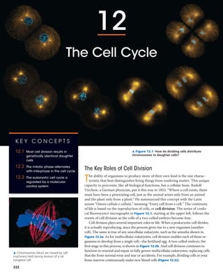
▲ Figure 12.1 How do dividing cells distribute
chromosomes to daughter cells?
The Key Roles of Cell Division
The ability of organisms to produce more of their own kind is the one charac-
teristic that best distinguishes living things from nonliving matter. This unique
capacity to procreate, like all biological functions, has a cellular basis. Rudolf
Virchow, a German physician, put it this way in 1855: “Where a cell exists, there
must have been a preexisting cell, just as the animal arises only from an animal
and the plant only from a plant.” He summarized this concept with the Latin
axiom “Omnis cellula e cellula,” meaning “Every cell from a cell.” The continuity
of life is based on the reproduction of cells, or cell division. The series of confo-
cal fluorescence micrographs in Figure 12.1, starting at the upper left, follows the
events of cell division as the cells of a two-celled embryo become four.
Cell division plays several important roles in life. When a prokaryotic cell divides,
it is actually reproducing, since the process gives rise to a new organism (another
cell). The same is true of any unicellular eukaryote, such as the amoeba shown in
Figure 12.2a. As for multicellular eukaryotes, cell division enables each of these or-
ganisms to develop from a single cell—the fertilized egg. A two-celled embryo, the
first stage in this process, is shown in Figure 12.2b. And cell division continues to
function in renewal and repair in fully grown multicellular eukaryotes, replacing cells
that die from normal wear and tear or accidents. For example, dividing cells in your
bone marrow continuously make new blood cells (Figure 12.2c).
K E Y C O N C E P T S
12.1 Most cell division results in
genetically identical daughter
cells
12.2 The mitotic phase alternates
with interphase in the cell cycle
12.3 The eukaryotic cell cycle is
regulated by a molecular
control system
▲ Chromosomes (blue) are moved by cell
machinery (red) during division of a rat
kangaroo cell.
232
2. CHA PTER 12 The Cell Cycle 233
The cell division process is an integral part of the cell
cycle, the life of a cell from the time it is first formed dur-
ing division of a parent cell until its own division into two
daughter cells. (Our use of the words daughter or sister in
relation to cells is not meant to imply gender.) Passing identi-
cal genetic material to cellular offspring is a crucial function
of cell division. In this chapter, you will learn how this process
occurs. After studying the cellular mechanics of cell division
in eukaryotes and bacteria, you will learn about the molecular
control system that regulates progress through the eukaryotic
cell cycle and what happens when the control system mal-
functions. Because a breakdown in cell cycle control plays a
major role in cancer development, this aspect of cell biology is
an active area of research.
100 μm
50 μm
20 μm
◀ (c) Tissue renewal. These
dividing bone marrow cells
will give rise to new blood
cells (LM).
◀ (a)
(b) Growth and develop-
ment. This micrograph
shows a sand dollar
embryo shortly after the
fertilized egg divided,
forming two cells (LM).
▶
(a) Reproduction. An amoeba,
a single-celled eukaryote, is
dividing into two cells. Each
new cell will be an
individual organism (LM).
▲ Figure 12.2 The functions of cell division.
Cellular Organization of the Genetic Material
A cell’s endowment of DNA, its genetic information, is
called its genome. Although a prokaryotic genome is often
a single DNA molecule, eukaryotic genomes usually consist
of a number of DNA molecules. The overall length of DNA
in a eukaryotic cell is enormous. A typical human cell, for
example, has about 2 m of DNA—a length about 250,000
times greater than the cell’s diameter. Before the cell can
divide to form genetically identical daughter cells, all of this
DNA must be copied, or replicated, and then the two copies
must be separated so that each daughter cell ends up with a
complete genome.
The replication and distribution of so much DNA is
manageable because the DNA molecules are packaged
into structures called chromosomes, so named because
they take up certain dyes used in microscopy (from the
Greek chroma, color, and soma, body; Figure 12.3).
Each eukaryotic chromosome consists of one very long,
linear DNA molecule associated with many proteins (see
Figure 6.9). The DNA molecule carries several hundred to a
few thousand genes, the units of information that specify an
organism’s inherited traits. The associated proteins main-
tain the structure of the chromosome and help control the
activity of the genes. Together, the entire complex of DNA
and proteins that is the building material of chromosomes
is referred to as chromatin. As you will soon see, the chro-
matin of a chromosome varies in its degree of condensation
during the process of cell division.
Every eukaryotic species has a characteristic number of
chromosomes in each cell’s nucleus. For example, the nuclei
of human somatic cells (all body cells except the reproduc-
tive cells) each contain 46 chromosomes, made up of two
sets of 23, one set inherited from each parent. Reproductive
C O N C E P T 12.1
Most cell division results in genetically
identical daughter cells
The reproduction of a cell, with all of its complexity, cannot
occur by a mere pinching in half; a cell is not like a soap bub-
ble that simply enlarges and splits in two. In both prokaryotes
and eukaryotes, most cell division involves the distribution of
identical genetic material—DNA—to two daughter cells. (The
exception is meiosis, the special type of eukaryotic cell divi-
sion that can produce sperm and eggs.) What is most remark-
able about cell division is the fidelity with which the DNA is
passed from one generation of cells to the next. A dividing cell
replicates its DNA, allocates the two copies to opposite ends
of the cell, and only then splits into daughter cells.
20 μm
▲ Figure 12.3 Eukaryotic chromosomes. Chromosomes (stained
purple) are visible within the nucleus of this cell from an African blood
lily. The thinner red threads in the surrounding cytoplasm are the cyto-
skeleton. The cell is preparing to divide (LM).
3. 234 UNIT TWO The Cell
cells, or gametes—sperm and eggs—have one set, or half as
many chromosomes as somatic cells; in our example, human
gametes have one set of 23 chromosomes. The number of
chromosomes in somatic cells varies widely among species:
18 in cabbage plants, 48 in chimpanzees, 56 in elephants, 90
in hedgehogs, and 148 in one species of alga. We’ll now con-
sider how these chromosomes behave during cell division.
Distribution of Chromosomes During
Eukaryotic Cell Division
When a cell is not dividing, and even as it replicates its DNA
in preparation for cell division, each chromosome is in the
form of a long, thin chromatin fiber. After DNA replica-
tion, however, the chromosomes condense as a part of cell
division: Each chromatin fiber becomes densely coiled and
folded, making the chromosomes much shorter and so thick
that we can see them with a light microscope.
Each duplicated chromosome has two sister chromatids,
which are joined copies of the original chromosome
(Figure 12.4). The two chromatids, each containing an
identical DNA molecule, are initially
attached all along their lengths by pro-
tein complexes called cohesins; this at-
tachment is known as sister chromatid
cohesion. Each sister chromatid has a
centromere, a region of the chromo-
somal DNA where the chromatid is
attached most closely to its sister chro-
matid. This attachment is mediated
by proteins bound to the centromeric
DNA; other bound proteins condense
the DNA, giving the duplicated chro-
mosome a narrow “waist.” The portion
of a chromatid to either side of the
centromere is referred to as an arm
of the chromatid. (An unduplicated
chromosome has a single centromere,
distinguished by the proteins that bind
there, and two arms.)
Later in the cell division process, the
two sister chromatids of each dupli-
cated chromosome separate and move
into two new nuclei, one forming at
each end of the cell. Once the sister
chromatids separate, they are no longer
called sister chromatids but are con-
sidered individual chromosomes; this
step essentially doubles the number of
chromosomes in the cell. Thus, each
new nucleus receives a collection of
chromosomes identical to that of the
parent cell (Figure 12.5). Mitosis, the
division of the genetic material in the nucleus, is usually fol-
lowed immediately by cytokinesis, the division of the cyto-
plasm. One cell has become two, each the genetic equivalent
of the parent cell.
From a fertilized egg, mitosis and cytokinesis produced
the 200 trillion somatic cells that now make up your body,
and the same processes continue to generate new cells to
Sister
chromatids
Centromere 0.5 μm
▲ Figure 12.4 A highly condensed, duplicated human chromo-
some (SEM).
D R AW I T Circle one sister chromatid of the chromosome in this
micrograph.
Chromosome duplication
(including DNA replication)
and condensation
Chromosome
arm
Centromere
Chromosomes
Chromosomal
DNA molecules
Sister
chromatids
Separation of sister
chromatids into
two chromosomes
One of the multiple chromosomes
in a eukaryotic cell is represented
here, not yet duplicated. Normally
it would be a long, thin chromatin
fiber containing one DNA molecule
and associated proteins; here its
condensed form is shown for
illustration purposes only.
1
Once duplicated, a chromosome
consists of two sister chroma-
tids connected along their entire
lengths by sister chromatid
cohesion. Each chromatid contains
a copy of the DNA molecule.
2
Molecular and mechanical
processes separate the sister
chromatids into two chromosomes
and distribute them to two
daughter cells.
3
▲ Figure 12.5 Chromosome duplication and distribution during cell division.
? How many chromatid arms does the chromosome in 2 have?
4. CHA PTER 12 The Cell Cycle 235
replace dead and damaged ones. In contrast, you produce
gametes—eggs or sperm—by a variation of cell division
called meiosis, which yields daughter cells with only one set
of chromosomes, half as many chromosomes as the parent
cell. Meiosis in humans occurs only in special cells in the
ovaries or testes (the gonads). Generating gametes, meio-
sis reduces the chromosome number from 46 (two sets) to
23 (one set). Fertilization fuses two gametes together and
returns the chromosome number to 46 (two sets). Mitosis
then conserves that number in every somatic cell nucleus of
the new human individual. In Chapter 13, we will examine
the role of meiosis in reproduction and inheritance in more
detail. In the remainder of this chapter, we focus on mitosis
and the rest of the cell cycle in eukaryotes.
subphases of interphase, in fact, a cell grows by producing
proteins and cytoplasmic organelles such as mitochondria
and endoplasmic reticulum. Duplication of the chromo-
somes, crucial for eventual division of the cell, occurs en-
tirely during the S phase. (We will discuss synthesis of DNA
in Chapter 16.) Thus, a cell grows (G1), continues to grow as
it copies its chromosomes (S), grows more as it completes
preparations for cell division (G2), and divides (M). The
daughter cells may then repeat the cycle.
A particular human cell might undergo one division in
24 hours. Of this time, the M phase would occupy less than
1 hour, while the S phase might occupy about 10–12 hours,
or about half the cycle. The rest of the time would be appor-
tioned between the G1 and G2 phases. The G2 phase usually
takes 4–6 hours; in our example, G1 would occupy about
5–6 hours. G1 is the most variable in length in different
types of cells. Some cells in a multicellular organism divide
very infrequently or not at all. These cells spend their time
in G1 (or a related phase called G0) doing their job in the
organism—a nerve cell carries impulses, for example.
Mitosis is conventionally broken down into five stages:
prophase, prometaphase, metaphase, anaphase, and
telophase. Overlapping with the latter stages of mitosis,
cytokinesis completes the mitotic phase. Figure 12.7 de-
scribes these stages in an animal cell. Study this figure thor-
oughly before progressing to the next two sections, which
examine mitosis and cytokinesis more closely.
The Mitotic Spindle: A Closer Look
Many of the events of mitosis depend on the mitotic spindle,
which begins to form in the cytoplasm during prophase.
This structure consists of fibers made of microtubules and
C O N C E P T C H E C K 1 2 . 1
1. How many chromosomes are drawn in each part of Fig-
ure 12.5? (Ignore the micrograph in part 2.)
2. W H AT I F ? A chicken has 78 chromosomes in its so-
matic cells. How many chromosomes did the chicken
inherit from each parent? How many chromosomes
are in each of the chicken’s gametes? How many chro-
mosomes will be in each somatic cell of the chicken’s
offspring?
For suggested answers, see Appendix A.
C O N C E P T 12.2
The mitotic phase alternates with
interphase in the cell cycle
In 1882, a German anatomist named Walther Flemming
developed dyes that allowed him to observe, for the first
time, the behavior of chromosomes during mitosis and cy-
tokinesis. (In fact, Flemming coined the terms mitosis and
chromatin.) During the period between one cell division and
the next, it appeared to Flemming that the cell was simply
growing larger. But we now know that many critical events
occur during this stage in the life of a cell.
Phases of the Cell Cycle
Mitosis is just one part of the cell cycle (Figure 12.6). In fact,
the mitotic (M) phase, which includes both mitosis and
cytokinesis, is usually the shortest part of the cell cycle. The
mitotic phase alternates with a much longer stage called
interphase, which often accounts for about 90% of the cycle.
Interphase can be divided into subphases: the G1 phase
(“first gap”), the S phase (“synthesis”), and the G2 phase
(“second gap”). The G phases were misnamed as “gaps”
when they were first observed because the cells appeared
inactive, but we now know that intense metabolic activity
and growth occur throughout interphase. During all three
Cytokinesis
M
itosis
G1
G2
S
(DNA synthesis)
MITOTIC
INTERPHASE
(M) PHASE
▲ Figure 12.6 The cell cycle. In a dividing cell, the mitotic (M)
phase alternates with interphase, a growth period. The first part of
interphase (G1) is followed by the S phase, when the chromosomes du-
plicate; G2 is the last part of interphase. In the M phase, mitosis distrib-
utes the daughter chromosomes to daughter nuclei, and cytokinesis
divides the cytoplasm, producing two daughter cells.
5. Centrosomes
(with centriole pairs)
Early mitotic
spindle
Fragments
of nuclear
envelope
Centromere Nonkinetochore
microtubules
Kinetochore
microtubule
KinetochoreNucleolus Nuclear
envelope
Plasma
membrane
Two sister chromatids
of one chromosome
Aster
Chromosomes
(duplicated,
uncondensed)
Prophase
-
Prometaphase
-
? How many molecules of DNA are in the
prometaphase drawing? How many molecules per
chromosome? How many double helices are there
per chromosome? Per chromatid?
G2 of Interphase
nucleolus
-
-
The fluorescence micrographs show divid-
ing lung cells from a newt; this species has
22 chromosomes. Chromosomes appear
blue, microtubules green, and intermedi-
ate filaments red. For simplicity, the draw-
ings show only 6 chromosomes.
G2 of Interphase Prophase Prometaphase
236 UNIT TWO The Cell
▼ Figure 12.7
Exploring Mitosis in an Animal Cell
7. 238 UNIT TWO The Cell
associated proteins. While the mitotic spindle assembles, the
other microtubules of the cytoskeleton partially disassemble,
providing the material used to construct the spindle. The
spindle microtubules elongate (polymerize) by incorporat-
ing more subunits of the protein tubulin (see Table 6.1) and
shorten (depolymerize) by losing subunits.
In animal cells, the assembly of spindle microtubules
starts at the centrosome, a subcellular region containing
material that functions throughout the cell cycle to organize
the cell’s microtubules. (It is also a type of microtubule-
organizing center.) A pair of centrioles is located at the
center of the centrosome, but they are not essential for cell
division: If the centrioles are destroyed with a laser micro-
beam, a spindle nevertheless forms during mitosis. In fact,
centrioles are not even present in plant cells, which do form
mitotic spindles.
During interphase in animal cells, the single centrosome
duplicates, forming two centrosomes, which remain near
the nucleus. The two centrosomes move apart during pro-
phase and prometaphase of mitosis as spindle microtubules
grow out from them. By the end of prometaphase, the two
centrosomes, one at each pole of the spindle, are at opposite
ends of the cell. An aster, a radial array of short microtu-
bules, extends from each centrosome. The spindle includes
the centrosomes, the spindle microtubules, and the asters.
Each of the two sister chromatids of a duplicated chro-
mosome has a kinetochore, a structure made up of proteins
that have assembled on specific sections of DNA at each
centromere. The chromosome’s two kinetochores face in
opposite directions. During prometaphase, some of the
spindle microtubules attach to the kinetochores; these are
called kinetochore microtubules. (The number of microtu-
bules attached to a kinetochore varies among species, from
one microtubule in yeast cells to 40 or so in some mam-
malian cells.) When one of a chromosome’s kinetochores
is “captured” by microtubules, the chromosome begins to
move toward the pole from which those microtubules ex-
tend. However, this movement is checked as soon as micro-
tubules from the opposite pole attach to the kinetochore on
the other chromatid. What happens next is like a tug-of-war
that ends in a draw. The chromosome moves first in one
direction, then in the other, back and forth, finally settling
midway between the two ends of the cell. At metaphase,
the centromeres of all the duplicated chromosomes are on
a plane midway between the spindle’s two poles. This plane
is called the metaphase plate, which is an imaginary plate
rather than an actual cellular structure (Figure 12.8). Mean-
while, microtubules that do not attach to kinetochores have
been elongating, and by metaphase they overlap and interact
with other nonkinetochore microtubules from the opposite
pole of the spindle. By metaphase, the microtubules of the
asters have also grown and are in contact with the plasma
membrane. The spindle is now complete.
The structure of the spindle correlates well with its func-
tion during anaphase. Anaphase begins suddenly when the
cohesins holding together the sister chromatids of each
chromosome are cleaved by an enzyme called separase.
Once separated, the chromatids become full-fledged chro-
mosomes that move toward opposite ends of the cell.
How do the kinetochore microtubules function in this
poleward movement of chromosomes? Apparently, two
Sister
chromatids
Aster
Centrosome
Metaphase
plate
(imaginary)
Kineto-
chores
Kinetochore
microtubules
Overlapping
nonkinetochore
microtubules
0.5 μm
Chromosomes
1 μm
Centrosome
Microtubules
▲ Figure 12.8 The mitotic spindle at metaphase. The kineto-
chores of each chromosome’s two sister chromatids face in opposite
directions. Here, each kinetochore is attached to a cluster of kineto-
chore microtubules extending from the nearest centrosome. Non-
kinetochore microtubules overlap at the metaphase plate (TEMs).
D R AW I T On the lower micrograph, draw a line indicating the position
of the metaphase plate. Circle an aster. Draw arrows indicating the direc-
tions of chromosome movement once anaphase begins.
8. CHA PTER 12 The Cell Cycle 239
mechanisms are in play, both involving motor proteins.
(To review how motor proteins move an object along a mi-
crotubule, see Figure 6.21.) Results of a cleverly designed
experiment suggested that motor proteins on the kineto-
chores “walk” the chromosomes along the microtubules,
which depolymerize at their kinetochore ends after the
motor proteins have passed (Figure 12.9). (This is referred
to as the “Pac-man” mechanism because of its resemblance
to the arcade game character that moves by eating all the
dots in its path.) However, other researchers, working with
different cell types or cells from other species, have shown
that chromosomes are “reeled in” by motor proteins at the
spindle poles and that the microtubules depolymerize after
they pass by these motor proteins. The general consensus
now is that both mechanisms are used and that their relative
contributions vary among cell types.
In a dividing animal cell, the nonkinetochore microtu-
bules are responsible for elongating the whole cell during
anaphase. Nonkinetochore microtubules from opposite
poles overlap each other extensively during metaphase (see
Figure 12.8). During anaphase, the region of overlap is re-
duced as motor proteins attached to the microtubules walk
them away from one another, using energy from ATP. As
the microtubules push apart from each other, their spindle
poles are pushed apart, elongating the cell. At the same
time, the microtubules lengthen somewhat by the addition
of tubulin subunits to their overlapping ends. As a result, the
microtubules continue to overlap.
At the end of anaphase, duplicate groups of chromo-
somes have arrived at opposite ends of the elongated parent
cell. Nuclei re-form during telophase. Cytokinesis generally
begins during anaphase or telophase, and the spindle even-
tually disassembles by depolymerization of microtubules.
Cytokinesis: A Closer Look
In animal cells, cytokinesis occurs by a process known as
cleavage. The first sign of cleavage is the appearance of a
cleavage furrow, a shallow groove in the cell surface near
the old metaphase plate (Figure 12.10a). On the cytoplas-
mic side of the furrow is a contractile ring of actin microfila-
ments associated with molecules of the protein myosin. The
actin microfilaments interact with the myosin molecules,
causing the ring to contract. The contraction of the dividing
cell’s ring of microfilaments is like the pulling of a draw-
string. The cleavage furrow deepens until the parent cell is
pinched in two, producing two completely separated cells,
each with its own nucleus and its own share of cytosol, or-
ganelles, and other subcellular structures.
Cytokinesis in plant cells, which have cell walls, is mark-
edly different. There is no cleavage furrow. Instead, during
telophase, vesicles derived from the Golgi apparatus move
along microtubules to the middle of the cell, where they
Inquiry
At which end do kinetochore microtubules
shorten during anaphase?
▼ Figure 12.9
Experiment Gary Borisy and colleagues at the University of Wisconsin
wanted to determine whether kinetochore microtubules depolymerize
at the kinetochore end or the pole end as chromosomes move toward
the poles during mitosis. First they labeled the microtubules of a pig
kidney cell in early anaphase with a yellow fluorescent dye.
Kinetochore
Spindle
pole
Then they marked a region of the kinetochore microtubules between
one spindle pole and the chromosomes by using a laser to eliminate
the fluorescence from that region, while leaving the microtubules
intact (see below). As anaphase proceeded, they monitored the
changes in microtubule length on either side of the mark.
Mark
Results As the chromosomes moved poleward, the microtubule
segments on the kinetochore side of the mark shortened, while those
on the spindle pole side stayed the same length.
Conclusion During anaphase in this cell type, chromosome
movement is correlated with kinetochore microtubules shortening
at their kinetochore ends and not at their spindle pole ends. This
experiment supports the hypothesis that during anaphase, a
chromosome is walked along a microtubule as the microtubule
depolymerizes at its kinetochore end, releasing tubulin subunits.
Chromosome
movement
Kinetochore
Tubulin
subunits
Chromosome
Motor
proteinMicrotubule
Source: G. J. Gorbsky, P. J. Sammak, and G. G. Borisy, Chromosomes move poleward
in anaphase along stationary microtubules that coordinately disassemble from their
kinetochore ends, Journal of Cell Biology 104:9–18 (1987).
W H AT I F ? If this experiment had been done on a cell type in which
“reeling in” at the poles was the main cause of chromosome movement,
how would the mark have moved relative to the poles? How would the
microtubule lengths have changed?
9. 240 UNIT TWO The Cell
coalesce, producing a cell plate (Figure 12.10b). Cell wall
materials carried in the vesicles collect inside the cell plate
as it grows. The cell plate enlarges until its surrounding
membrane fuses with the plasma membrane along the pe-
rimeter of the cell. Two daughter cells result, each with its
own plasma membrane. Meanwhile, a new cell wall arising
from the contents of the cell plate has formed between the
daughter cells.
Figure 12.11 is a series of micrographs of a dividing plant
cell. Examining this figure will help you review mitosis and
cytokinesis.
Binary Fission in Bacteria
Prokaryotes (bacteria and archaea) can undergo a type of re-
production in which the cell grows to roughly double its size
and then divides to form two cells. The term binary fission,
meaning “division in half,” refers to this process and to the
asexual reproduction of single-celled eukaryotes, such as the
amoeba in Figure 12.2a. However, the process in eukaryotes
involves mitosis, while that in prokaryotes does not.
In bacteria, most genes are carried on a single bacte-
rial chromosome that consists of a circular DNA molecule
and associated proteins. Although bacteria are smaller and
simpler than eukaryotic cells, the challenge of replicating
their genomes in an orderly fashion and distributing the
copies equally to two daughter cells is still formidable. The
chromosome of the bacterium Escherichia coli, for example,
when it is fully stretched out, is about 500 times as long as
the cell. For such a long chromosome to fit within the cell
requires that it be highly coiled and folded.
In E. coli, the process of cell division is initiated when
the DNA of the bacterial chromosome begins to replicate
at a specific place on the chromosome called the origin of
replication, producing two origins. As the chromosome
continues to replicate, one origin moves rapidly toward the
opposite end of the cell (Figure 12.12). While the chromo-
some is replicating, the cell elongates. When replication is
complete and the bacterium has reached about twice its ini-
tial size, its plasma membrane pinches inward, dividing the
parent E. coli cell into two daughter cells. In this way, each
cell inherits a complete genome.
Using the techniques of modern DNA technology to tag
the origins of replication with molecules that glow green in
fluorescence microscopy (see Figure 6.3), researchers have
directly observed the movement of bacterial chromosomes.
This movement is reminiscent of the poleward movements
of the centromere regions of eukaryotic chromosomes
during anaphase of mitosis, but bacteria don’t have visible
mitotic spindles or even microtubules. In most bacterial spe-
cies studied, the two origins of replication end up at oppo-
site ends of the cell or in some other very specific location,
possibly anchored there by one or more proteins. How bac-
terial chromosomes move and how their specific location is
(a) Cleavage of an animal cell (SEM)
(b) Cell plate formation in a plant cell (TEM)
Daughter cells
Cleavage furrow
Contractile ring of
microfilaments
Daughter cells
Cell plate
Wall of
parent cell
Vesicles
forming
cell plate New cell wall
100 μm
1 μm
▼ Figure 12.10 Cytokinesis in animal and plant cells.
10. CHAPTER 12 The Cell Cycle 241
established and maintained are active areas of research. Sev-
eral proteins have been identified that play important roles.
Polymerization of one protein resembling eukaryotic actin
apparently functions in bacterial chromosome movement
during cell division, and another protein that is related to
tubulin helps pinch the plasma membrane inward, separat-
ing the two bacterial daughter cells.
The Evolution of Mitosis
E VO L U T I O N Given that prokaryotes preceded eukaryotes
on Earth by more than a billion years, we might hypothesize
that mitosis evolved from simpler prokaryotic mechanisms
of cell reproduction. The fact that some of the proteins in-
volved in bacterial binary fission are related to eukaryotic
proteins that function in mitosis supports that hypothesis.
As eukaryotes with nuclear envelopes and larger genomes
evolved, the ancestral process of binary fission, seen today
in bacteria, somehow gave rise to mitosis. Variations on cell
division exist in different groups of organisms. These variant
processes may be similar to mechanisms used by ancestral
species and thus may resemble steps in the evolution of mi-
tosis from a binary fission-like process presumably carried
out by very early bacteria. Possible intermediate stages are
suggested by two unusual types of nuclear division found
today in certain unicellular eukaryotes—dinoflagellates,
diatoms, and some yeasts (Figure 12.13). These two modes
of nuclear division are thought to be cases where ancestral
mechanisms have remained relatively unchanged over evo-
lutionary time. In both types, the nuclear envelope remains
intact, in contrast to what happens in most eukaryotic cells.
Nucleus Chromosomes
condensing ChromosomesNucleolus
Prophase. The chromo-
somes are condensing
and the nucleolus is
beginning to disappear.
Although not yet visible
in the micrograph, the
mitotic spindle is starting
to form.
Prometaphase. Discrete
chromosomes are now
visible; each consists of
two aligned, identical
sister chromatids. Later
in prometaphase, the
nuclear envelope will
fragment.
Anaphase. The
chromatids of each
chromosome have
separated, and the
daughter chromosomes
are moving to the ends
of the cell as their
kinetochore micro-
tubules shorten.
Telophase. Daughter
nuclei are forming.
Meanwhile, cytokinesis
has started: The cell
plate, which will divide
the cytoplasm in two, is
growing toward the
perimeter of the parent
cell.
Metaphase. The spindle
is complete, and the
chromosomes, attached
to microtubules at their
kinetochores, are all at
the metaphase plate.
10 μm
Cell plate
1 2 3 4 5
▲ Figure 12.11 Mitosis in a plant cell. These light micrographs show mitosis in cells of an onion root.
Chromosome
replication begins.
Soon after, one copy
of the origin moves
rapidly toward the
other end of the cell by
a mechanism involving
an actin-like protein.
1
Replication continues.
One copy of the origin
is now at each end of
the cell. Meanwhile,
the cell elongates.
2
Replication finishes.
The plasma membrane
is pinched inward by
a tubulin-like protein,
and a new cell wall is
deposited.
3
Two daughter
cells result.
4
Origin of
replication
E. coli cell Bacterial
chromosome
Plasma
membrane
Cell wall
Two copies
of origin
Origin Origin
▲ Figure 12.12 Bacterial cell division by binary fission. The
bacterium E. coli, shown here, has a single, circular chromosome.
11. 242 UNIT TWO The Cell
Bacteria. During binary fission in bacteria, the origins of the
daughter chromosomes move to opposite ends of the cell. The
mechanism involves polymerization of actin-like molecules, and
possibly proteins that may anchor the daughter chromosomes to
specific sites on the plasma membrane.
(a)
Diatoms and some yeasts. In these two other groups of
unicellular eukaryotes, the nuclear envelope also remains intact
during cell division. In these organisms, the microtubules form a
spindle within the nucleus. Microtubules separate the
chromosomes, and the nucleus splits into two daughter nuclei.
(c)
Most eukaryotes. In most other eukaryotes, including plants and
animals, the spindle forms outside the nucleus, and the nuclear
envelope breaks down during mitosis. Microtubules separate the
chromosomes, and two nuclear envelopes then form.
(d)
Microtubules
Chromosomes
Kinetochore
microtubule
Fragments of
nuclear envelope
Kinetochore
microtubule
Intact nuclear
envelope
Intact nuclear
envelope
Bacterial
chromosome
Dinoflagellates. In unicellular protists called dinoflagellates, the
chromosomes attach to the nuclear envelope, which remains
intact during cell division. Microtubules pass through the nucleus
inside cytoplasmic tunnels, reinforcing the spatial orientation of
the nucleus, which then divides in a process reminiscent of
bacterial binary fission.
(b)
▲ Figure 12.13 Mechanisms of cell division in several groups
of organisms. Some unicellular eukaryotes existing today have
mechanisms of cell division that may resemble intermediate steps in
the evolution of mitosis. Except for (a), these schematic diagrams do
not show cell walls.
C O N C E P T 12.3
The eukaryotic cell cycle is regulated by
a molecular control system
The timing and rate of cell division in different parts of a
plant or animal are crucial to normal growth, development,
and maintenance. The frequency of cell division varies
with the type of cell. For example, human skin cells divide
frequently throughout life, whereas liver cells maintain the
ability to divide but keep it in reserve until an appropriate
need arises—say, to repair a wound. Some of the most spe-
cialized cells, such as fully formed nerve cells and muscle
cells, do not divide at all in a mature human. These cell cycle
differences result from regulation at the molecular level. The
mechanisms of this regulation are of great interest, not only
to understand the life cycles of normal cells but also to learn
how cancer cells manage to escape the usual controls.
The Cell Cycle Control System
What controls the cell cycle? In the early 1970s, a variety
of experiments led to the hypothesis that the cell cycle is
driven by specific signaling molecules present in the cyto-
plasm. Some of the first strong evidence for this hypothesis
came from experiments with mammalian cells grown in
culture. In these experiments, two cells in different phases of
the cell cycle were fused to form a single cell with two nuclei
(Figure 12.14). If one of the original cells was in the S phase
and the other was in G1, the G1 nucleus immediately entered
the S phase, as though stimulated by signaling molecules
present in the cytoplasm of the first cell. Similarly, if a cell
undergoing mitosis (M phase) was fused with another cell in
any stage of its cell cycle, even G1, the second nucleus imme-
diately entered mitosis, with condensation of the chromatin
and formation of a mitotic spindle.
The experiment shown in Figure 12.14 and other experi-
ments on animal cells and yeasts demonstrated that the
C O N C E P T C H E C K 1 2 . 2
1. How many chromosomes are drawn in Figure 12.8? Are
they duplicated? How many chromatids are shown?
2. Compare cytokinesis in animal cells and plant cells.
3. During which stages of the cell cycle does a chromo-
some consist of two identical chromatids?
4. Compare the roles of tubulin and actin during eukaryotic
cell division with the roles of tubulin-like and actin-like
proteins during bacterial binary fission.
5. A kinetochore has been compared to a coupling device
that connects a motor to the cargo that it moves. Explain.
6. M A K E C O N N E C T I O N S What other functions do actin
and tubulin carry out? Name the proteins they interact
with to do so. (Review Figures 6.21a and 6.26a.)
For suggested answers, see Appendix A.
12. CHA PTER 12 The Cell Cycle 243
sequential events of the cell cycle are directed by a distinct
cell cycle control system, a cyclically operating set of mole-
cules in the cell that both triggers and coordinates key events
in the cell cycle. The cell cycle control system has been com-
pared to the control device of an automatic washing machine
(Figure 12.15). Like the washer’s timing device, the cell cycle
control system proceeds on its own, according to a built-in
clock. However, just as a washer’s cycle is subject to both
internal control (such as the sensor that detects when the tub
is filled with water) and external adjustment (such as starting
or stopping the machine), the cell cycle is regulated at certain
checkpoints by both internal and external signals that stop or
restart the machine. A checkpoint is a control point in the
cell cycle where stop and go-ahead signals can regulate the
cycle. Three important checkpoints are found in the G1, G2,
and M phases (the red gates in Figure 12.15).
To understand how cell cycle checkpoints work, we first
need to see what kinds of molecules make up the cell cycle
control system (the molecular basis for the cell cycle clock)
and how a cell progresses through the cycle. Then we will
consider the internal and external checkpoint signals that
can make the clock either pause or continue.
The Cell Cycle Clock: Cyclins and Cyclin-Dependent
Kinases
Rhythmic fluctuations in the abundance and activity of cell
cycle control molecules pace the sequential events of the
cell cycle. These regulatory molecules are mainly proteins
of two types: protein kinases and cyclins. Protein kinases are
enzymes that activate or inactivate other proteins by phos-
phorylating them (see Chapter 11).
Many of the kinases that drive the cell cycle are actually
present at a constant concentration in the growing cell, but
much of the time they are in an inactive form. To be active,
such a kinase must be attached to a cyclin, a protein that
gets its name from its cyclically fluctuating concentration in
the cell. Because of this requirement, these kinases are called
cyclin-dependent kinases, or Cdks. The activity of a Cdk
rises and falls with changes in the concentration of its cy-
clin partner. Figure 12.16a shows the fluctuating activity of
MPF, the cyclin-Cdk complex that was discovered first (in
frog eggs). Note that the peaks of MPF activity correspond
to the peaks of cyclin concentration. The cyclin level rises
during the S and G2 phases and then falls abruptly during
M phase.
The initials MPF stand for “maturation-promoting fac-
tor,” but we can think of MPF as “M-phase-promoting fac-
tor” because it triggers the cell’s passage into the M phase,
Inquiry
Do molecular signals in the cytoplasm regulate
the cell cycle?
▼ Figure 12.14
Experiment Researchers at the University of Colorado wondered
whether a cell’s progression through the cell cycle is controlled by
cytoplasmic molecules. To investigate this, they selected cultured
mammalian cells that were at different phases of the cell cycle and
induced them to fuse. Two such experiments are shown here.
When a cell in the
S phase was fused with
a cell in G1, the G1
nucleus immediately
entered the S
phase—DNA was
synthesized.
When a cell in the
M phase was fused with
a cell in G1, the G1
nucleus immediately
began mitosis—a spindle
formed and the chromo-
somes condensed, even
though the chromosomes
had not been duplicated.
S S M M
S
Experiment 1 Experiment 2
G1 M G1
Conclusion The results of fusing a G1 cell with a cell in the S or
M phase of the cell cycle suggest that molecules present in the cyto-
plasm during the S or M phase control the progression to those phases.
Source: R. T. Johnson and P. N. Rao, Mammalian cell fusion: Induction of premature
chromosome condensation in interphase nuclei, Nature 226:717–722 (1970).
W H AT I F ? If the progression of phases did not depend on cytoplas-
mic molecules and, instead, each phase automatically began when the
previous one was complete, how would the results have differed?
G1 checkpoint
M checkpoint
G2 checkpoint
G1
G2M
S
Control
system
▲ Figure 12.15 Mechanical analogy for the cell cycle control
system. In this diagram of the cell cycle, the flat “stepping stones”
around the perimeter represent sequential events. Like the control de-
vice of an automatic washer, the cell cycle control system proceeds on
its own, driven by a built-in clock. However, the system is subject to in-
ternal and external regulation at various checkpoints; three important
checkpoints are shown (red).
13. 244 UNIT TWO The Cell
lamina (see Figure 6.9), which promotes fragmentation of
the nuclear envelope during prometaphase of mitosis. There
is also evidence that MPF contributes to molecular events
required for chromosome condensation and spindle forma-
tion during prophase.
During anaphase, MPF helps switch itself off by initiating
a process that leads to the destruction of its own cyclin. The
noncyclin part of MPF, the Cdk, persists in the cell, inactive
until it becomes part of MPF again by associating with new
cyclin molecules synthesized during the S and G2 phases of
the next round of the cycle.
The fluctuating activities of different cyclin-Cdk com-
plexes are of major importance in controlling all the stages
of the cell cycle and give the go-ahead signals at some
checkpoints as well. As mentioned above, MPF controls the
cell’s passage through the G2 checkpoint. Cell behavior at
the G1 checkpoint is also regulated by the activity of cyclin-
Cdk protein complexes. Animal cells appear to have at least
three Cdk proteins and several different cyclins that operate
at this checkpoint. Next, let’s consider checkpoints in more
detail.
Stop and Go Signs: Internal and External Signals at the
Checkpoints
Animal cells generally have built-in stop signals that halt the
cell cycle at checkpoints until overridden by go-ahead sig-
nals. (The signals are transmitted within the cell by the kinds
of signal transduction pathways discussed in Chapter 11.)
Many signals registered at checkpoints come from cellular
surveillance mechanisms inside the cell. These signals report
whether crucial cellular processes that should have occurred
by that point have in fact been completed correctly and thus
whether or not the cell cycle should proceed. Checkpoints
also register signals from outside the cell. Three important
checkpoints are those in G1, G2, and M phases, shown in
Figure 12.15.
For many cells, the G1 checkpoint—dubbed the “restric-
tion point” in mammalian cells—seems to be the most
important. If a cell receives a go-ahead signal at the G1
checkpoint, it will usually complete the G1, S, G2, and
M phases and divide. If it does not receive a go-ahead signal
at that point, it may exit the cycle, switching into a nondi-
viding state called the G0 phase (Figure 12.17a). Most cells
of the human body are actually in the G0 phase. As men-
tioned earlier, mature nerve cells and muscle cells never
divide. Other cells, such as liver cells, can be “called back”
from the G0 phase to the cell cycle by external cues, such as
growth factors released during injury.
Biologists are currently working out the pathways that
link signals originating inside and outside the cell with the
responses by cyclin-dependent kinases and other proteins.
An example of an internal signal occurs at the third impor-
tant checkpoint, the M phase checkpoint (Figure 12.17b).
past the G2 checkpoint (Figure 12.16b). When cyclins that
accumulate during G2 associate with Cdk molecules, the
resulting MPF complex phosphorylates a variety of proteins,
initiating mitosis. MPF acts both directly as a kinase and
indirectly by activating other kinases. For example, MPF
causes phosphorylation of various proteins of the nuclear
G
1
S
G1 S G2
MPF activity
Cyclin
concentration
Time
(a) Fluctuation of MPF activity and cyclin concentration during
the cell cycle
M G1 S G1G2 MM
Cyclinaccumulation
Cdk
Degraded
cyclin
Cyclin is
degraded
Cdk
(b) Molecular mechanisms that help regulate the cell cycle
Cyclin
MPF
G2
checkpoint
M
G2
During G1, the degradation
of cyclin continues, and
the Cdk component of
MPF is recycled.
5
1
Cyclin combines
with Cdk, producing
MPF. When enough
MPF molecules
accumulate, the cell
passes the G2
checkpoint and
begins mitosis.
2MPF promotes
mitosis by phos-
phorylating
various proteins.
MPF‘s activity
peaks during
metaphase.
3During
anaphase, the
cyclin com-
ponent of MPF is
degraded,
terminating the
M phase. The
cell enters the
G1 phase.
4
Synthesis of cyclin
begins in late S
phase and continues
through G2. Because
cyclin is protected
from degradation
during this stage, it
accumulates.
▲ Figure 12.16 Molecular control of the cell cycle at the G2
checkpoint. The steps of the cell cycle are timed by rhythmic fluctua-
tions in the activity of cyclin-dependent kinases (Cdks). Here we focus
on a cyclin-Cdk complex in animal cells called MPF, which acts at the
G2 checkpoint as a go-ahead signal, triggering the events of mitosis.
? Explain how the events in the diagram in (b) are related to the
“Time” axis of the graph in (a), beginning at the left end.
14. CHA PTER 12 The Cell Cycle 245
Anaphase, the separation of sister chromatids,
does not begin until all the chromosomes are
properly attached to the spindle at the meta-
phase plate. Researchers have learned that
as long as some kinetochores are unattached
to spindle microtubules, the sister chromatids
remain together, delaying anaphase. Only when
the kinetochores of all the chromosomes are properly
attached to the spindle does the appropriate regulatory
protein complex become activated. (In this case, the regula-
tory molecule is not a cyclin-Cdk complex but, instead, a
different complex made up of several proteins.) Once acti-
vated, the complex sets off a chain of molecular events that
activates the enzyme separase, which cleaves the cohesins,
allowing the sister chromatids to separate. This mechanism
ensures that daughter cells do not end up with missing or
extra chromosomes.
Studies using animal cells in culture have led to the
identification of many external factors, both chemical and
physical, that can influence cell division. For example, cells
fail to divide if an essential nutrient is lacking in the cul-
ture medium. (This is analogous to trying to run a washing
machine without the water supply hooked up; an internal
sensor won’t allow the machine to continue past the point
where water is needed.) And even if all other conditions are
favorable, most types of mammalian cells divide in culture
only if the growth medium includes specific growth factors.
As mentioned in Chapter 11, a growth factor is a protein
released by certain cells that stimulates other cells to divide.
Different cell types respond specifically to different growth
factors or combinations of growth factors.
Consider, for example, platelet-derived growth factor
(PDGF), which is made by blood cell fragments called plate-
lets. The experiment illustrated in Figure 12.18 demonstrates
that PDGF is required for the division of cultured fibroblasts,
a type of connective tissue cell. Fibroblasts have PDGF recep-
tors on their plasma membranes. The binding of PDGF mol-
ecules to these receptors (which are receptor tyrosine kinases;
see Figure 11.8) triggers a signal transduction pathway that
allows the cells to pass the G1 checkpoint and divide. PDGF
stimulates fibroblast division not only in the artificial condi-
tions of cell culture, but also in an animal’s body. When an
injury occurs, platelets release PDGF in the vicinity. The re-
sulting proliferation of fibroblasts helps heal the wound.
The effect of an external physical factor on cell division
is clearly seen in density-dependent inhibition, a phenom-
enon in which crowded cells stop dividing (Figure 12.19a).
As first observed many years ago, cultured cells normally
divide until they form a single layer of cells on the inner
surface of the culture flask, at which point the cells stop di-
viding. If some cells are removed, those bordering the open
G1
G2M
S
G1 G1
G0
G1 checkpoint
If a cell receives a go-ahead signal, the
cell continues on in the cell cycle.
In the absence of a go-ahead signal,
a cell exits the cell cycle and enters
G0, a nondividing state.
(a) G1 checkpoint
When all chromosomes are attached
to spindle fibers from both poles,
a go-ahead signal allows the cell to
proceed into anaphase.
A cell in mitosis receives a stop signal
when any of its chromosomes are not
attached to spindle fibers.
(b) M checkpoint
G2M
G1
M checkpoint
Prometaphase Metaphase
G2
checkpointAnaphase
G2
M
G1
▶ Figure 12.17 Two impor-
tant checkpoints. At certain
checkpoints in the cell cycle (red
gates), cells do different things
depending on the signals they
receive. Events of the (a) G1 and
(b) M checkpoints are shown. In
(b), the G2 checkpoint has already
been passed by the cell.
W H AT I F ? What might be
the result if the cell ignored ei-
ther checkpoint and progressed
through the cell cycle?
15. 246 UNIT TWO The Cell
Loss of Cell Cycle Controls in Cancer Cells
Cancer cells do not heed the normal signals that regulate the
cell cycle. In culture, they do not stop dividing when growth
factors are depleted. A logical hypothesis is that cancer
cells do not need growth factors in their culture medium to
grow and divide. They may make a required growth factor
themselves, or they may have an abnormality in the signal-
ing pathway that conveys the growth factor’s signal to the
cell cycle control system even in the absence of that factor.
Another possibility is an abnormal cell cycle control system.
In these scenarios, the underlying basis of the abnormality is
almost always a change in one or more genes (for example, a
mutation) that alters the function of their protein products,
resulting in faulty cell cycle control.
space begin dividing again and continue until the vacancy is
filled. Follow-up studies revealed that the binding of a cell-
surface protein to its counterpart on an adjoining cell sends
a cell division-inhibiting signal forward in the cell cycle,
even in the presence of growth factors.
Most animal cells also exhibit anchorage dependence
(see Figure 12.19a). To divide, they must be attached to a
substratum, such as the inside of a culture flask or the extra-
cellular matrix of a tissue. Experiments suggest that like cell
density, anchorage is signaled to the cell cycle control sys-
tem via pathways involving plasma membrane proteins and
elements of the cytoskeleton linked to them.
Density-dependent inhibition and anchorage dependence
appear to function not only in cell culture but also in the
body’s tissues, checking the growth of cells at some optimal
density and location during embryonic development and
throughout an organism’s life. Cancer cells, which we dis-
cuss next, exhibit neither density-dependent inhibition nor
anchorage dependence (Figure 12.19b).
When cells have formed a
complete single layer, they stop
dividing (density-dependent
inhibition).
If some cells are scraped away,
the remaining cells divide to fill
the gap and then stop once they
contact each other (density-
dependent inhibition).
Cells anchor to dish surface and
divide (anchorage dependence).
(b)
(a) Normal mammalian cells. Contact with neighboring cells and
the availability of nutrients, growth factors, and a substratum for
attachment limit cell density to a single layer.
Cancer cells. Cancer cells usually continue to divide well beyond
a single layer, forming a clump of overlapping cells. They do not
exhibit anchorage dependence or density-dependent inhibition.
20 μm
20 μm
▲ Figure 12.19 Density-dependent inhibition and anchorage
dependence of cell division. Individual cells are shown dispropor-
tionately large in the drawings.
Cells are transferred to
culture vessels containing
a basic growth medium
consisting of glucose,
amino acids, salts, and
antibiotics (to prevent
bacterial growth).
A sample of human
connective tissue is
cut up into small
pieces.
1
2 Enzymes are used to
digest the extracellular
matrix in the tissue
pieces, resulting in a
suspension of free
fibroblasts.
3
PDGF is added to half
the vessels. The culture
vessels are incubated
at 37°C for 24 hours.
4
Scalpels
Petri
dish
With PDGF
In the basic growth medium plus
PDGF, the cells proliferate. The
SEM shows cultured fibroblasts.
Without PDGF
In the basic growth medium
without PDGF (the control),
the cells fail to divide.
10μm
◀ Figure 12.18
The effect of
platelet-derived
growth factor
(PDGF) on cell
division.
M A K E C O N N E C T I O N S
PDGF signals cells by binding
to a cell-surface receptor
tyrosine kinase. If you added
a chemical that blocked
phosphorylation, how would
the results differ? (See
Figure 11.8.)
16. CHA PTER 12 The Cell Cycle 247
tumor is said to have cancer; Figure 12.20 shows the develop-
ment of breast cancer, as well as a typical breast cancer cell.
The changes that have occurred in cells of malignant tu-
mors show up in many ways besides excessive proliferation.
These cells may have unusual numbers of chromosomes,
though whether this is a cause or an effect of transforma-
tion is a topic of debate. Their metabolism may be altered,
and they may cease to function in any constructive way.
Abnormal changes on the cell surface cause cancer cells to
lose attachments to neighboring cells and the extracellular
matrix, allowing them to spread into nearby tissues. Cancer
cells may also secrete signaling molecules that cause blood
vessels to grow toward the tumor. A few tumor cells may
separate from the original tumor, enter blood vessels and
lymph vessels, and travel to other parts of the body. There,
they may proliferate and form a new tumor. This spread of
cancer cells to locations distant from their original site is
called metastasis (see Figure 12.20).
A tumor that appears to be localized may be treated with
high-energy radiation, which damages DNA in cancer cells
much more than it does in normal cells, apparently because
the majority of cancer cells have lost the ability to repair
such damage. To treat known or suspected metastatic tu-
mors, chemotherapy is used, in which drugs that are toxic to
actively dividing cells are administered through the circula-
tory system. As you might expect, chemotherapeutic drugs
interfere with specific steps in the cell cycle. For example, the
drug Taxol freezes the mitotic spindle by preventing micro-
tubule depolymerization, which stops actively dividing cells
from proceeding past metaphase and leads to their destruc-
tion. The side effects of chemotherapy are due to the effects
of the drugs on normal cells that divide often, due to the
function of that cell type in the organism. For example, nau-
sea results from chemotherapy’s effects on intestinal cells,
hair loss from effects on hair follicle cells, and susceptibility
to infection from effects on immune system cells. You’ll work
There are other important differences between normal
cells and cancer cells that reflect derangements of the cell
cycle. If and when they stop dividing, cancer cells do so at
random points in the cycle, rather than at the normal check-
points. Moreover, cancer cells can go on dividing indefinitely
in culture if they are given a continual supply of nutrients;
in essence, they are “immortal.” A striking example is a cell
line that has been reproducing in culture since 1951. Cells of
this line are called HeLa cells because their original source
was a tumor removed from a woman named Henrietta Lacks.
Cells in culture that acquire the ability to divide indefinitely
are said to have undergone transformation, the process that
causes them to behave like cancer cells. By contrast, nearly all
normal, nontransformed mammalian cells growing in culture
divide only about 20 to 50 times before they stop dividing,
age, and die. Finally, cancer cells evade the normal controls
that trigger a cell to undergo apoptosis when something is
wrong—for example, when an irreparable mistake has oc-
curred during DNA replication preceding mitosis.
The abnormal behavior of cancer cells can be catastrophic
when it occurs in the body. The problem begins when a single
cell in a tissue undergoes the first changes of the multistep
process that converts a normal cell to a cancer cell. Such a
cell often has altered proteins on its surface, and the body’s
immune system normally recognizes the cell as “nonself”—
an insurgent—and destroys it. However, if the cell evades
destruction, it may proliferate and form a tumor, a mass of
abnormal cells within otherwise normal tissue. The abnor-
mal cells may remain at the original site if they have too few
genetic and cellular changes to survive at another site. In that
case, the tumor is called a benign tumor. Most benign tumors
do not cause serious problems and can be removed by surgery.
In contrast, a malignant tumor includes cells whose genetic
and cellular changes enable them to spread to new tissues and
impair the functions of one or more organs; these cells are also
considered transformed cells. An individual with a malignant
2 Cancer cells invade
neighboring tissue.
1 3A tumor grows from
a single cancer cell.
Tumor
Cancer cells spread through
lymph and blood vessels to
other parts of the body.
4 A small percentage of cancer
cells may metastasize to
another part of the body.
Lymph
vessel
Blood
vessel
Cancer
cell
Metastatic
tumor
Breast cancer cell (colorized
SEM)
Glandular
tissue
5μm
▼ Figure 12.20 The growth and metastasis of a malignant breast
tumor. A series of genetic and cellular changes contribute to a tumor be-
coming malignant (cancerous). The cells of malignant tumors grow in an
uncontrolled way and can spread to neighboring tissues and, via lymph
and blood vessels, to other parts of the body. The spread of cancer cells
beyond their original site is called metastasis.
17. 248 UNIT TWO The Cell
with data from an experiment involving a potential chemo-
therapeutic agent in the Scientific Skills Exercise.
Over the past several decades, researchers have produced
a flood of valuable information about cell-signaling path-
ways and how their malfunction contributes to the develop-
ment of cancer through effects on the cell cycle. Coupled
with new molecular techniques, such as the ability to rapidly
sequence the DNA of cells in a particular tumor, medical
treatments for cancer are beginning to become more “per-
sonalized” to a particular patient’s tumor (see Figure 18.27).
For example, the cells of roughly 20% of breast cancer tu-
mors show abnormally high amounts of a cell-surface recep-
tor tyrosine kinase called HER2, and many show an increase
in the number of estrogen receptor (ER) molecules, intra-
cellular receptors that can trigger cell division. Based on
lab findings, a physician can prescribe chemotherapy with
a molecule that blocks the function of the specific protein
(Herceptin for HER2 and tamoxifen for ERs). Treatment
using these agents, when appropriate, has led to increased
survival rates and fewer cancer recurrences.
S C I E N T I F I C S K I L L S E X E R C I S E
At What Phase Is the Cell Cycle Arrested by an Inhibitor?
Many medical treatments are aimed at stopping cancer cell proliferation
by blocking the cell cycle of cancerous tumor cells. One potential treat-
ment is a cell cycle inhibitor derived from human umbilical cord stem
cells. In this exercise, you will compare two histograms to determine
where in the cell cycle the inhibitor blocks the division of cancer cells.
How the Experiment Was Done In the treated sample, human glio-
blastoma (brain cancer) cells were grown in tissue culture in the presence
of the inhibitor, while control sample cells were grown in its absence.
After 72 hours of growth, the two cell samples were harvested. To get a
“snapshot” of the phase of the cell cycle each cell was in at that time, the
samples were treated with a fluorescent chemical that binds to DNA and
then run through a flow cytometer, an instrument that records the fluores-
cence level of each cell. Computer software then graphed the number of
cells in each sample with a particular fluorescence level, as shown below.
Data from the Experiment
200
0
40
A B C A B C
Control Treated
80
120
160
0 200 400 600
Amount of fluorescence per cell (fluorescence units)
Numberofcells
0 200 400 600
The data are plotted in a type of graph called a histogram (above),
which groups values for a numeric variable on the x-axis into intervals.
A histogram allows you to see how all the experimental subjects (cells,
in this case) are distributed along a continuous variable (amount of
fluorescence). In these histograms, the bars are so narrow that the data
Interpreting Histograms
appear to follow a curve for which you
can detect peaks and dips. Each narrow
bar represents the number of cells ob-
served to have a level of fluorescence in
the range of that interval. This in turn
indicates the relative amount of DNA
in those cells. Overall, comparing the two histograms allows you to see
how the DNA content of this cell population is altered by the treatment.
Interpret the Data
1. Familiarize yourself with the data shown in the histograms. (a) Which
axis indirectly shows the relative amount of DNA per cell? Explain
your answer. (b) In the control sample, compare the first peak in
the histogram (in region A) to the second peak (in region C). Which
peak shows the population of cells with the higher amount of DNA
per cell? Explain. (For additional information about graphs, see
the Scientific Skills Review in Appendix F and in the Study Area in
MasteringBiology.)
2. (a) In the control sample histogram, identify the phase of the cell cycle
(G1, S, or G2) of the population of cells in each region delineated by
vertical lines. Label the histogram with these phases and explain your
answer. (b) Does the S phase population of cells show a distinct peak
in the histogram? Why or why not?
3. The histogram representing the treated sample shows the effect of
growing the cancer cells alongside human umbilical cord stem cells
that produce the potential inhibitor. (a) Label the histogram with
the cell cycle phases. Which phase of the cell cycle has the great-
est number of cells in the treated sample? Explain. (b) Compare the
distribution of cells among G1, S, and G2 phases in the control and
treated samples. What does this tell you about the cells in the treated
sample? (c) Based on what you learned in Concept 12.3, propose a
mechanism by which the stem cell-derived inhibitor might arrest the
cancer cell cycle at this stage. (More than one answer is possible.)
A version of this Scientific Skills Exercise can be assigned in
MasteringBiology.
Data from K. K. Velpula et al., Regulation of glioblastoma progression by cord blood
stem cells is mediated by downregulation of cyclin D1, PLoS ONE 6(3): e18017 (2011).
C O N C E P T C H E C K 1 2 . 3
1. In Figure 12.14, why do the nuclei resulting from experi-
ment 2 contain different amounts of DNA?
2. How does MPF allow a cell to pass the G2 phase check-
point and enter mitosis? (See Figure 12.16.)
3. M A K E C O N N E C T I O N S Explain in general how recep-
tor tyrosine kinases and intracellular receptors might
function in triggering cell division. (Review Figures 11.8
and 11.9 and Chapter 11.)
For suggested answers, see Appendix A.
18. CHA PTER 12 The Cell Cycle 249
The mitotic spindle, made up of microtubules, controls chro-
mosome movement during mitosis. In animal cells, it arises
from the centrosomes and includes spindle microtubules and
asters. Some spindle microtubules attach to the kinetochores
of chromosomes and move the chromosomes to the metaphase
plate. After sister chromatids separate, motor proteins move
them along kinetochore microtubules toward opposite ends of
the cell. The cell elongates when motor proteins push nonkinet-
ochore microtubules from opposite poles away from each other.
Mitosis is usually followed by cytokinesis. Animal cells carry out
cytokinesis by cleavage, and plant cells form a cell plate.
During binary fission in bacteria, the chromosome replicates
and the daughter chromosomes actively move apart. Some of
the proteins involved in bacterial binary fission are related to
eukaryotic actin and tubulin.
Since prokaryotes preceded eukaryotes by more than a billion
years, it is likely that mitosis evolved from prokaryotic cell divi-
sion. Certain unicellular eukaryotes exhibit mechanisms of cell
division that may be similar to those of ancestors of existing eu-
karyotes. Such mechanisms might represent intermediate steps
in the evolution of mitosis.
? In which of the three subphases of interphase and the stages of
mitosis do chromosomes exist as single DNA molecules?
C O N C E P T 12.3
The eukaryotic cell cycle is regulated by a molecular control
system (pp. 242–248)
Signaling molecules present in the cytoplasm regulate progress
through the cell cycle.
The cell cycle control system is molecularly based. Cyclic
changes in regulatory proteins work as a cell cycle clock. The key
molecules are cyclins and cyclin-dependent kinases (Cdks).
The clock has specific checkpoints where the cell cycle stops
until a go-ahead signal is received; important checkpoints occur
in G1, G2, and M phases. Cell culture has enabled researchers to
study the molecular details of cell division. Both internal signals
and external signals control the cell cycle checkpoints via signal
transduction pathways. Most cells exhibit density-dependent
inhibition of cell division as well as anchorage dependence.
Cancer cells elude normal cell cycle regulation and divide un-
checked, forming tumors. Malignant tumors invade nearby tis-
sues and can undergo metastasis, exporting cancer cells to other
sites, where they may form secondary tumors. Recent cell cycle
and cell signaling research, and new techniques for sequencing
DNA, have led to improved cancer treatments.
? Explain the significance of the G1, G2, and M checkpoints and the
go-ahead signals involved in the cell cycle control system.
TEST YOUR UNDERSTANDING
LEVEL 1: KNOWLEDGE/COMPREHENSION
1. Through a microscope, you can see a cell plate beginning to
develop across the middle of a cell and nuclei forming on ei-
ther side of the cell plate. This cell is most likely
a. an animal cell in the process of cytokinesis.
b. a plant cell in the process of cytokinesis.
c. a bacterial cell dividing.
d. a plant cell in metaphase.
SUMMARY OF KEY CONCEPTS
Unicellular organisms reproduce by cell division; multicellular
organisms depend on cell division for their development from a
fertilized egg and for growth and repair. Cell division is part of
the cell cycle, an ordered sequence of events in the life of a cell.
C O N C E P T 12.1
Most cell division results in genetically identical daughter
cells (pp. 233–235)
The genetic material (DNA) of a cell—its genome—is parti-
tioned among chromosomes. Each eukaryotic chromosome
consists of one DNA molecule associated with many proteins.
Together, the complex of DNA and associated proteins is called
chromatin. The chromatin of a chromosome exists in different
states of condensation at different times. In animals, gametes
have one set of chromosomes and somatic cells have two sets.
Cells replicate their genetic material before they divide, each
daughter cell receiving a copy of the DNA. Prior to cell divi-
sion, chromosomes are duplicated. Each one then consists of
two identical sister chromatids joined along their lengths by
sister chromatid cohesion and held most tightly together at a
constricted region at the centromeres. When this cohesion is
broken, the chromatids separate during cell division, becoming
the chromosomes of the daughter cells. Eukaryotic cell division
consists of mitosis (division of the nucleus) and cytokinesis
(division of the cytoplasm).
? Differentiate between these terms: chromosome, chromatin, and
chromatid.
C O N C E P T 12.2
The mitotic phase alternates with interphase in the cell cycle
(pp. 235–242)
Between divisions, a cell is in interphase: the G1, S, and G2
phases. The cell grows throughout interphase, with DNA being
replicated only during the synthesis (S) phase. Mitosis and cyto-
kinesis make up the mitotic (M) phase of the cell cycle.
Chapter Review12
G1
Cytokinesis
Telophase and
Cytokinesis
Anaphase
Metaphase
Prometaphase
Prophase
MITOTIC(M)PHASE
Mitosis G2
S
INTERPHASE
19. 250 UNIT TWO The Cell
2. Vinblastine is a standard chemotherapeutic drug used to treat
cancer. Because it interferes with the assembly of microtu-
bules, its effectiveness must be related to
a. disruption of mitotic spindle formation.
b. suppression of cyclin production.
c. myosin denaturation and inhibition of cleavage furrow
formation.
d. inhibition of DNA synthesis.
3. One difference between cancer cells and normal cells is that
cancer cells
a. are unable to synthesize DNA.
b. are arrested at the S phase of the cell cycle.
c. continue to divide even when they are tightly packed
together.
d. cannot function properly because they are affected by
density-dependent inhibition.
4. The decline of MPF activity at the end of mitosis is due to
a. the destruction of the protein kinase Cdk.
b. decreased synthesis of Cdk.
c. the degradation of cyclin.
d. the accumulation of cyclin.
5. In the cells of some organisms, mitosis occurs without cytoki-
nesis. This will result in
a. cells with more than one nucleus.
b. cells that are unusually small.
c. cells lacking nuclei.
d. cell cycles lacking an S phase.
6. Which of the following does not occur during mitosis?
a. condensation of the chromosomes
b. replication of the DNA
c. separation of sister chromatids
d. spindle formation
LEVEL 2: APPLICATION/ANALYSIS
7. A particular cell has half as much DNA as some other cells in a
mitotically active tissue. The cell in question is most likely in
a. G1.
b. G2.
Students Go to MasteringBiology for assignments, the eText, and the
Study Area with practice tests, animations, and activities.
Instructors Go to MasteringBiology for automatically graded tutorials and
questions that you can assign to your students, plus Instructor Resources.
8. The drug cytochalasin B blocks the function of actin. Which
of the following aspects of the animal cell cycle would be most
disrupted by cytochalasin B?
a. spindle formation
b. spindle attachment to kinetochores
c. cell elongation during anaphase
d. cleavage furrow formation and cytokinesis
9. In the light micrograph below of dividing cells near the tip of
an onion root, identify a cell in each of the following stages:
prophase, prometaphase, metaphase, anaphase, and telophase.
Describe the major events occurring at each stage.
c. prophase.
d. metaphase.
Shown here are two HeLa cancer cells that are just completing
cytokinesis. Explain how the cell division of cancer cells like
these is misregulated. What genetic and other changes might
have caused these cells to escape normal cell cycle regulation?
For selected answers, see Appendix A.
10. D R AW I T Draw one eukaryotic chromosome as it would ap-
pear during interphase, during each of the stages of mitosis,
and during cytokinesis. Also draw and label the nuclear enve-
lope and any microtubules attached to the chromosome(s).
LEVEL 3: SYNTHESIS/EVALUATION
11. EVOLUTION CONNECTION
The result of mitosis is that the daughter cells end up with
the same number of chromosomes that the parent cell had.
Another way to maintain the number of chromosomes would
be to carry out cell division first and then duplicate the chro-
mosomes in each daughter cell. Do you think this would be an
equally good way of organizing the cell cycle? Why do you sup-
pose that evolution has not led to this alternative?
12. SCIENTIFIC INQUIRY
Although both ends of a microtubule can gain or lose subunits,
one end (called the plus end) polymerizes and depolymerizes
at a higher rate than the other end (the minus end). For spindle
microtubules, the plus ends are in the center of the spindle,
and the minus ends are at the poles. Motor proteins that move
along microtubules specialize in walking either toward the plus
end or toward the minus end; the two types are called plus
end–directed and minus end–directed motor proteins, respec-
tively. Given what you know about chromosome movement
and spindle changes during anaphase, predict which type of
motor proteins would be present on (a) kinetochore microtu-
bules and (b) nonkinetochore microtubules.
13. WRITE ABOUT A THEME: INFORMATION
Continuity of life is based on heritable information in the form
of DNA. In a short essay (100–150 words), explain how the
process of mitosis faithfully parcels out exact copies of this
heritable information in the production of genetically identical
daughter cells.
14. SYNTHESIZE YOUR KNOWLEDGE