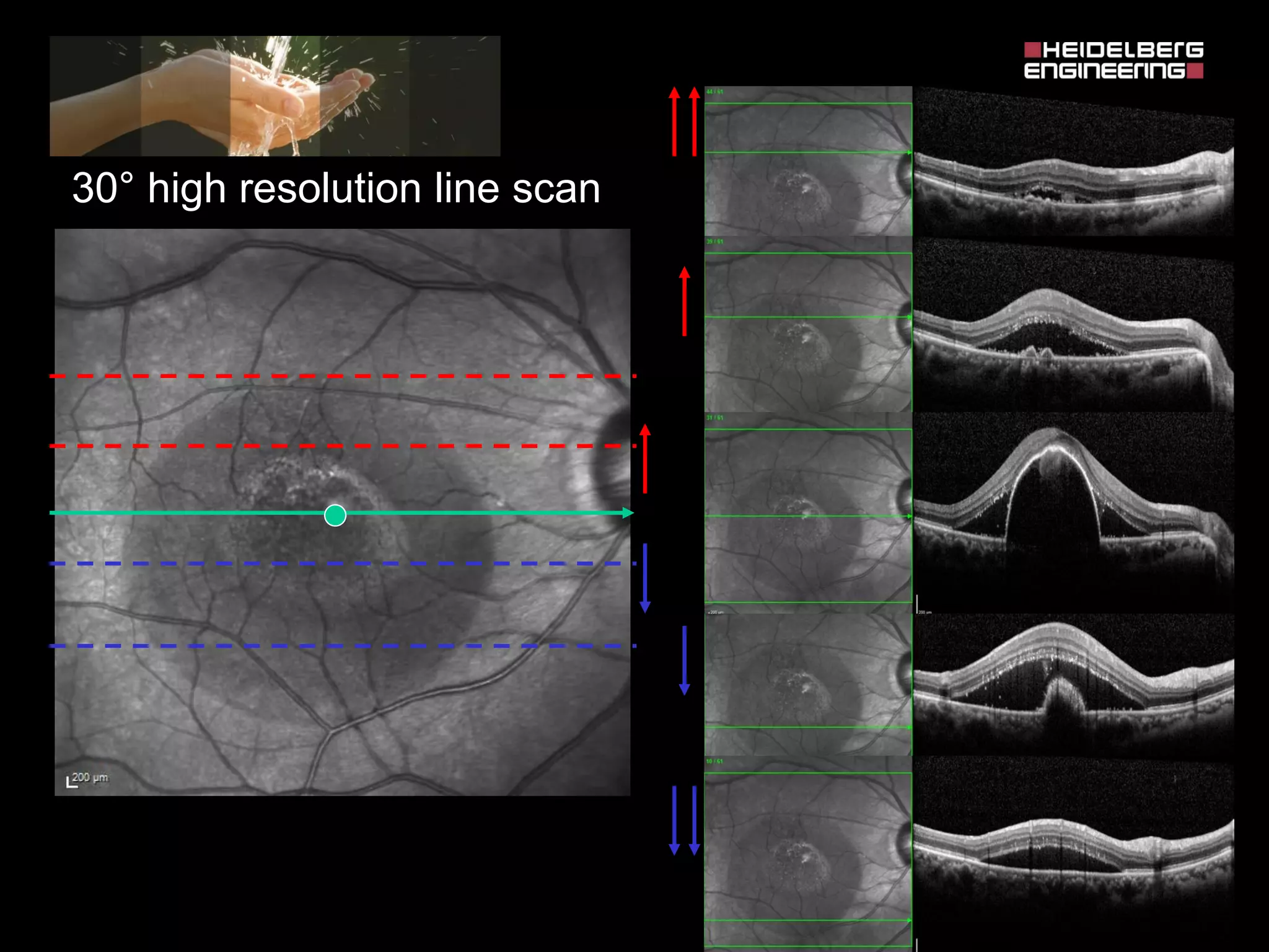
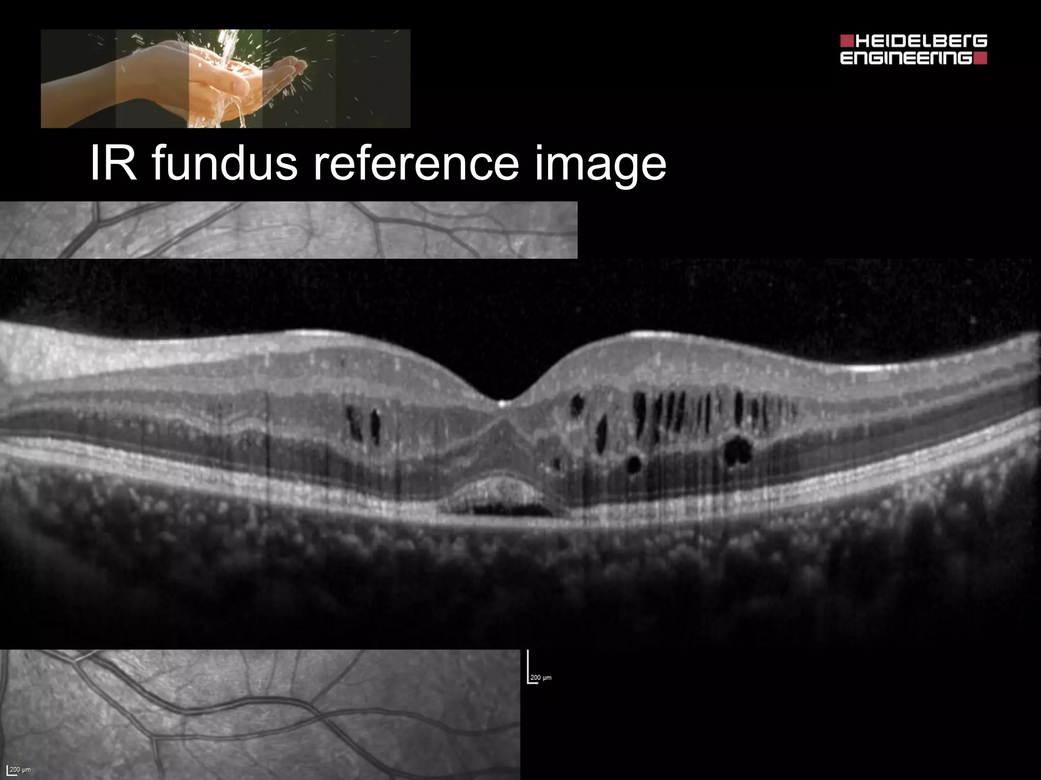
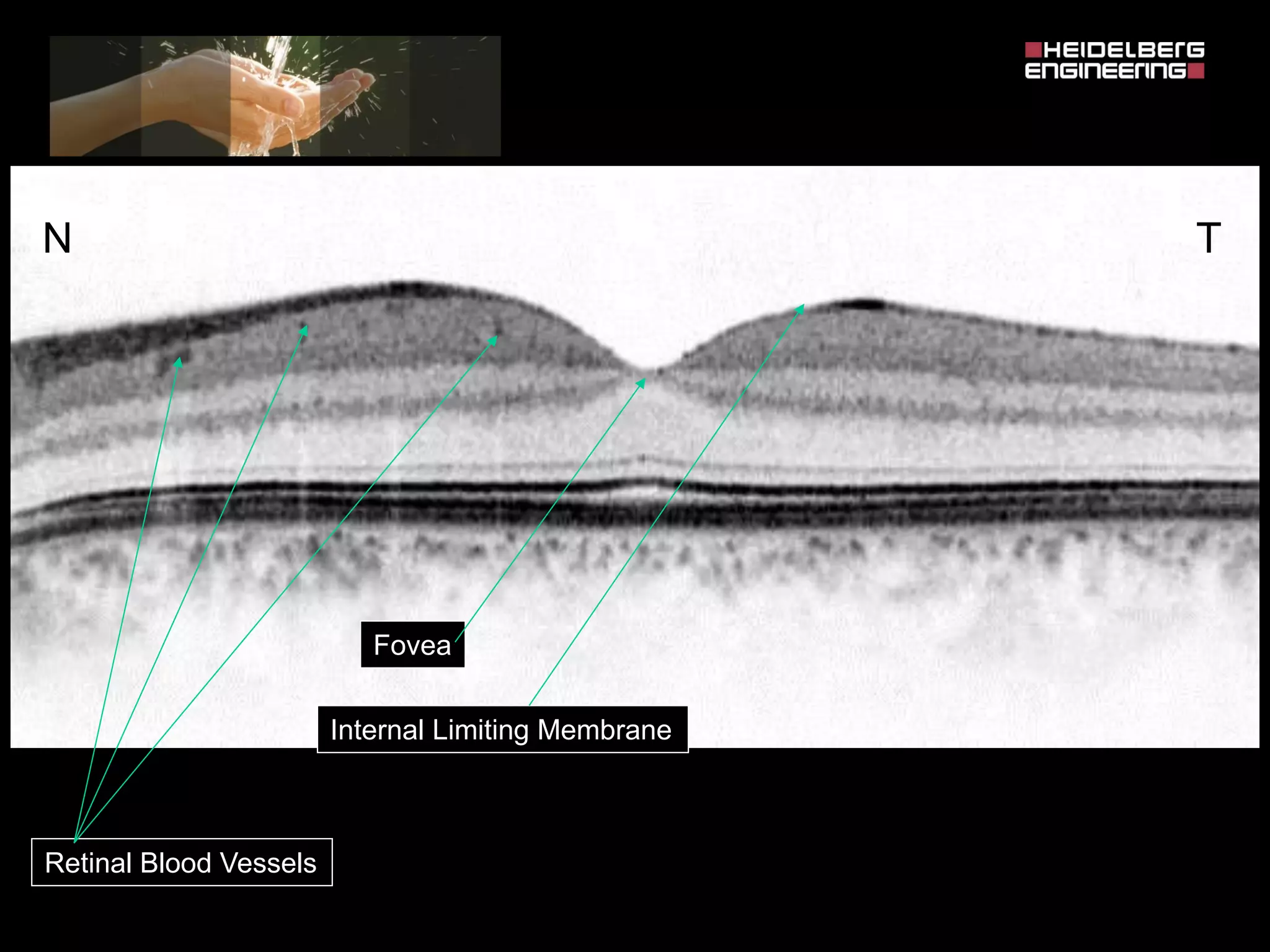
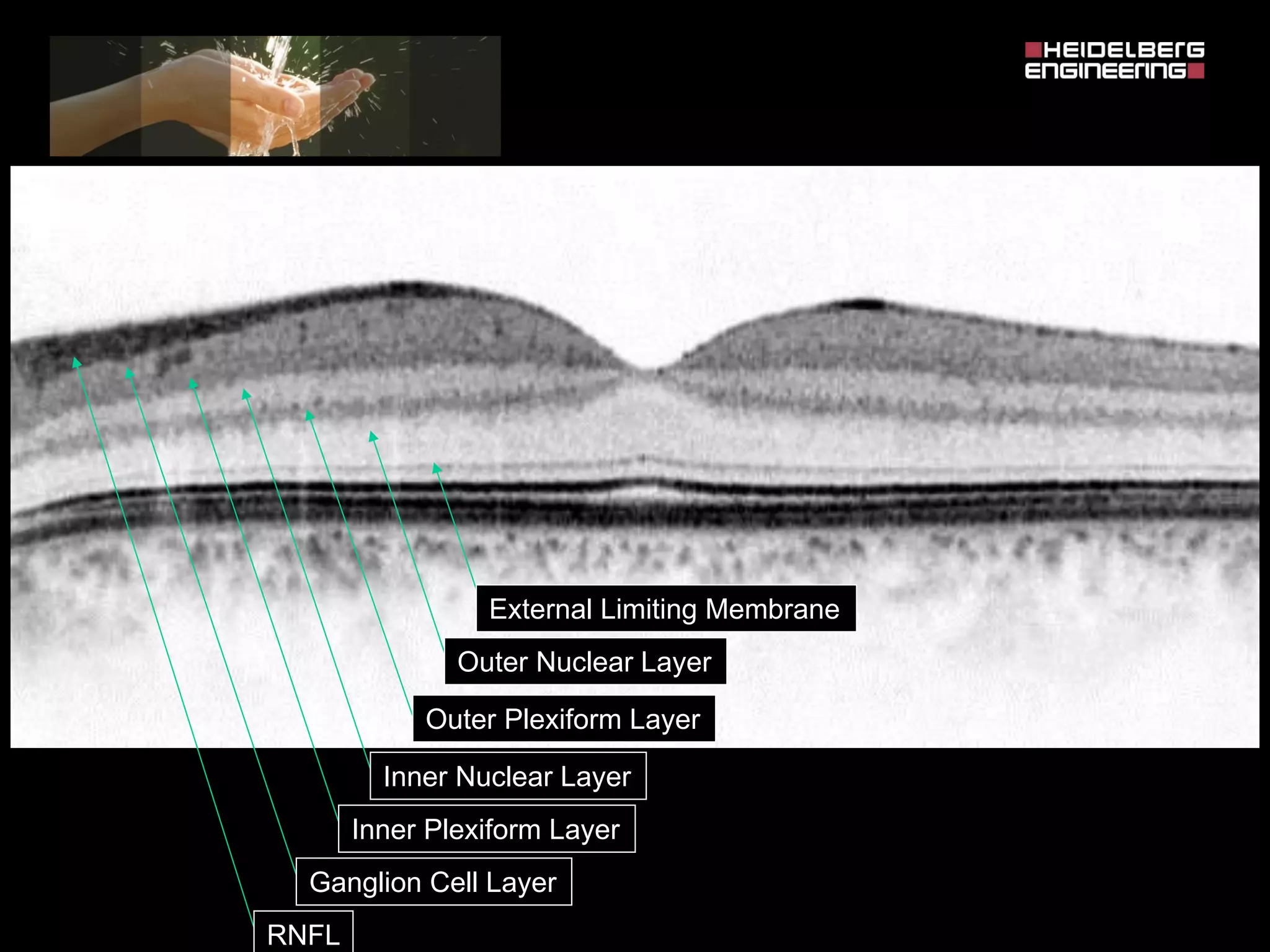
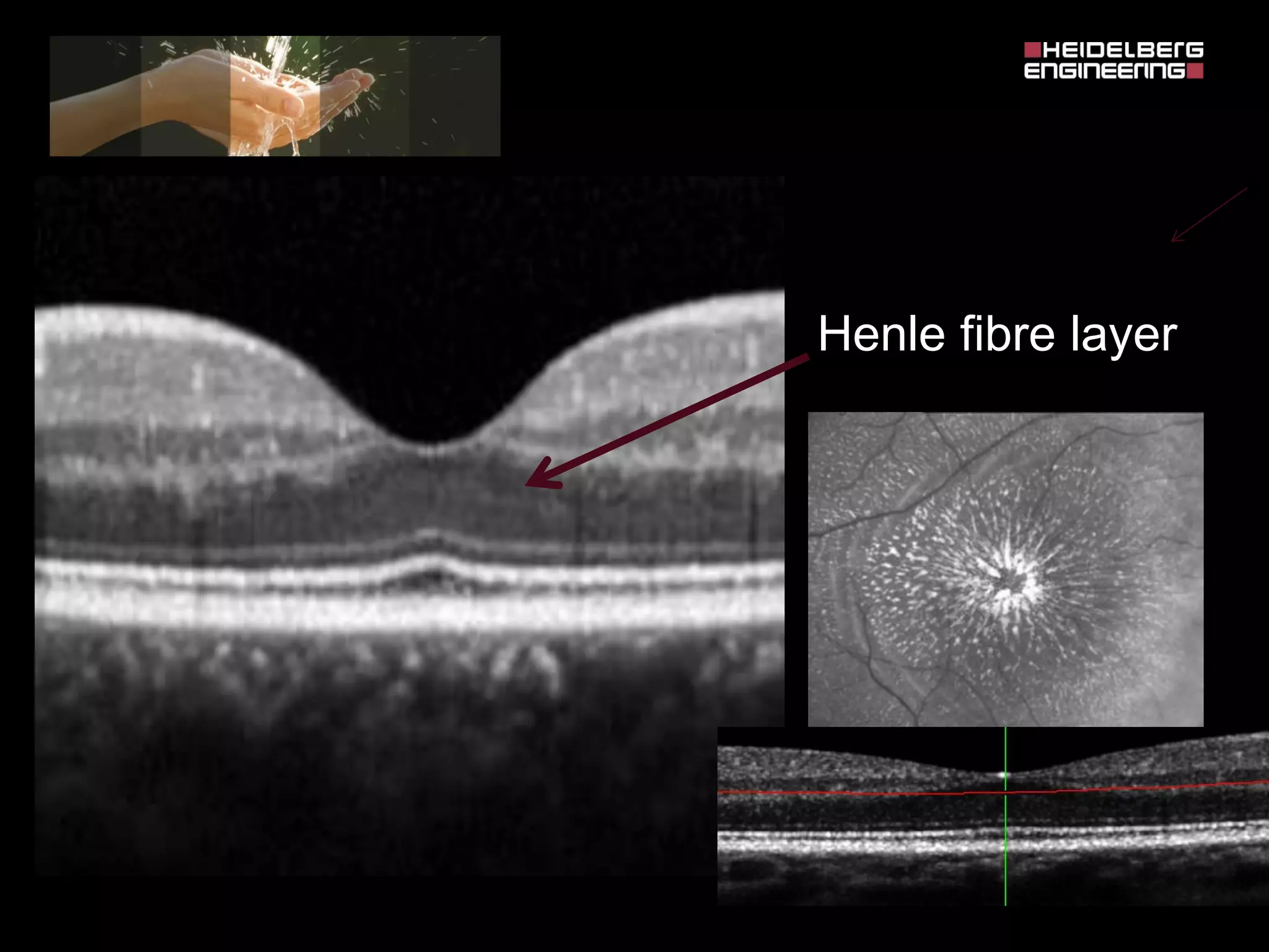
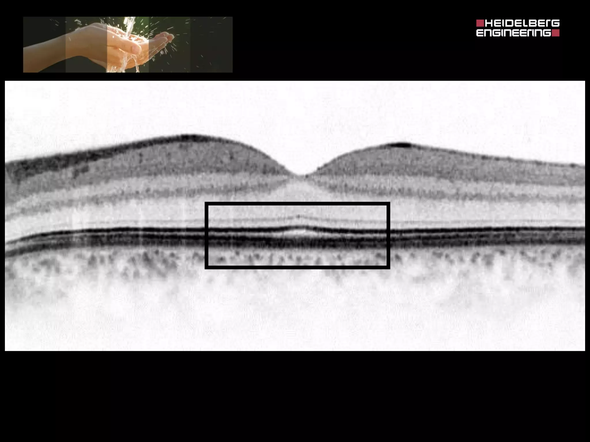
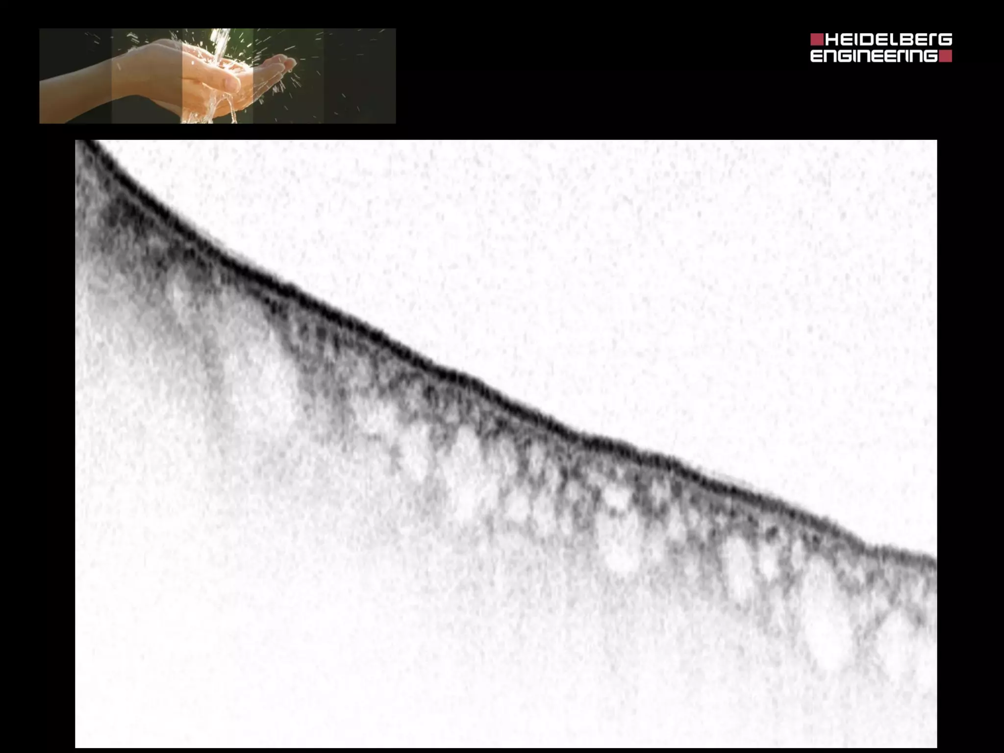
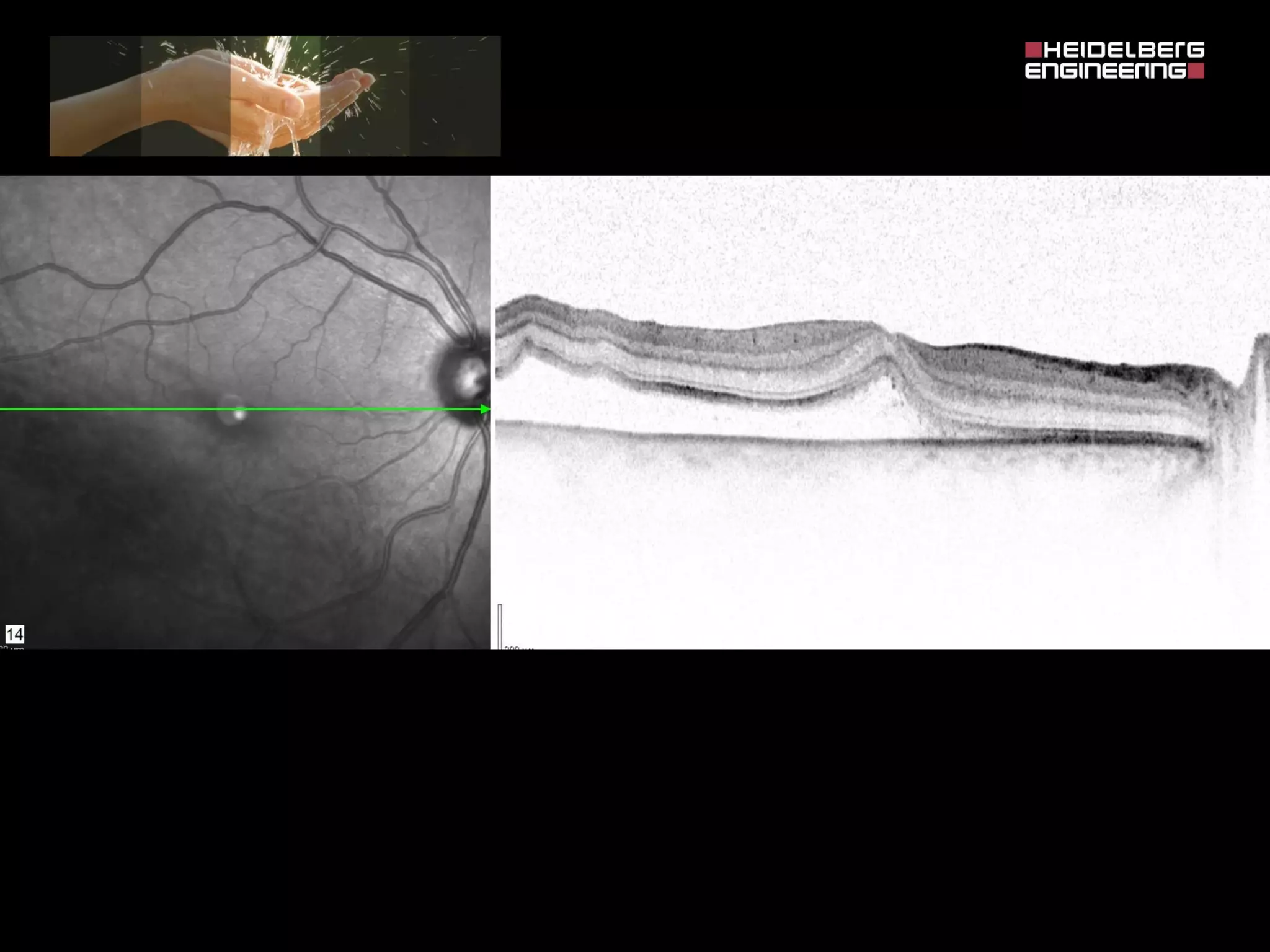
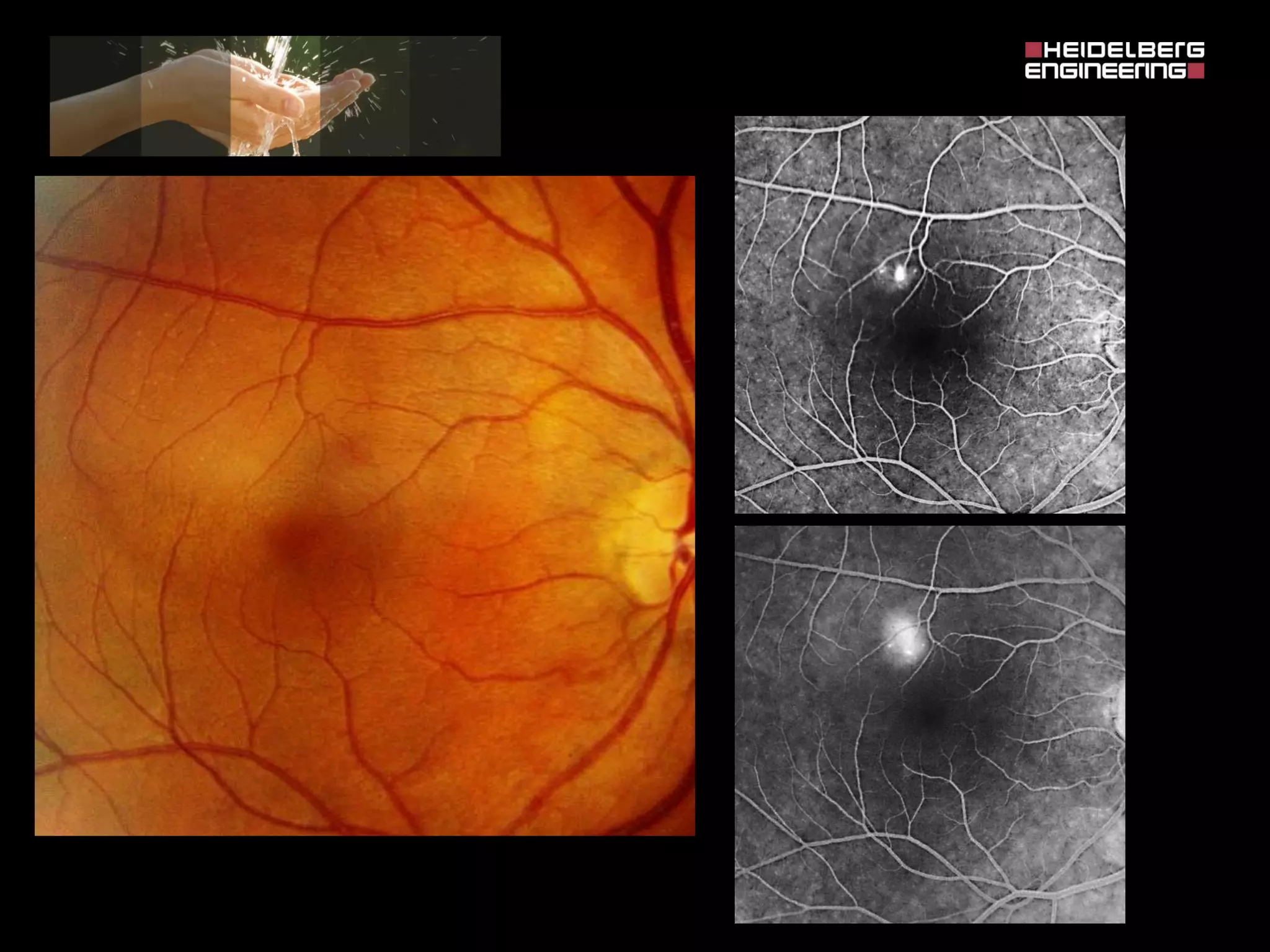
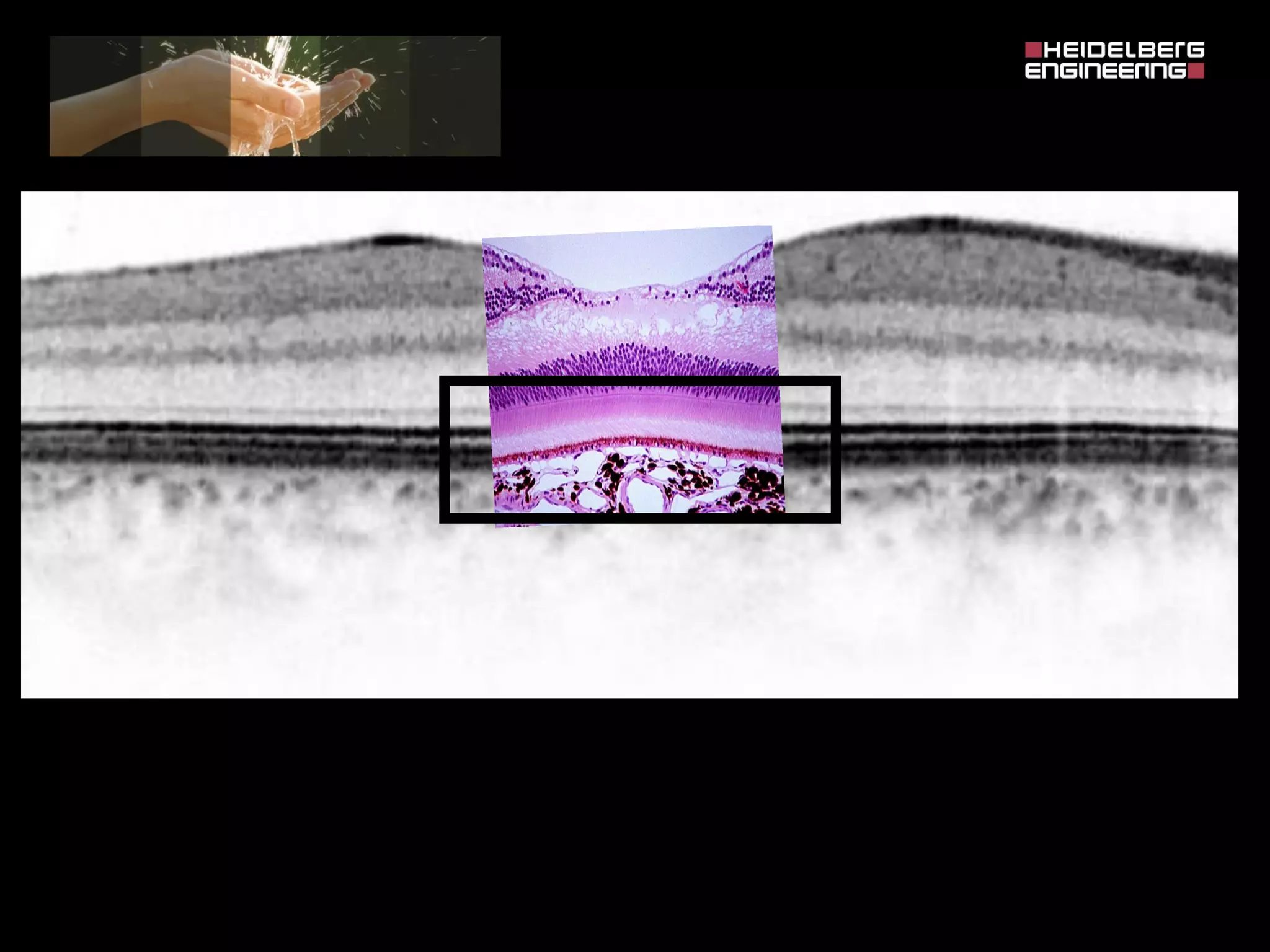
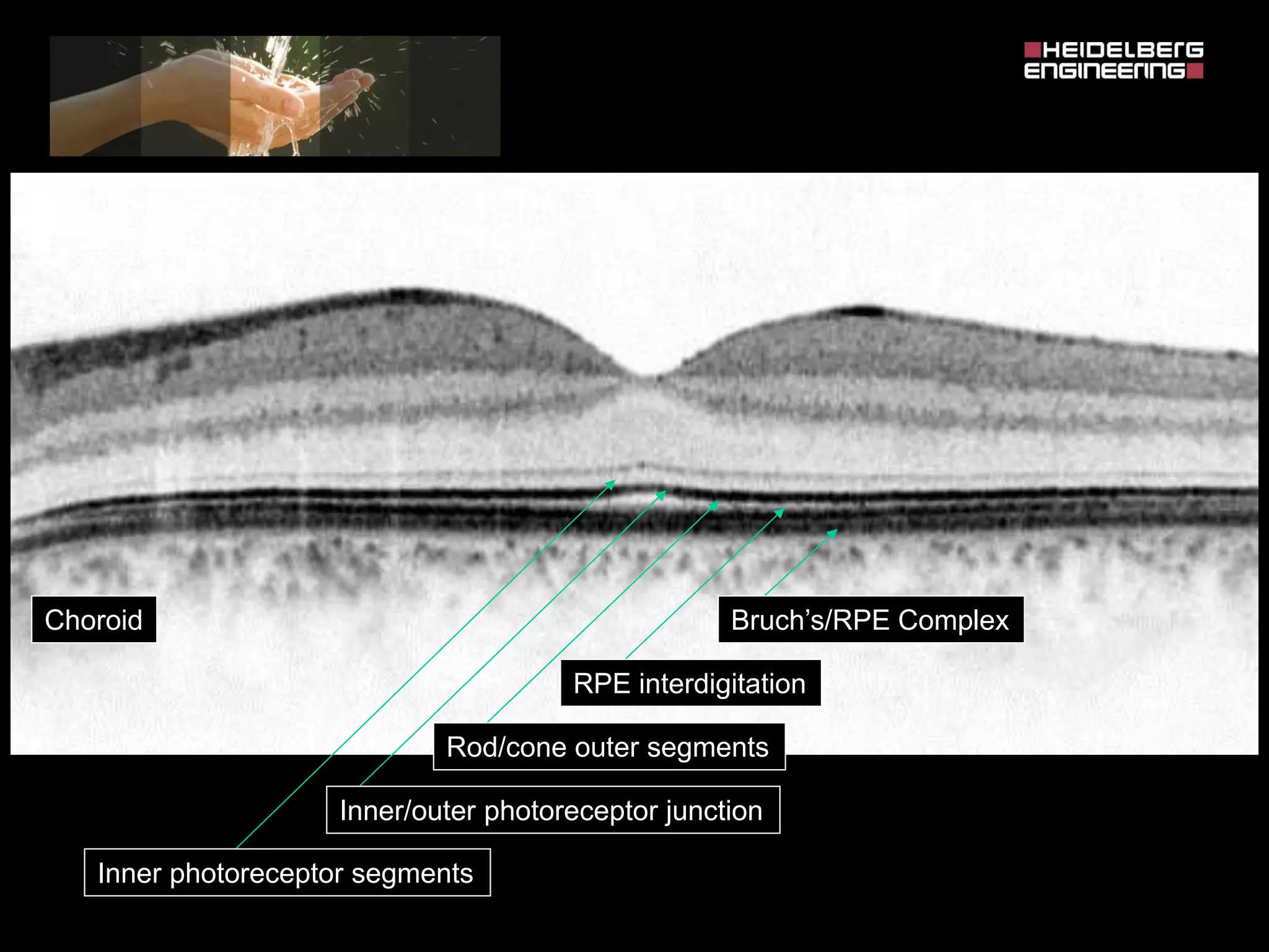
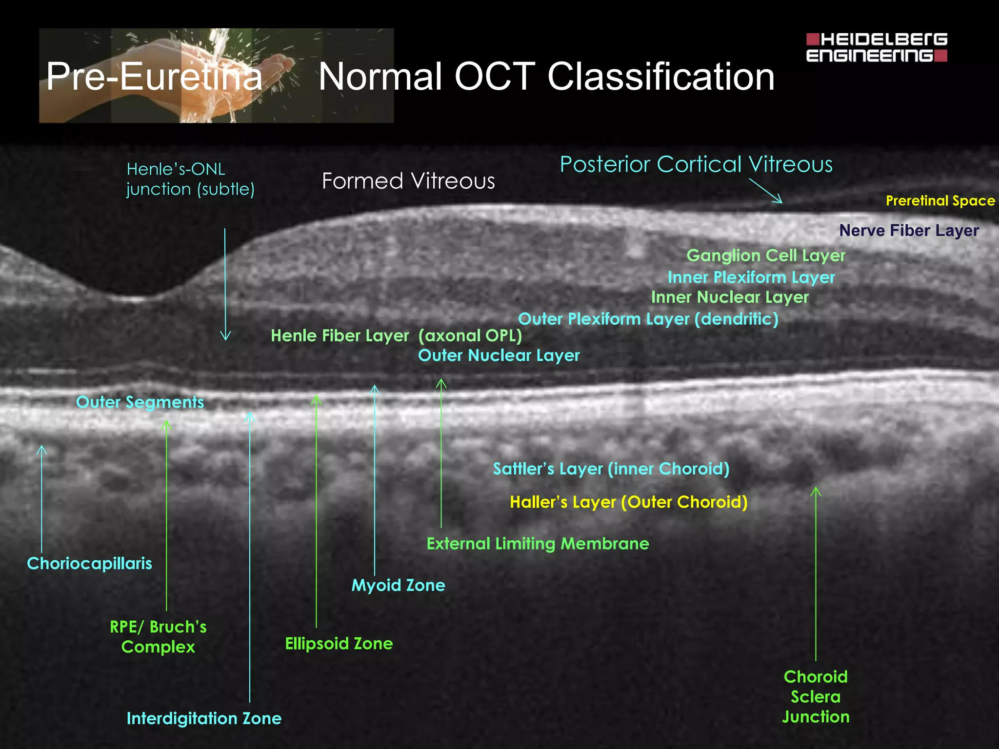
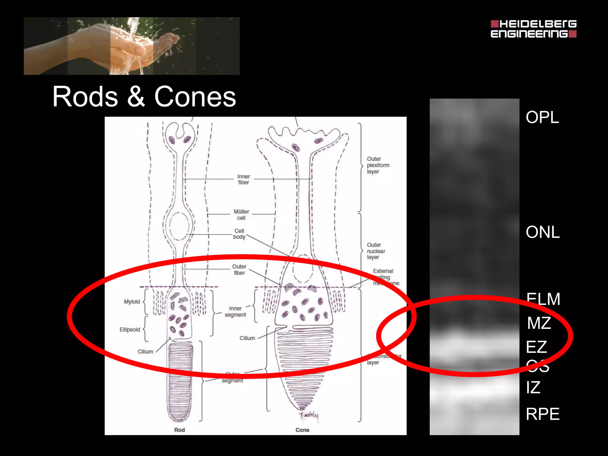
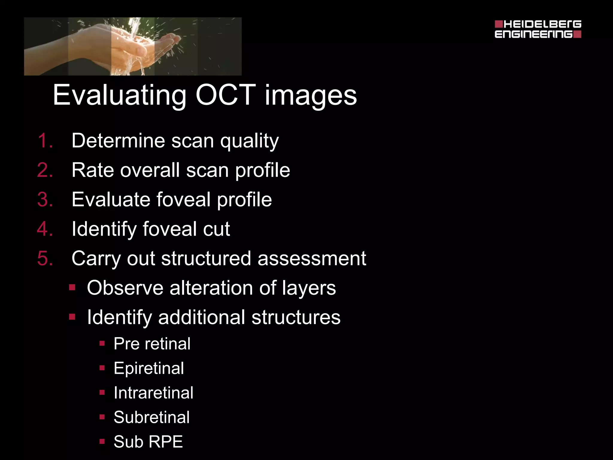
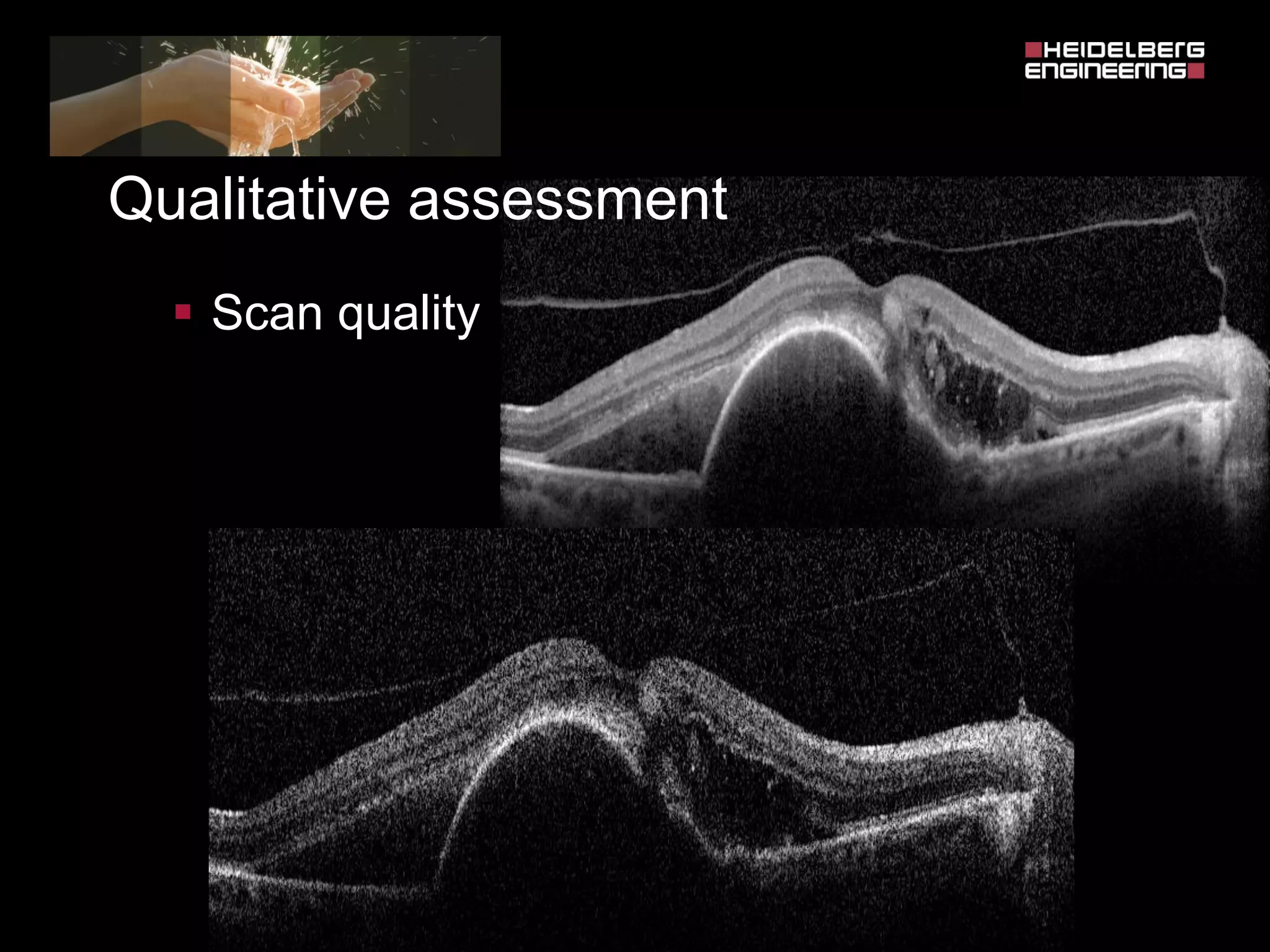
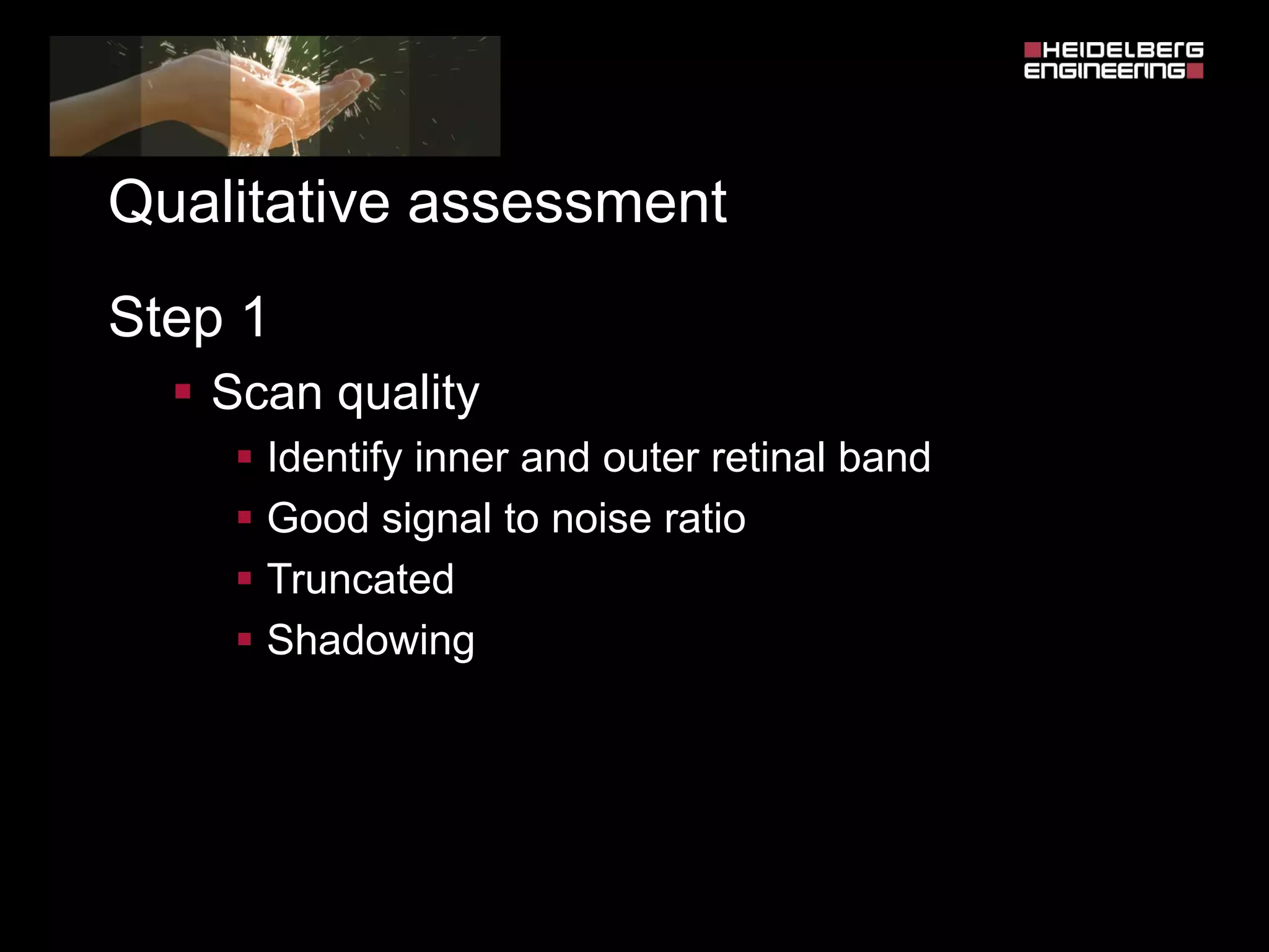
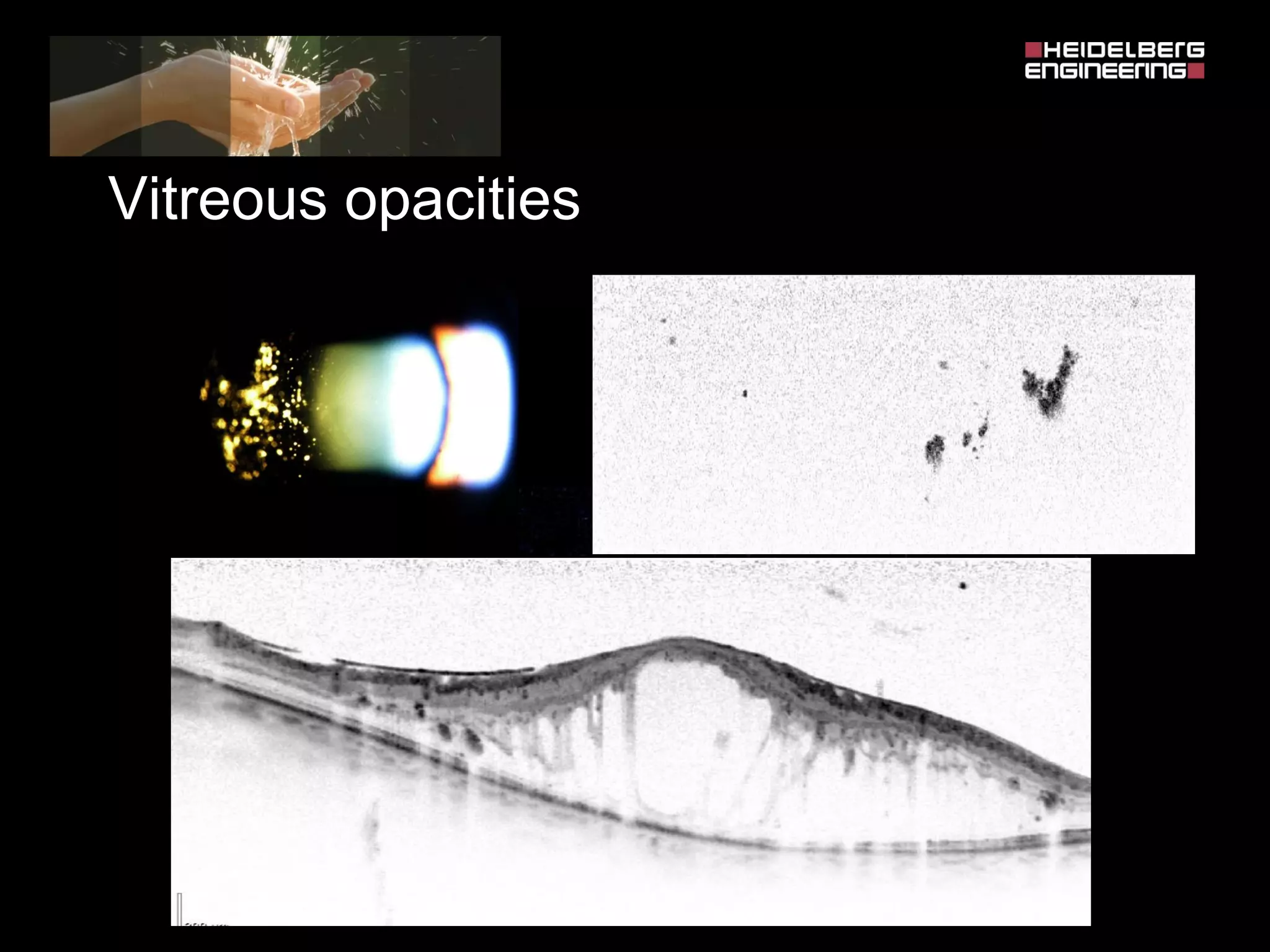
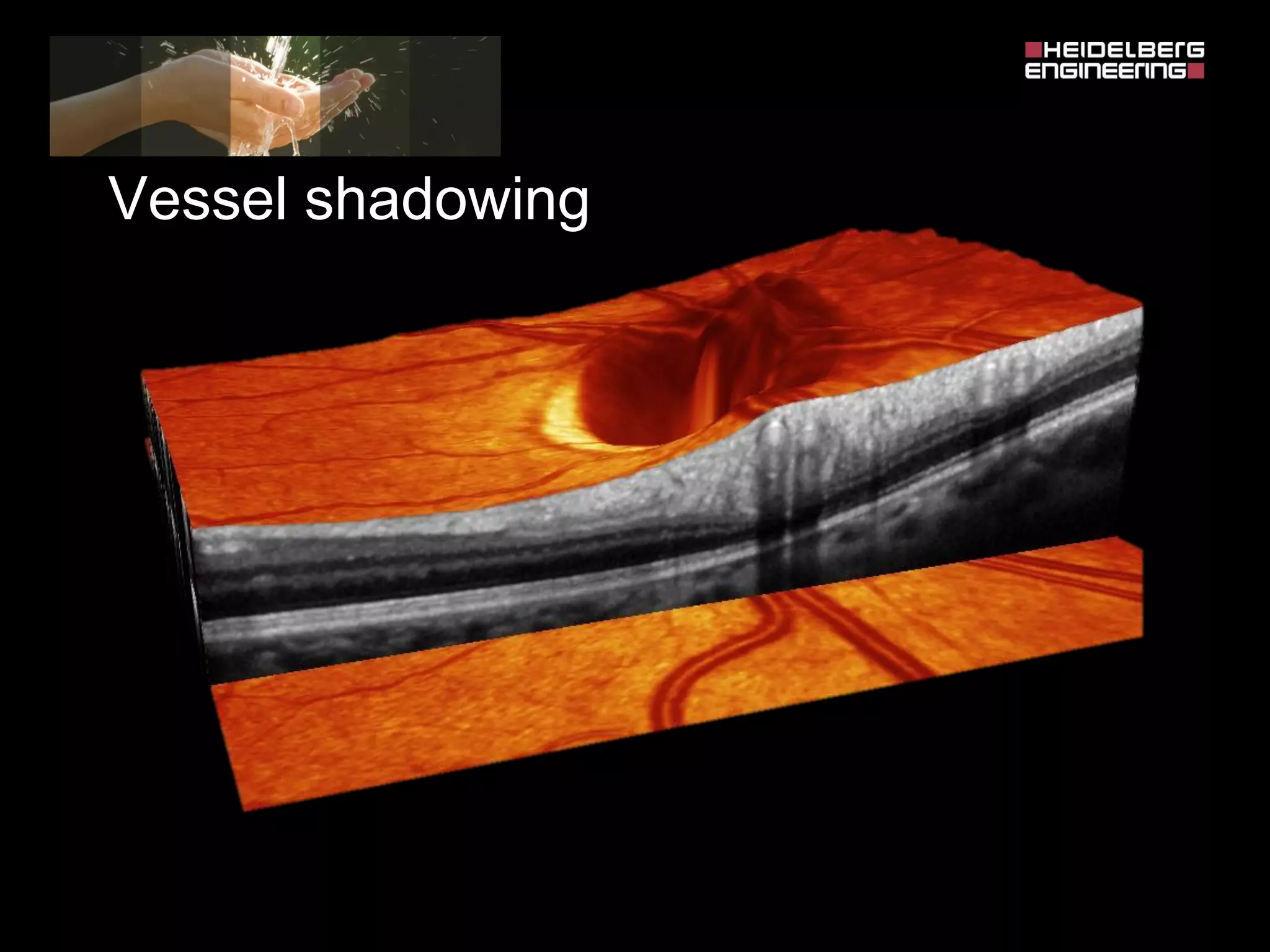
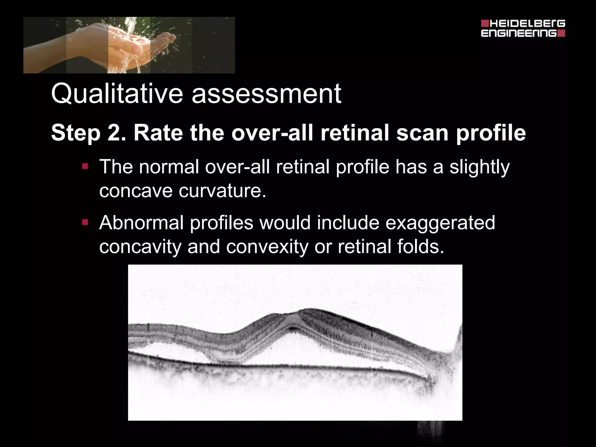
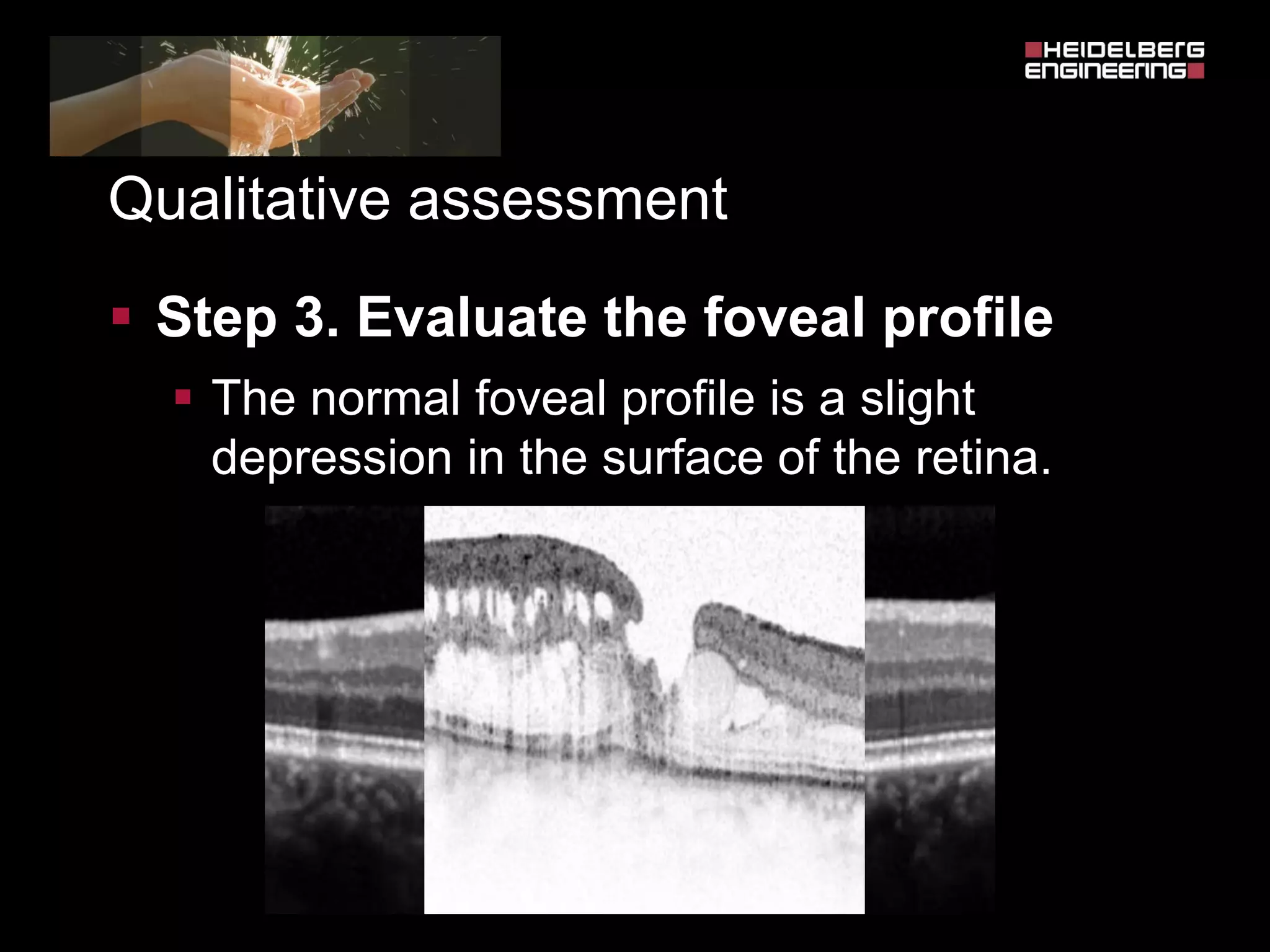
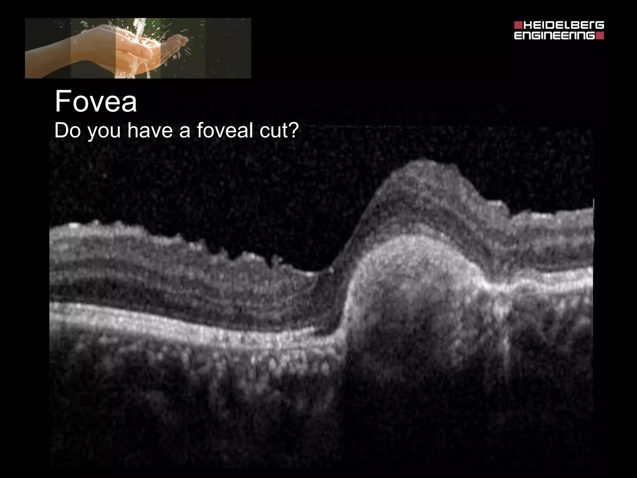
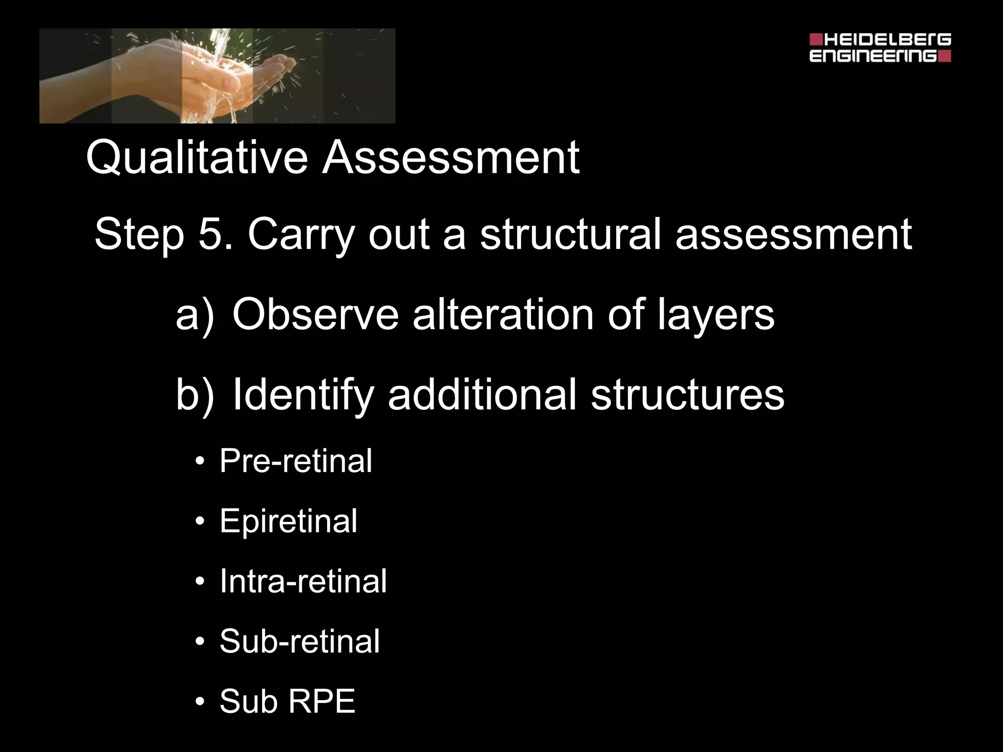
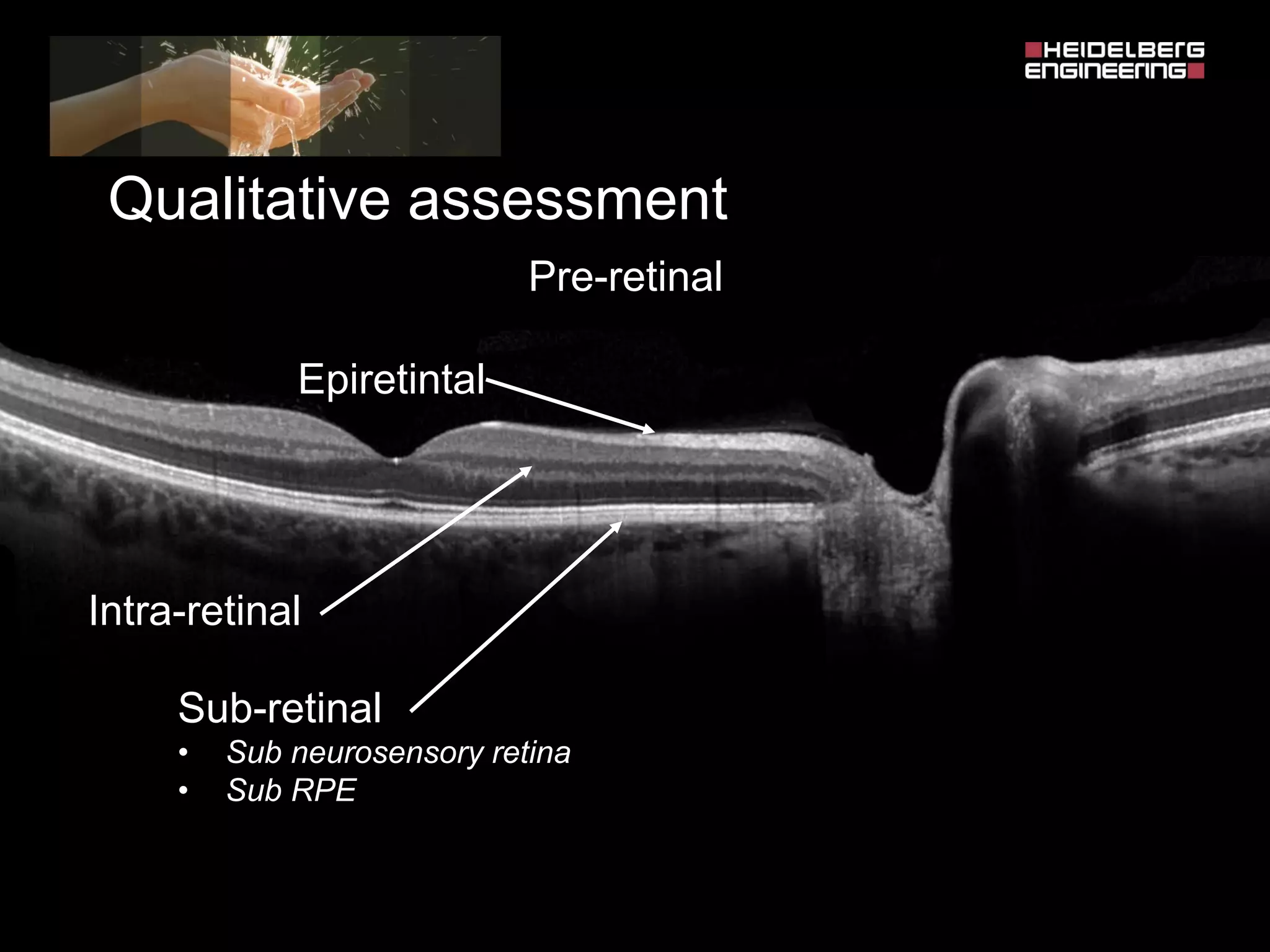
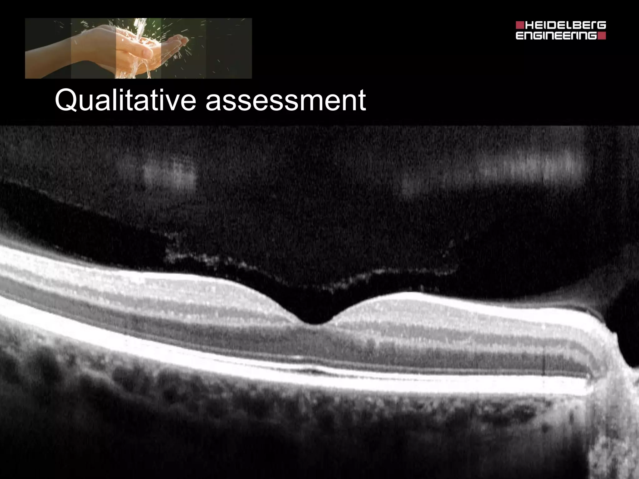
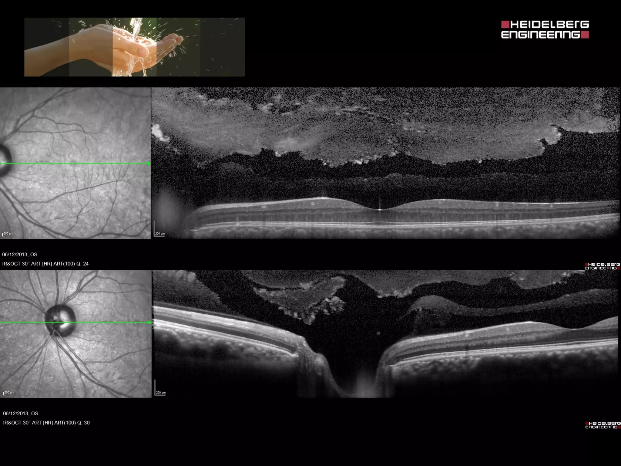
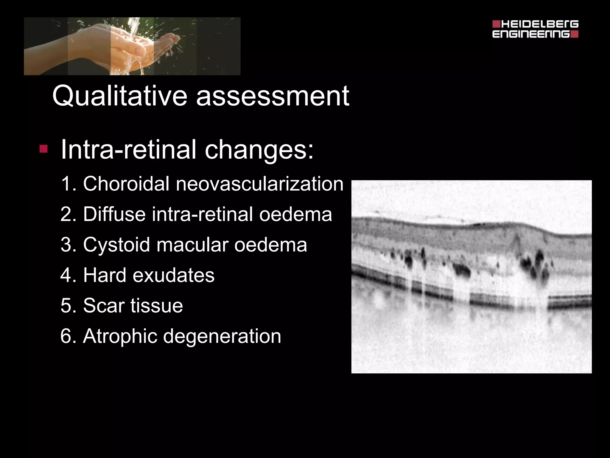
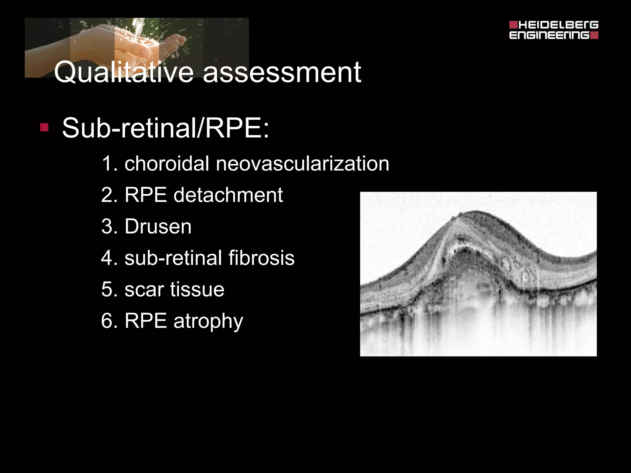
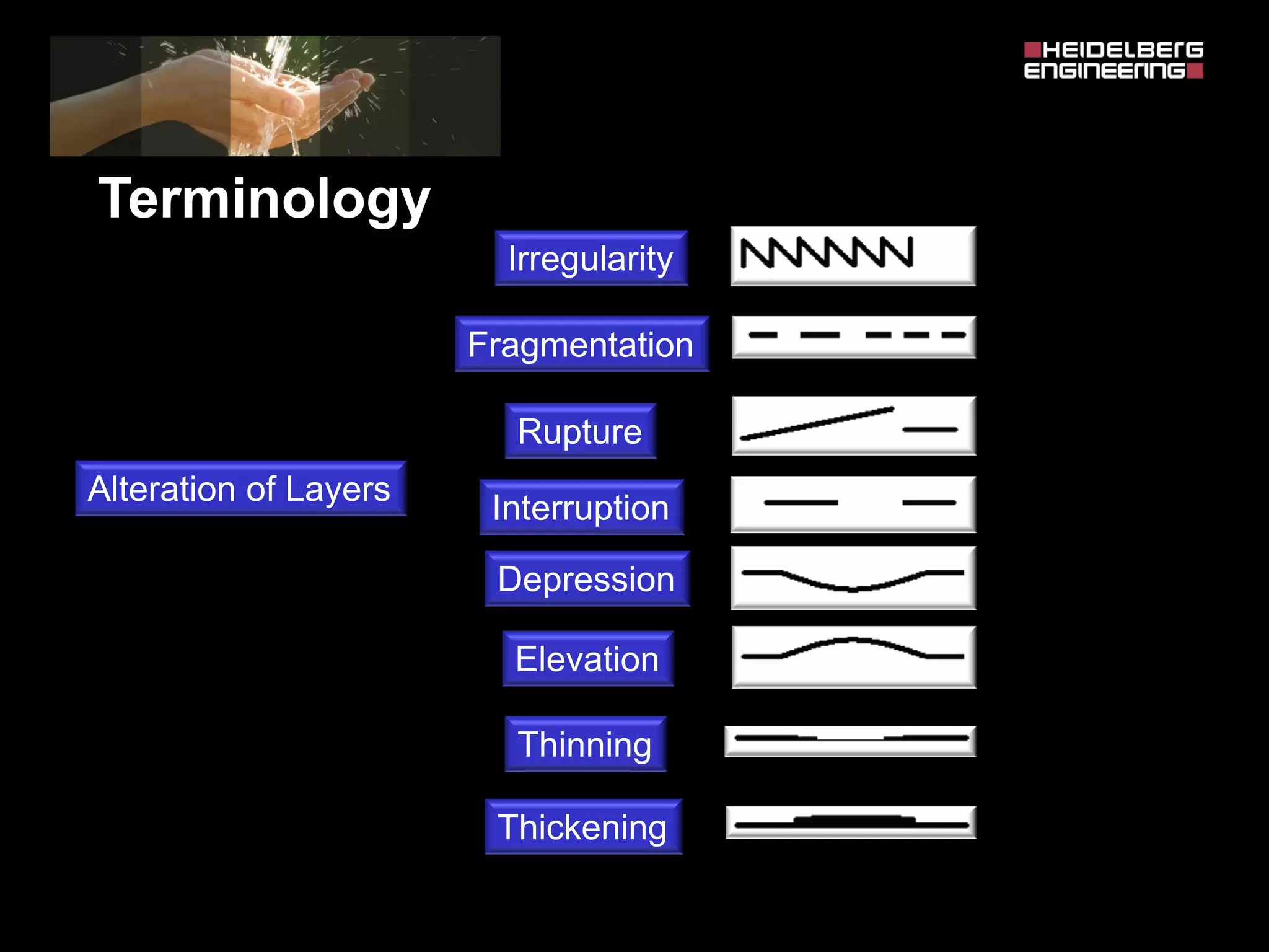
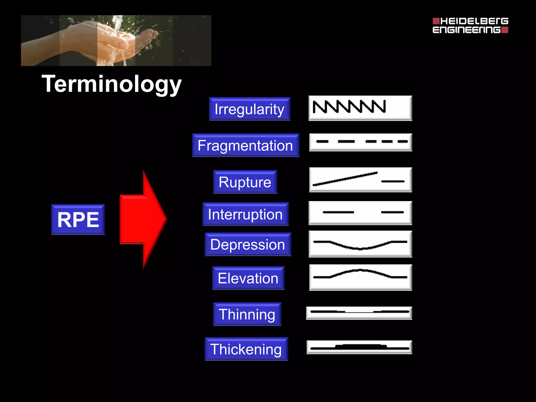
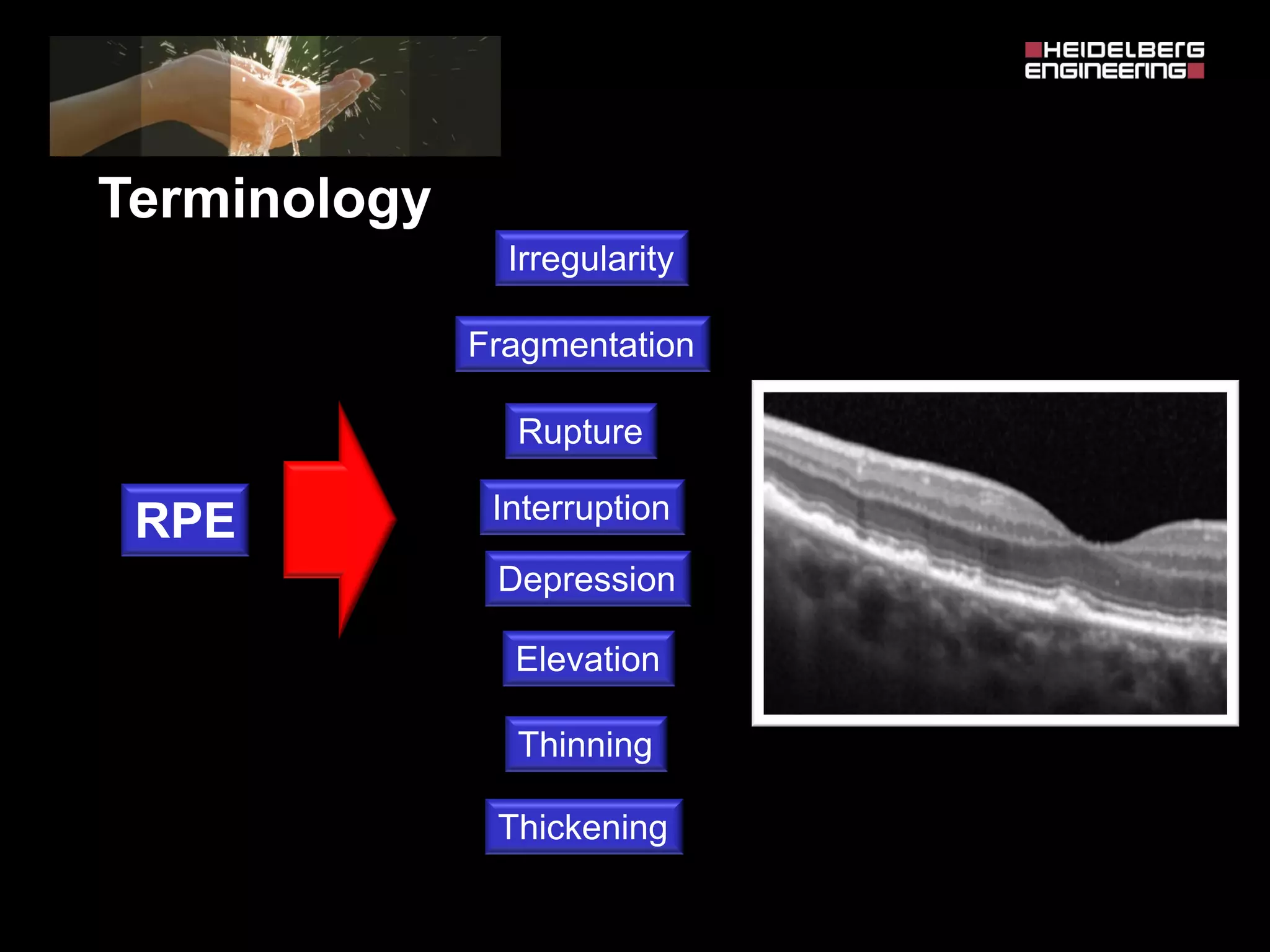
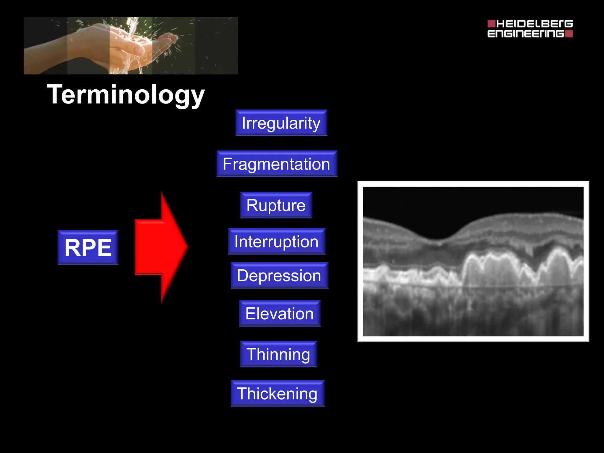
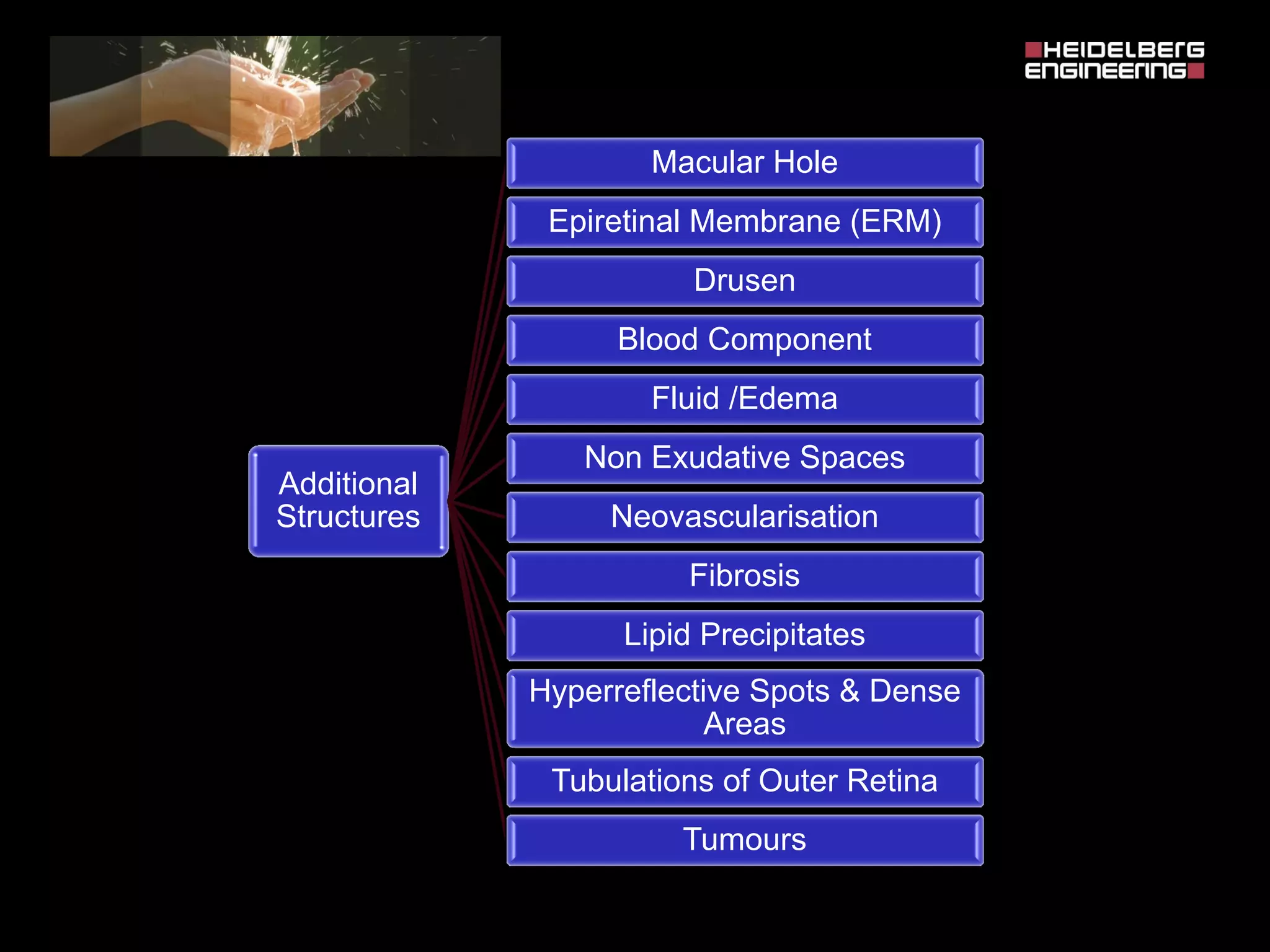
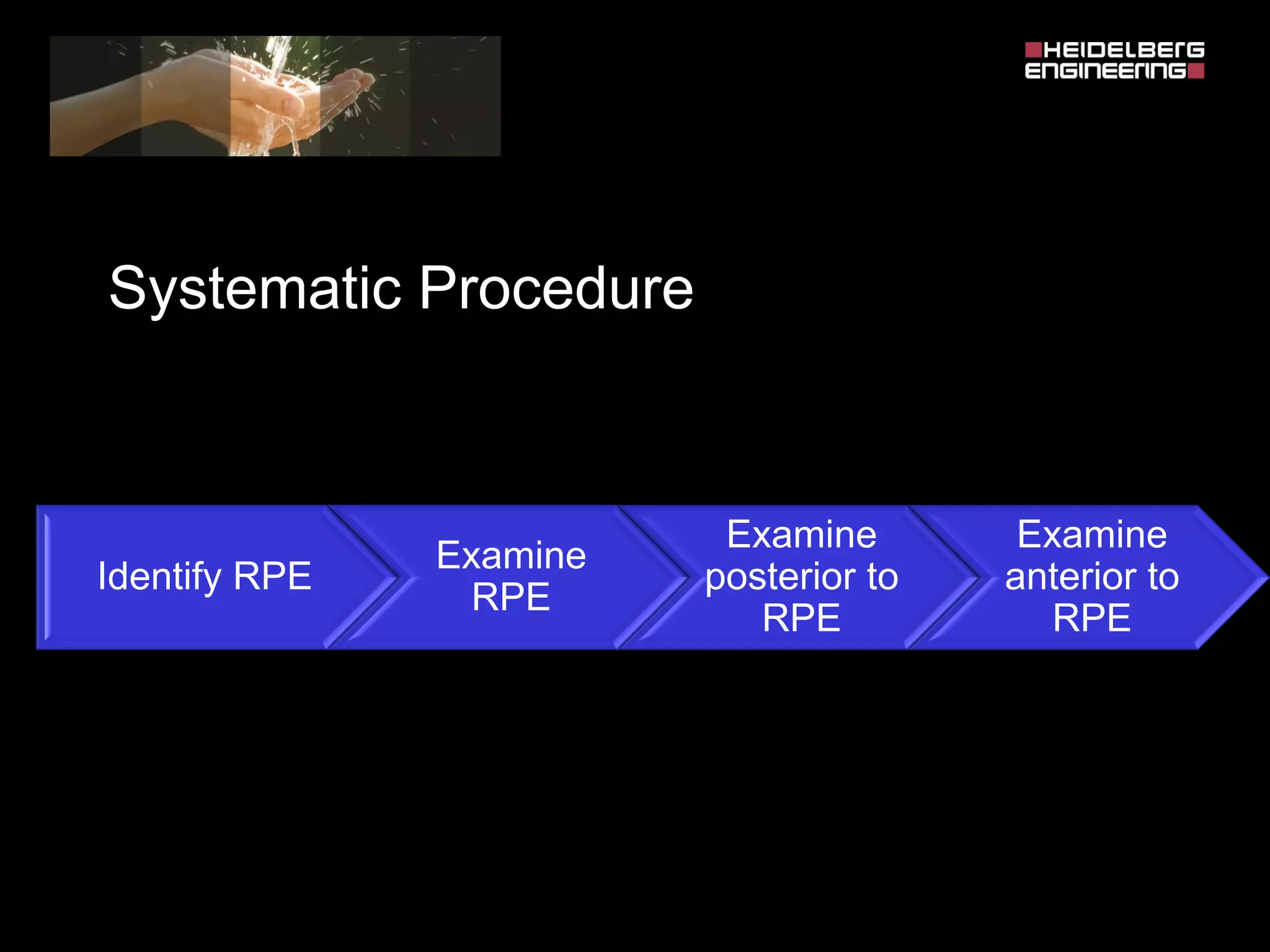
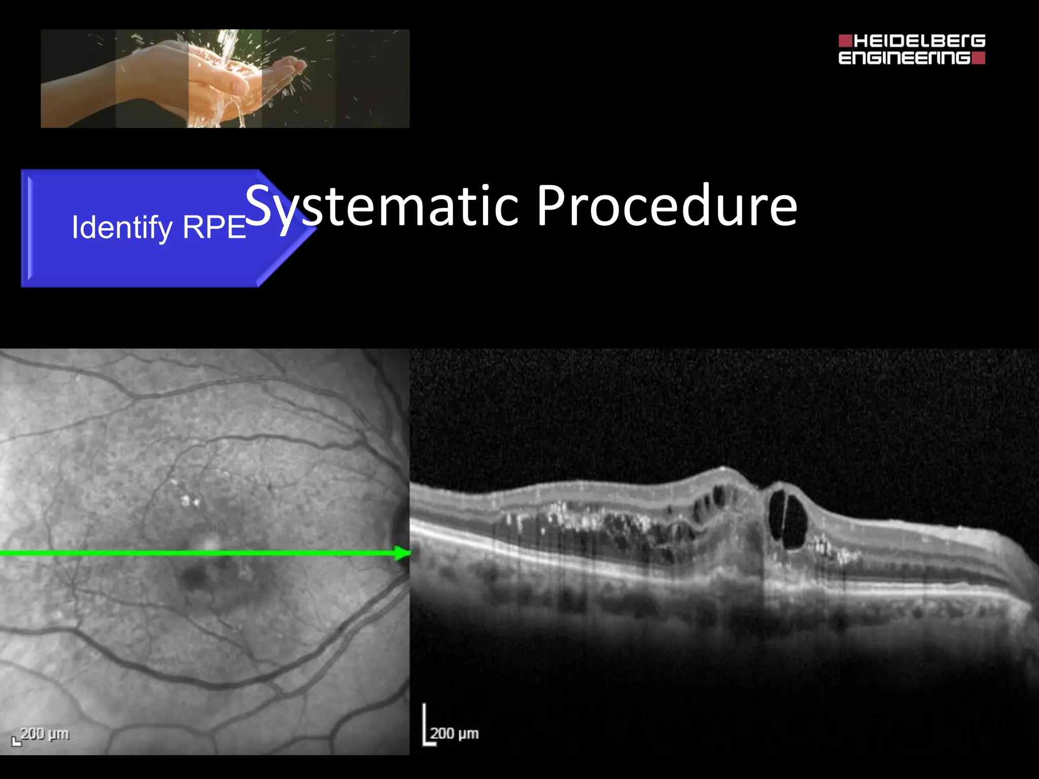
This document provides guidance on interpreting optical coherence tomography (OCT) scans of the retina. It begins by outlining key principles, such as utilizing fundus images and understanding the significance of OCT findings. It then details a 5-step process for evaluating scans: 1) assessing scan quality, 2) rating the overall retinal profile, 3) evaluating the foveal profile, 4) identifying any foveal cut, and 5) carrying out a structural assessment. This includes observing layer alterations, identifying additional structures, and using standardized terminology to describe pathological features. Key pathological structures and findings are defined, including changes affecting the retinal pigment epithelium, sub-RPE space, and intraretinal and subretinal spaces.