This document discusses fundus autofluorescence, which maps the metabolic function of the retinal pigment epithelium (RPE). It explains that lipofuscin, a byproduct of phagocytosis in the RPE, is a major source of autofluorescence. Increased lipofuscin accumulation is implicated in aging and retinal diseases. Fundus autofluorescence imaging allows evaluation of spatial and temporal changes in lipofuscin distribution, providing insight into disease progression and prognosis. Specific autofluorescence patterns are described for various retinal conditions, and its utility for monitoring treatment effects is discussed.
Update knowledge about Muntifocal IOL made by Asaduzzaman
Working as Associate Optometrist in Ispahani Islamia Eye Institute &Hospita, Dhaka 1215
Email:asad.optom92@yaho. com
Update knowledge about Muntifocal IOL made by Asaduzzaman
Working as Associate Optometrist in Ispahani Islamia Eye Institute &Hospita, Dhaka 1215
Email:asad.optom92@yaho. com
Pachychoroid spectrum of disease now also include central serous chorioretinopathy. The presentation include history, pathogenesis, clinical features, diffrential and treatment of CSCR
Central Serous Retinopathy is known to be an idiopathic, sporadic, self-limiting collection of fluid at posterior pole which causes mild to moderate visual loss.
- Video recording of this lecture in English language: https://youtu.be/lK81BzxMqdo
- Video recording of this lecture in Arabic language: https://youtu.be/Ve4P0COk9OI
- Link to download the book free: https://nephrotube.blogspot.com/p/nephrotube-nephrology-books.html
- Link to NephroTube website: www.NephroTube.com
- Link to NephroTube social media accounts: https://nephrotube.blogspot.com/p/join-nephrotube-on-social-media.html
The prostate is an exocrine gland of the male mammalian reproductive system
It is a walnut-sized gland that forms part of the male reproductive system and is located in front of the rectum and just below the urinary bladder
Function is to store and secrete a clear, slightly alkaline fluid that constitutes 10-30% of the volume of the seminal fluid that along with the spermatozoa, constitutes semen
A healthy human prostate measures (4cm-vertical, by 3cm-horizontal, 2cm ant-post ).
It surrounds the urethra just below the urinary bladder. It has anterior, median, posterior and two lateral lobes
It’s work is regulated by androgens which are responsible for male sex characteristics
Generalised disease of the prostate due to hormonal derangement which leads to non malignant enlargement of the gland (increase in the number of epithelial cells and stromal tissue)to cause compression of the urethra leading to symptoms (LUTS
Ethanol (CH3CH2OH), or beverage alcohol, is a two-carbon alcohol
that is rapidly distributed in the body and brain. Ethanol alters many
neurochemical systems and has rewarding and addictive properties. It
is the oldest recreational drug and likely contributes to more morbidity,
mortality, and public health costs than all illicit drugs combined. The
5th edition of the Diagnostic and Statistical Manual of Mental Disorders
(DSM-5) integrates alcohol abuse and alcohol dependence into a single
disorder called alcohol use disorder (AUD), with mild, moderate,
and severe subclassifications (American Psychiatric Association, 2013).
In the DSM-5, all types of substance abuse and dependence have been
combined into a single substance use disorder (SUD) on a continuum
from mild to severe. A diagnosis of AUD requires that at least two of
the 11 DSM-5 behaviors be present within a 12-month period (mild
AUD: 2–3 criteria; moderate AUD: 4–5 criteria; severe AUD: 6–11 criteria).
The four main behavioral effects of AUD are impaired control over
drinking, negative social consequences, risky use, and altered physiological
effects (tolerance, withdrawal). This chapter presents an overview
of the prevalence and harmful consequences of AUD in the U.S.,
the systemic nature of the disease, neurocircuitry and stages of AUD,
comorbidities, fetal alcohol spectrum disorders, genetic risk factors, and
pharmacotherapies for AUD.
New Directions in Targeted Therapeutic Approaches for Older Adults With Mantl...i3 Health
i3 Health is pleased to make the speaker slides from this activity available for use as a non-accredited self-study or teaching resource.
This slide deck presented by Dr. Kami Maddocks, Professor-Clinical in the Division of Hematology and
Associate Division Director for Ambulatory Operations
The Ohio State University Comprehensive Cancer Center, will provide insight into new directions in targeted therapeutic approaches for older adults with mantle cell lymphoma.
STATEMENT OF NEED
Mantle cell lymphoma (MCL) is a rare, aggressive B-cell non-Hodgkin lymphoma (NHL) accounting for 5% to 7% of all lymphomas. Its prognosis ranges from indolent disease that does not require treatment for years to very aggressive disease, which is associated with poor survival (Silkenstedt et al, 2021). Typically, MCL is diagnosed at advanced stage and in older patients who cannot tolerate intensive therapy (NCCN, 2022). Although recent advances have slightly increased remission rates, recurrence and relapse remain very common, leading to a median overall survival between 3 and 6 years (LLS, 2021). Though there are several effective options, progress is still needed towards establishing an accepted frontline approach for MCL (Castellino et al, 2022). Treatment selection and management of MCL are complicated by the heterogeneity of prognosis, advanced age and comorbidities of patients, and lack of an established standard approach for treatment, making it vital that clinicians be familiar with the latest research and advances in this area. In this activity chaired by Michael Wang, MD, Professor in the Department of Lymphoma & Myeloma at MD Anderson Cancer Center, expert faculty will discuss prognostic factors informing treatment, the promising results of recent trials in new therapeutic approaches, and the implications of treatment resistance in therapeutic selection for MCL.
Target Audience
Hematology/oncology fellows, attending faculty, and other health care professionals involved in the treatment of patients with mantle cell lymphoma (MCL).
Learning Objectives
1.) Identify clinical and biological prognostic factors that can guide treatment decision making for older adults with MCL
2.) Evaluate emerging data on targeted therapeutic approaches for treatment-naive and relapsed/refractory MCL and their applicability to older adults
3.) Assess mechanisms of resistance to targeted therapies for MCL and their implications for treatment selection
Tom Selleck Health: A Comprehensive Look at the Iconic Actor’s Wellness Journeygreendigital
Tom Selleck, an enduring figure in Hollywood. has captivated audiences for decades with his rugged charm, iconic moustache. and memorable roles in television and film. From his breakout role as Thomas Magnum in Magnum P.I. to his current portrayal of Frank Reagan in Blue Bloods. Selleck's career has spanned over 50 years. But beyond his professional achievements. fans have often been curious about Tom Selleck Health. especially as he has aged in the public eye.
Follow us on: Pinterest
Introduction
Many have been interested in Tom Selleck health. not only because of his enduring presence on screen but also because of the challenges. and lifestyle choices he has faced and made over the years. This article delves into the various aspects of Tom Selleck health. exploring his fitness regimen, diet, mental health. and the challenges he has encountered as he ages. We'll look at how he maintains his well-being. the health issues he has faced, and his approach to ageing .
Early Life and Career
Childhood and Athletic Beginnings
Tom Selleck was born on January 29, 1945, in Detroit, Michigan, and grew up in Sherman Oaks, California. From an early age, he was involved in sports, particularly basketball. which played a significant role in his physical development. His athletic pursuits continued into college. where he attended the University of Southern California (USC) on a basketball scholarship. This early involvement in sports laid a strong foundation for his physical health and disciplined lifestyle.
Transition to Acting
Selleck's transition from an athlete to an actor came with its physical demands. His first significant role in "Magnum P.I." required him to perform various stunts and maintain a fit appearance. This role, which he played from 1980 to 1988. necessitated a rigorous fitness routine to meet the show's demands. setting the stage for his long-term commitment to health and wellness.
Fitness Regimen
Workout Routine
Tom Selleck health and fitness regimen has evolved. adapting to his changing roles and age. During his "Magnum, P.I." days. Selleck's workouts were intense and focused on building and maintaining muscle mass. His routine included weightlifting, cardiovascular exercises. and specific training for the stunts he performed on the show.
Selleck adjusted his fitness routine as he aged to suit his body's needs. Today, his workouts focus on maintaining flexibility, strength, and cardiovascular health. He incorporates low-impact exercises such as swimming, walking, and light weightlifting. This balanced approach helps him stay fit without putting undue strain on his joints and muscles.
Importance of Flexibility and Mobility
In recent years, Selleck has emphasized the importance of flexibility and mobility in his fitness regimen. Understanding the natural decline in muscle mass and joint flexibility with age. he includes stretching and yoga in his routine. These practices help prevent injuries, improve posture, and maintain mobilit
Explore natural remedies for syphilis treatment in Singapore. Discover alternative therapies, herbal remedies, and lifestyle changes that may complement conventional treatments. Learn about holistic approaches to managing syphilis symptoms and supporting overall health.
TEST BANK for Operations Management, 14th Edition by William J. Stevenson, Ve...kevinkariuki227
TEST BANK for Operations Management, 14th Edition by William J. Stevenson, Verified Chapters 1 - 19, Complete Newest Version.pdf
TEST BANK for Operations Management, 14th Edition by William J. Stevenson, Verified Chapters 1 - 19, Complete Newest Version.pdf
Prix Galien International 2024 Forum ProgramLevi Shapiro
June 20, 2024, Prix Galien International and Jerusalem Ethics Forum in ROME. Detailed agenda including panels:
- ADVANCES IN CARDIOLOGY: A NEW PARADIGM IS COMING
- WOMEN’S HEALTH: FERTILITY PRESERVATION
- WHAT’S NEW IN THE TREATMENT OF INFECTIOUS,
ONCOLOGICAL AND INFLAMMATORY SKIN DISEASES?
- ARTIFICIAL INTELLIGENCE AND ETHICS
- GENE THERAPY
- BEYOND BORDERS: GLOBAL INITIATIVES FOR DEMOCRATIZING LIFE SCIENCE TECHNOLOGIES AND PROMOTING ACCESS TO HEALTHCARE
- ETHICAL CHALLENGES IN LIFE SCIENCES
- Prix Galien International Awards Ceremony
Anti ulcer drugs and their Advance pharmacology ||
Anti-ulcer drugs are medications used to prevent and treat ulcers in the stomach and upper part of the small intestine (duodenal ulcers). These ulcers are often caused by an imbalance between stomach acid and the mucosal lining, which protects the stomach lining.
||Scope: Overview of various classes of anti-ulcer drugs, their mechanisms of action, indications, side effects, and clinical considerations.
3. Autofluorescence
Some materials contain a naturally
autofluorescent component that can be
visualised when excited with a light of
particular wavelength
5. RPE and lipofuscin
RPE constitutes a monolayer of
polygonal cells between the
choroid and neuro-sensory
retina
Multiple functions
RPE dysfunction implicated in
variety of retinal diseases
LF is a byproduct of
accumulation of sheded outer
segments of the photoreceptors
6. Lipofuscin accumulates as a byproduct
of phagocytosis of photoreceptors’ outer
segment
In advanced age: It may occupy 20% of
free cytoplasmic space of RPE cells
The older we grow the more we glow
7. lipofuscin
WHY IS IT DANGEROUS ?
A2-E (N-retinylidene-N-retinylethanol-
amine) the dominant fluorophore possess
toxic properties
Interferes with the normal cell function
Precursors of A2-E are also toxic
Products of photo-oxidation of RPE
lipofuscin serves as trigger for
complement activation inflammation
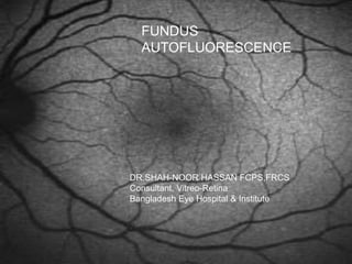
9. Fundus autofluorescence
Metabolically mapping the RPE
Developed as a tool to evaluate the RPE
during aging and ocular disease
Andrea von Ruckmann, Fredrick W. Fitzke and Alan C. Bird- Moorfield’s
eye hospital
10. Scanning Laser
Ophthalmoscope
Webb and co-workers
55° of field in one frame
Low power laser source
Scan in x and y axis
Confocality ensures that light fluorescence
and reflectance is derived from same ocular
plane
HRA2: Excitation 488nm
Emitted light above 500nm
12. FAF image acquisition
Align the camera using the IR
illumination.
Spectralis HRA+OCT only: Once you
see a sharp well-focused image,
change to Redfree illumination to fine-
tune focus.
Change to the FA illumination. The
image will now be considerably darker.
Automatic (recommended!) or
manual (Spectralis HRA+OCT only)
sensitivity control will outline the
retinal blood vessels.
Turn
or
automatic
13. FAF image acquisition – Mean
function
Activate the ART (Automatic Real
Time) Mean function to generate a ‘live
Mean’ Autofluorescence image online
and view it as it is created.
Note: Following the injection of
fluorescein dye, it will be impossible to
perform FAF imaging.
Press
14. Normal Autofluorescence
distribution
Optic nerve head
Absence of
autofluorescent pigment
Retinal blood vessels
Absorption by blood
vessels
Foveal area
Absorption by luteal
pigment
Parafoveal area
Mildly decreased
intensity due to high
melanin content and
lower density of
lipofuscin granules in
central RPE
15. In disease state
Excessive accumulation of lipofuscin in
the lysosomal compartment of the RPE
is the downstream process in many
hereditary and age related diseases
16.
17. AF and ARMD- Basic
Considerations
RPE is thought to play a key role in the
early and late phases of the disease
Hallmark of aging is the accumulation of
lipofuscin granules in the cytoplasm of
the RPE cells
Lipofuscin accumulation is the common
downstream process
18. Ability to document spatial distribution of
lipofuscin and its changes over time.
The amount of autofluorescence is signature for
previous or possible future oxidative injury
Hyperfluorescence in FAF FA 36 sec FA 69 sec
19. Geographic Atrophy
Seen as hypo-
autofluorescent areas
No RPE, NO
LIPOFUSCIN NO
AUTOFLUORESCENCE
Measure the atrophic
area to see for the
progression
20. Geographic atrophy
Identification of peri-lesional abnormalities
Hyperautofl signals bordering denote sick
RPE
The damage marches in areas of high signals
Predictor of future trouble
21. Pigment Epithelial Detachment
Serous PED:
Increased FAF signal
corresponding to area of
detachmet
Underlying CNVM: No
specific findings
Surrounding area: Hypo AF
signal
22. Choroidal
neovascularization
Irregular FAF in areas of CNV
High FAF outside the edge of the
lesion
FAF intensity decreased over the
disciform scars
In early stages, preservation of
FAF
Extent of abnormal areas on FAF
is more than that on FA
24. CSR
Leaks at the level of RPE
leading to central serous
detachment
Chronic disease associated
with atrophic changes at the
level of RPE and retina
FA and ICG – hemodynamics
and fluid dynamics
OCT- clues to size and
elevation of detachment
Autofluorescence- functional
status
27. Macular dystrophies-
Stargradt’s disease
Areas of atrophy on fundus
corresponded to hypo-
autofluorescence
Flecks seen as
depigmented lesion
appeared as areas of hypo-
autofl
Predictive value is yet to be
determined
28. Macular dystrophies: Best’s
disease
Autofl characters: central round
area of increased FAF
Pseudohypopyon stage:
increased FAF in the lower part
Late stages: irregular FAF
within the lesion with
disseminated spots of
increased FAF
29. Macular dystrophies: Best’s
disease
Pattern of spread on FAF: centrifugal
Atrophic regions are associated with low
levels of background FAF, lower visual
acuity, abnormal colour vision, central
scotomas and poorer
electrophysiological results
FAF appears more striking and
widespread
Spoke like, diffuse or combination
30. X-linked retinoschisis
FAF finding reflect typical radiating
cystic changes
The changes on FAF are most likely due
to altered passage of exciting and
emitted light from the retinal folds
31. Retinitis Pigmentosa
In dominant and recessive
and rod-cone dystrophies
Absent FAF in areas of outer
retinal atrophy
Normal FAF in adjacent
regions of surviving retina
High FAF in surviving areas in
some cases
Macular oedema of more than
4 months high FAF
32. Retinitis Pigmentosa
Parafoveal ring of increased FAF
Correlation exists between these areas
of high FAF and photopic and scotopic
sensitivity
33. Serpigenous Choroiditis
In SC: Autofluorescence is
detected within 2-5 days
after the appearance of
lesion
Provides a clear
delineation of the area of
RPE damage
Progressive decrease in
autofluorescence was seen
during the scarring phase
35. Macular Hole
Bright fluorescence of macular holes
similar to images on FA
Pseudoholes: no such high
autofluorescence
Attached operculum shows focal
decreased autofluorescence
37. VKH
Hypo AF in the areas of serous
detachments
NIR AF: hyper AF at the macula and
hypo AF in the areas of serous
detachment
With treatment: BL-FAF: subtle FAF
NIR FAF: more wide spread FAF
39. Applications for therapeutic
interventions
In advanced atrophic AMD: useful to
develop and assess the therapeutic
interventions
Fenritidine, an oral medicine shown to
reduce the production of toxic fluophores
In retinal dystrophies: useful to assess
the functional preservation of the outer
retina
In Leber’s: normal or slightly reduced FAF
40. RPE FAF & therapeutic
outcome
The RPE-FAF of exudative AMD lesions varies
greatly.
FAF differences have a great influence on the
chances of antivascular endothelial growth factor
(VEGF) therapy success.
Development of visual acuity is less favorable in
eyes with initially increased central FAF.
Heimes et al. - Foveal RPE FAF as a prognostic factor for anti-VEGF therapy in exudative AMD - GraefesArch 2008
41. CONCLUSION
Non invasive ,Easy to perform
Provides a novel prognostic marker for
disease progression
Metabolic changes and loss of RPE
integrity corresponds to visual function
In combination with SD OCT it adds to
our understanding of retinal diseases
from a broad point of view.
Ability to visualize the biochemistry and look into the RPE cells
Classification abnormal autofluorescence patterns in early age-related macular disease with fundus
photography and autofluorescence images introduced by Bindewald et al.12 Eight phenotypic patterns are
differentiated: NORMAL (A, B) -- homogenous background FAF and a gradual decrease in the inner macula toward
the fovea due to the masking effect of macular pigment. Only small hard drusen are visible in the corresponding
fundus photograph. MINIMAL CHANGE (C, D) -- only minimal variations from normal background FAF. There is
limited irregular increase or decrease in FAF intensity due to multiple small hard drusen. FOCAL (E, F ) -- several well definied
spots with markedly increased FAF. Fundus photograph of the same eye with multiple including hard and soft
drusen. PATCHY (G, H) -- multiple large areas (O200 mm diameter) of increased FAF corresponding to large, soft
drusen and/or hyperpigmentation on the fundus photograph LINEAR (I, J ) -- characterized by the presence of at least
one linear areas with markedly increased FAF. A corresponding hyperpigmented line is visible in the fundus
photograph. LACE-LIKE (K, L) -- multiple branching linear structures of increased FAF. This pattern may correspond
to hyperpigmentation on the fundus photograph or to no visible abnormalities. RETICULAR (M, N) -- multiple,
specific small areas of decreased FAF with brighter lines in-between. The reticular pattern not only occurs in the
macular area but is found more typically in a superotemporal location. There may be visible reticular drusen in the
corresponding fundus photograph. SPECKLED (O, P) -- variety of FAF abnormalities in a larger area of the FAF image.
There seem to be fewer pathologic areas in the corresponding fundus.
The RPE is blocked by membrane in classic
However, the pathobiology of many findings in
central serous chorioretinopathy has remained elusive,
because of our inability to image physiologic changes
induced by the disease. Autofluorescence photography
provides functional images of the fundus by employing
the stimulated emission of light from naturally occurring
fluorophores, the most significant being lipofuscin. In the
case of retinal pigment epithelial cells, the buildup of
lipofuscin is related in large part to the phagocytosis of
photoreceptor outer segments containing damage accumulated
through use, and indigestible altered molecules
are retained within lysosomes and eventually become
lipofuscin.
NIR-Near infra red
BL-Blue light
but other retinal fluorophores that may occur
in pathological conditions such as fluid, hemorrhages,
or melanin deposition must be differentiated.