
Recommended
More Related Content
What's hot
What's hot (20)
Similar to Wrist joint
Similar to Wrist joint (20)
More from Neeta Chhabra
Recently uploaded
Recently uploaded (20)
Wrist joint
- 1. Wrist Joint & Joints of Hand
- 2. Wrist Joint SYNONYMN RADIO CARPAL JOINT Type: Synovial joint Sub type: Ellipsoid variety Biaxial Wrist complex • Radiocarpal joint • Midcarpal joint
- 3. Above: Distal end of the radius Articular disc of inferior RU joint Below: Scaphoid, Lunate, and Triquetral bones • The proximal articular surface forms an ellipsoid concave surface, in which fits the distal ellipsoid convex surface. Articulating Surfaces
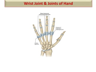
- 4. Capsular ligament Palmar ligaments Dorsal ligament Collateral ligaments Ligaments Ligaments
- 5. Encloses the joint and is attached above to the distal ends of the radius and ulna and below to the proximal row of carpal bones. Capsular ligament
- 6. • Lines the capsule and is attached to the margins of the articular surfaces. • The joint cavity does not communicates with that of the distal radio-ulnar joint or with the joint cavities of the intercarpal joints. • Prestyloid recess Synovial Membrane
- 7. • Palmar radiocarpal Ligament • Palmar ulnocarpal Ligament • Dorsal radiocarpal Ligament • Radial collateral ligament • Ulnar collateral ligament Ligaments Ligaments
- 8. Anteriorly: tendons of • Flexor digitorum superficialis • Flexor digitorum profundus • Flexor pollicis longus • Flexor carpi radialis • Flexor carpi ulnaris • Median and ulnar nerves Laterally: Radial artery Relations
- 9. Posteriorly: Tendons of • Abductor Pollicis Longus • Extensor Pollicis Brevis • Extensor Carpi Radialis Longus • Extensor Carpi Radialis Brevis • Extensor Pollicis Longus • Extensor Indicis • Extensor Digitorum • Extensor Digiti Minimi • Extensor Carpi Ulnaris Relations
- 10. • Blood Supply: Ant & Post carpal arches formed by branches of radial and ulnar arteries • Nerve supply: Anterior & posterior interosseous nerves Blood & Nerve Supply
- 11. Movements
- 12. Flexion: Mainly at midcarpal joint Range-0-60 degrees Muscles: Flexor Carpi Radialis Flexor Carpi Ulnaris Palmaris Longus. Assisted by: Flexor Digitorum Superficialis Flexor Digitorum Profundus Flexor Pollicis Longus. Movements
- 13. Extension: Mainly at wrist joint Range-0-50 degrees Muscles: Extensor Carpi Radialis Longus Extensor Carpi Radialis Brevis Extensor Carpi Ulnaris Assisted by: Extensor Digitorum Extensor Indicis Extensor Digiti Minimi Extensor Pollicis Longus Movements
- 14. Abduction: At Midcarpal joint Range:0-15 degrees Muscles : Flexor Carpi Radialis Extensor Carpi Radialis Longus Extensor Carpi Radialis Brevis Assisted by: Abductor Pollicis Longus Extensor Pollicis Longus Extensor Pollicis Brevis Movements
- 15. Adduction: Mainly at wrist joint Range:0-50 degree Muscles: Flexor Carpi Ulnaris Extensor Carpi Ulnaris Circumduction: Combination of flexion adduction, extension and abduction Movements
- 16. • Mid Carpal Joint Ellipsoid joint Articulation between proximal and distal carpal rows • Intercarpal Joints Plane synovial joints Between individual carpal bones of proximal row and individual carpal bones of distal row. Joints of Hand
- 17. • Carpometacarpal Joints (CMC) 1st : Saddle 2nd-5th : Plane joints • Intermetacarpal Joints Plane joints • Metacarpophalangeal joints Ellipsoid joints • Interphalangeal joints Hinge joints Joints of Hand
- 18. Type : Saddle variety of synovial joint Articulating surfaces : Proximal: distal surface of trapezium Distal: Proximal surface of base of 1st metacarpal 1st Carpometacarpal Joint
- 19. • Ligaments Capsular ligament Ant ligament Post ligaments Lateral ligament • Relations • Movements • Applied Anatomy • De Quervains Tenosynovitis 1st Carpometacarpal Joint THENAR MUSCLES-
- 21. • Rheumatoid Arthritis • Ganglion • Aspiration done from posterior surface between tendons of EPL and EI. • Immobilized in optimum position of 30 degrees(dorsiflexion) Clinical Anatomy
- 22. Fracture of the Scaphoid Cause :Fall on an outstretched hand Sign/Symptoms: tenderness in the anatomical snuffbox Complications : Avascular necrosis Arthritis Clinical Anatomy
- 23. Colle”s Fracture: Fracture of lower end of radius Distal fragment is displaced posteriorly Cause :Fall on an outstretched hand Signs: Dinner fork deformity Clinical Anatomy
- 24. THANKYOU