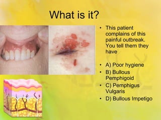
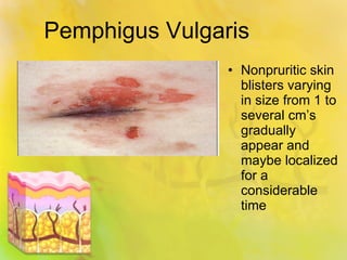
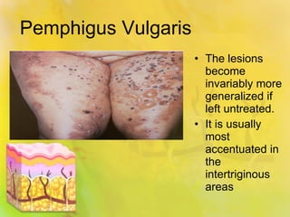
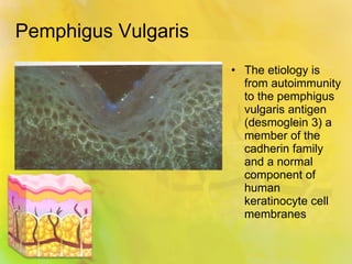
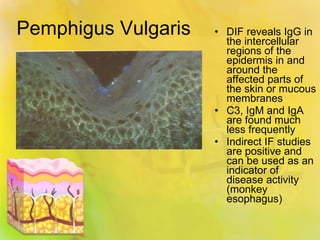
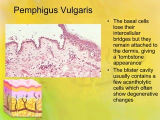
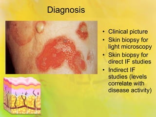
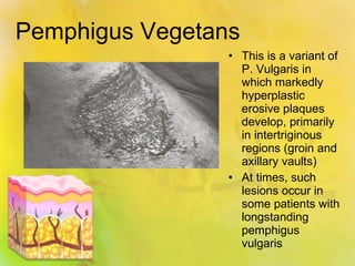
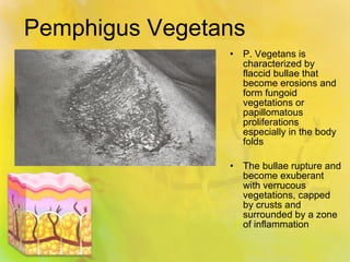
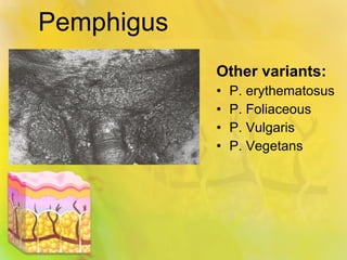
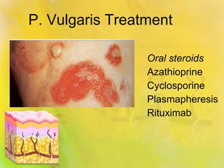
Pemphigus Vulgaris
What is it? This patient complains of this painful outbreak.  You tell them they have A) Poor hygiene B) Bullous Pemphigoid C) Pemphigus Vulgaris D) Bullous Impetigo
Pemphigus Vulgaris Pemphigus vulgaris is the most common form of pemphigus Oral lesions usually precede the skin blisters by weeks to months (80% present with oral lesions first)
Pemphigus Vulgaris The primary lesion is a flaccid blister/bullae that easily ruptures, leaving erosions and crusting, and eventual marked post-inflammatory changes
Pemphigus Vulgaris In severe cases the oral epithelium is completely denuded and is associated with intraoral pain that is particularly worsened by eating Infrequently other mucous membranes maybe involved (ocular and genitourinary)
Pemphigus Vulgaris Nonpruritic skin blisters varying in size from 1 to several cm’s gradually appear and maybe localized for a considerable time
Pemphigus Vulgaris The lesions become invariably more generalized if left untreated. It is usually most accentuated in the intertriginous areas
Pemphigus Vulgaris The etiology is from autoimmunity to the pemphigus vulgaris antigen (desmoglein 3) a member of the cadherin family and a normal component of human keratinocyte cell membranes
Pemphigus Vulgaris DIF reveals IgG in the intercellular regions of the epidermis in and around the affected parts of the skin or mucous membranes C3, IgM and IgA are found much less frequently Indirect IF studies are positive and can be used as an indicator of disease activity (monkey esophagus)
Pemphigus Vulgaris The basal cells lose their intercellular bridges but they remain attached to the dermis, giving a ‘tombstone appearance’ The blister cavity usually contains a few acantholytic cells which often show degenerative changes
Diagnosis Clinical picture Skin biopsy for light microscopy Skin biopsy for direct IF studies Indirect IF studies (levels correlate with disease activity)
Pemphigus Vegetans This is a variant of P. Vulgaris in which markedly hyperplastic erosive plaques develop, primarily in intertriginous regions (groin and axillary vaults) At times, such lesions occur in some patients with longstanding pemphigus vulgaris
Pemphigus Vegetans P. Vegetans is characterized by flaccid bullae that become erosions and form fungoid vegetations or papillomatous proliferations especially in the body folds The bullae rupture and become exuberant with verrucous vegetations, capped by crusts and surrounded by a zone of inflammation
Pemphigus  Other variants: P. erythematosus P. Foliaceous P. Vulgaris P. Vegetans
P. Vulgaris Treatment Oral steroids Azathioprine Cyclosporine Plasmapheresis Rituximab

Pemphigus vulgaris

  • 1.
  • 2.
    What is it?This patient complains of this painful outbreak. You tell them they have A) Poor hygiene B) Bullous Pemphigoid C) Pemphigus Vulgaris D) Bullous Impetigo
  • 3.
    Pemphigus Vulgaris Pemphigusvulgaris is the most common form of pemphigus Oral lesions usually precede the skin blisters by weeks to months (80% present with oral lesions first)
  • 4.
    Pemphigus Vulgaris Theprimary lesion is a flaccid blister/bullae that easily ruptures, leaving erosions and crusting, and eventual marked post-inflammatory changes
  • 5.
    Pemphigus Vulgaris Insevere cases the oral epithelium is completely denuded and is associated with intraoral pain that is particularly worsened by eating Infrequently other mucous membranes maybe involved (ocular and genitourinary)
  • 6.
    Pemphigus Vulgaris Nonpruriticskin blisters varying in size from 1 to several cm’s gradually appear and maybe localized for a considerable time
  • 7.
    Pemphigus Vulgaris Thelesions become invariably more generalized if left untreated. It is usually most accentuated in the intertriginous areas
  • 8.
    Pemphigus Vulgaris Theetiology is from autoimmunity to the pemphigus vulgaris antigen (desmoglein 3) a member of the cadherin family and a normal component of human keratinocyte cell membranes
  • 9.
    Pemphigus Vulgaris DIFreveals IgG in the intercellular regions of the epidermis in and around the affected parts of the skin or mucous membranes C3, IgM and IgA are found much less frequently Indirect IF studies are positive and can be used as an indicator of disease activity (monkey esophagus)
  • 10.
    Pemphigus Vulgaris Thebasal cells lose their intercellular bridges but they remain attached to the dermis, giving a ‘tombstone appearance’ The blister cavity usually contains a few acantholytic cells which often show degenerative changes
  • 11.
    Diagnosis Clinical pictureSkin biopsy for light microscopy Skin biopsy for direct IF studies Indirect IF studies (levels correlate with disease activity)
  • 12.
    Pemphigus Vegetans Thisis a variant of P. Vulgaris in which markedly hyperplastic erosive plaques develop, primarily in intertriginous regions (groin and axillary vaults) At times, such lesions occur in some patients with longstanding pemphigus vulgaris
  • 13.
    Pemphigus Vegetans P.Vegetans is characterized by flaccid bullae that become erosions and form fungoid vegetations or papillomatous proliferations especially in the body folds The bullae rupture and become exuberant with verrucous vegetations, capped by crusts and surrounded by a zone of inflammation
  • 14.
    Pemphigus Othervariants: P. erythematosus P. Foliaceous P. Vulgaris P. Vegetans
  • 15.
    P. Vulgaris TreatmentOral steroids Azathioprine Cyclosporine Plasmapheresis Rituximab