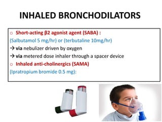
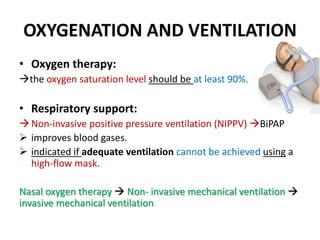
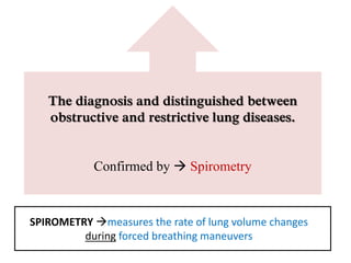
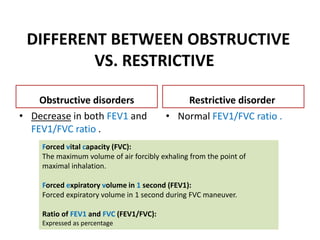
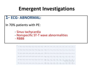
Clinical vignette 3
obstructive and
restrictive lung disease
Ghaida Al-Rashed
Objectives
Describe the treatment of acute exacerbation of
asthma and COPD ??
How can lung volumes be used to differentiate
between obstructive and restrictive lung
disease ?
 What are the emergent investigations are to be
performed on an emergency basis to reach the
diagnosis ?
Objectives:
Describe the treatment of acute exacerbation of
asthma and COPD ??
ACUTE EXACERBATION OF ASTHMA
Treatment
 Oxygen 40-60%
 High doses of inhaled bronchodilators .
 Systemic corticosteroids.
 Intravenous fluids.
 Subsequent management.
OXYGEN
The patient should supply by High concentrations
of oxygen:
Goal: SaO2 > 92%
Failure to achieve appropriate oxygenation  assisted
ventilation.
INHALED BRONCHODILATORS
o Short-acting β2 agonist agent (SABA) :
(Salbutamol 5 mg/hr) or (terbutaline 10mg/hr)
via nebulizer driven by oxygen
via metered dose inhaler through a spacer device
o Inhaled anti-cholinergics (SAMA)
(Ipratropium bromide 0.5 mg):
SYSTEMIC CORTICOSTEROIDS
• Hydrocortisone sodium succinate:
 Dose: 200 mg 4 hourly
 Rote: intravenous
 in patients who are unable to swallow or vomiting.
ARTERIAL BLOOD GASES
We should correct ABG especially If patients ..
o Initial PaCO2 measurement was raised ( >7 kPa)
o PaO2 was < 8 kPa (60 mmHg)
o the patient deteriorates.
SUBSEQUENT MANAGEMENT
If patients fail to improve:
o Intravenous magnesium sulphate (1.2–2 g over 20 min)
o Intravenous β2 agonists (e.g. Salbutamol)
o Intravenous aminophylline (5mg/kg loading dose over 20
minutes)
Chest x-ray
To exclude
pneumothorax
MANAGEMENT OF COPD
The goals of effective COPD management are to:
1. Prevent disease progression
2. Relieve symptoms
3. Improve exercise tolerance
4. Improve health status
5. Prevent and treat complication
OXYGENATION AND VENTILATION
• Oxygen therapy:
the oxygen saturation level should be at least 90%.
• Respiratory support:
Non-invasive positive pressure ventilation (NIPPV) BiPAP
 improves blood gases.
 indicated if adequate ventilation cannot be achieved using a
high-flow mask.
Nasal oxygen therapy  Non- invasive mechanical ventilation 
invasive mechanical ventilation
Con..
• SHORT-ACTING BRONCHODILATORS (nebulization)
1. Inhaled short-acting β2 agonist agent (SABA).
2. Inhaled anti-muscarinic (SAMA).
• CORTICOSTEROIDS:
Short courses of systemic corticosteroids.
• ANTIBIOTICS
with bacterial infection
PREVENTING FUTURE EXACERBATIONS
Appropriate
use of
inhaled
corticosteroid
+
inhaled
bronchodilator
s
Smoking
cessation
vaccination
Objectives:
How can lung volumes be used to differentiate
between obstructive and restrictive lung disease ?
OBSTRUCTIVE VS. RESTRICTIVE
Obstructive disorders
• Characterized by: reduction in
airflow.
• So, shortness of breath  in
exhaling air.
( the air will remain inside the
lung after full expiration )
1. COPD
2. Asthma
3. Bronchiectasis
Restrictive disorders
• Characterized by a reduction
in lung volume.
• So, Difficulty in taking air
inside the lung.
( DUE TO stiffness inside the lung tissue
or chest wall cavity )
1. Interstitial lung disease.
2. Scoliosis
3. Neuromuscular cause
4. Marked obesity
SPIROMETRY measures the rate of lung volume changes
during forced breathing maneuvers
The diagnosis and distinguished between
obstructive and restrictive lung diseases.
Confirmed by  Spirometry
DIFFERENT BETWEEN OBSTRUCTIVE
VS. RESTRICTIVE
Obstructive disorders
• Decrease in both FEV1 and
FEV1/FVC ratio .
Restrictive disorder
• Normal FEV1/FVC ratio .
Forced vital capacity (FVC):
The maximum volume of air forcibly exhaling from the point of
maximal inhalation.
Forced expiratory volume in 1 second (FEV1):
Forced expiratory volume in 1 second during FVC maneuver.
Ratio of FEV1 and FVC (FEV1/FVC):
Expressed as percentage
Objectives:
 What are the emergent investigations
are to be performed on an emergency basis
to reach the diagnosis ?
Emergent Investigations
1- ECG- ABNORMAL:
In 70% patients with PE:
- Sinus tachycardia
- Nonspecific ST-T wave abnormalities
- RBBB
2- ARTERIAL BLOOD GASES:
o hypoxemia, hypocapnia, and respiratory alkalosis due to
hyperventilation.
PO2 and A-a gradient
most often abnormal Profound hypoxia with normal chest X-ray
in the absence of preexisting lung disease is highly suspicious of
pulmonary embolism.
3- D-DIMER
• D-dimer: A degradation product of fibrin.
• is a substance in the blood that is often increased in people
with PE.
D-dimer levels are abnormal in patients with PE; a person
with a normal D-dimer level is unlikely to have a PE.
 Positive D-dimer indicate abnormal high level of fibrin
degradation product ( indicate significant blood clut)
Summary
References
• http://www.who.int/respiratory/copd/manag
ement/en/
• http://www.aafp.org/afp/2010/0301/p607.ht
ml
• http://emedicine.medscape.com/article/3009
01-workup#c9
obstructive & restrictive lung disease

obstructive & restrictive lung disease

  • 1.
    Clinical vignette 3 obstructiveand restrictive lung disease Ghaida Al-Rashed
  • 2.
    Objectives Describe the treatmentof acute exacerbation of asthma and COPD ?? How can lung volumes be used to differentiate between obstructive and restrictive lung disease ?  What are the emergent investigations are to be performed on an emergency basis to reach the diagnosis ?
  • 3.
    Objectives: Describe the treatmentof acute exacerbation of asthma and COPD ??
  • 4.
    ACUTE EXACERBATION OFASTHMA Treatment  Oxygen 40-60%  High doses of inhaled bronchodilators .  Systemic corticosteroids.  Intravenous fluids.  Subsequent management.
  • 5.
    OXYGEN The patient shouldsupply by High concentrations of oxygen: Goal: SaO2 > 92% Failure to achieve appropriate oxygenation  assisted ventilation.
  • 6.
    INHALED BRONCHODILATORS o Short-actingβ2 agonist agent (SABA) : (Salbutamol 5 mg/hr) or (terbutaline 10mg/hr) via nebulizer driven by oxygen via metered dose inhaler through a spacer device o Inhaled anti-cholinergics (SAMA) (Ipratropium bromide 0.5 mg):
  • 7.
    SYSTEMIC CORTICOSTEROIDS • Hydrocortisonesodium succinate:  Dose: 200 mg 4 hourly  Rote: intravenous  in patients who are unable to swallow or vomiting.
  • 8.
    ARTERIAL BLOOD GASES Weshould correct ABG especially If patients .. o Initial PaCO2 measurement was raised ( >7 kPa) o PaO2 was < 8 kPa (60 mmHg) o the patient deteriorates.
  • 9.
    SUBSEQUENT MANAGEMENT If patientsfail to improve: o Intravenous magnesium sulphate (1.2–2 g over 20 min) o Intravenous β2 agonists (e.g. Salbutamol) o Intravenous aminophylline (5mg/kg loading dose over 20 minutes) Chest x-ray To exclude pneumothorax
  • 10.
    MANAGEMENT OF COPD Thegoals of effective COPD management are to: 1. Prevent disease progression 2. Relieve symptoms 3. Improve exercise tolerance 4. Improve health status 5. Prevent and treat complication
  • 11.
    OXYGENATION AND VENTILATION •Oxygen therapy: the oxygen saturation level should be at least 90%. • Respiratory support: Non-invasive positive pressure ventilation (NIPPV) BiPAP  improves blood gases.  indicated if adequate ventilation cannot be achieved using a high-flow mask. Nasal oxygen therapy  Non- invasive mechanical ventilation  invasive mechanical ventilation
  • 12.
    Con.. • SHORT-ACTING BRONCHODILATORS(nebulization) 1. Inhaled short-acting β2 agonist agent (SABA). 2. Inhaled anti-muscarinic (SAMA). • CORTICOSTEROIDS: Short courses of systemic corticosteroids. • ANTIBIOTICS with bacterial infection
  • 13.
    PREVENTING FUTURE EXACERBATIONS Appropriate useof inhaled corticosteroid + inhaled bronchodilator s Smoking cessation vaccination
  • 14.
    Objectives: How can lungvolumes be used to differentiate between obstructive and restrictive lung disease ?
  • 15.
    OBSTRUCTIVE VS. RESTRICTIVE Obstructivedisorders • Characterized by: reduction in airflow. • So, shortness of breath  in exhaling air. ( the air will remain inside the lung after full expiration ) 1. COPD 2. Asthma 3. Bronchiectasis Restrictive disorders • Characterized by a reduction in lung volume. • So, Difficulty in taking air inside the lung. ( DUE TO stiffness inside the lung tissue or chest wall cavity ) 1. Interstitial lung disease. 2. Scoliosis 3. Neuromuscular cause 4. Marked obesity
  • 16.
    SPIROMETRY measures therate of lung volume changes during forced breathing maneuvers The diagnosis and distinguished between obstructive and restrictive lung diseases. Confirmed by  Spirometry
  • 17.
    DIFFERENT BETWEEN OBSTRUCTIVE VS.RESTRICTIVE Obstructive disorders • Decrease in both FEV1 and FEV1/FVC ratio . Restrictive disorder • Normal FEV1/FVC ratio . Forced vital capacity (FVC): The maximum volume of air forcibly exhaling from the point of maximal inhalation. Forced expiratory volume in 1 second (FEV1): Forced expiratory volume in 1 second during FVC maneuver. Ratio of FEV1 and FVC (FEV1/FVC): Expressed as percentage
  • 20.
    Objectives:  What arethe emergent investigations are to be performed on an emergency basis to reach the diagnosis ?
  • 21.
    Emergent Investigations 1- ECG-ABNORMAL: In 70% patients with PE: - Sinus tachycardia - Nonspecific ST-T wave abnormalities - RBBB
  • 22.
    2- ARTERIAL BLOODGASES: o hypoxemia, hypocapnia, and respiratory alkalosis due to hyperventilation. PO2 and A-a gradient most often abnormal Profound hypoxia with normal chest X-ray in the absence of preexisting lung disease is highly suspicious of pulmonary embolism.
  • 23.
    3- D-DIMER • D-dimer:A degradation product of fibrin. • is a substance in the blood that is often increased in people with PE. D-dimer levels are abnormal in patients with PE; a person with a normal D-dimer level is unlikely to have a PE.  Positive D-dimer indicate abnormal high level of fibrin degradation product ( indicate significant blood clut)
  • 24.
  • 25.

Editor's Notes

  • #7 Nebulization administration a drug by inhalation metered-dose inhaler (MDI) is a device that delivers a specific amount of medication to the lungs anti-cholinergicsblock the action of the neurotransmitter acetylcholine in the brain
  • #9 kilopascal  Millimeters of mercury
  • #12 Oxygen supplementation should be titrated to an oxygen saturation level of at least 90 percent BiPAP stands for Bilevel Positive Airway Pressur  example of Non-invasive positive pressure ventilation
  • #13 increase the time to subsequent exacerbation, decrease the rate of treatment failure
  • #16 So, the patient will present with shortness of breath  particularly in in exhaling air
  • #22 P- Pulmonal, RVH, RAD, RBBB  the right ventricle does  Right Bundle Branch Block
  • #23 Alveolar–arterial gradient
  • #24 Absence of D-dimer provides a strong evidence against thromboembolism