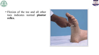
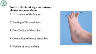
BABINSKI SIGN
Presented by – Shadab khan
Pediatric Physiotherapy
Department
• In superficial reflex it is one of the pathological reflex
• Babinski sign indicates the lesion in the corticospinal tract i.e. UMN
lesion
• Lesion anywhere between cerebral cortex and anterior horn cell
indicates UMN lesion
• Examples of UMN lesion includes Stroke, multiple sclerosis,
meningitis, brain tumor, traumatic brain injury, etc
Procedure
Position of the patient :
• The patient in the supine lying and should be relaxed
• Hip and knee must be extended and heel resting on the bed
• Instrument of Stimulation : Stimulate the plantar surface of the
foot with a blunt point e.g. handle of reflex hammer, blunt key
point, etc
PROCEDURE
The stimulus starts at the heel and the stimulator is moved up on lateral
side at a steady pace up to metatarsophalangeal joint and then turn
medially across the transverse arch of the foot.
• Flexion of the toe and all other
toes indicates normal plantar
reflex.
Positive Babinski sign or extensor
plantar response shows
1. Extension of the big toe
2. Fanning of the small toes
3. Dorsiflexion of the ankle
4. Contraction of tensor fascia lata
5. Flexion of knee and hip.
BABINSKI EQVALANCE
• Response is similar but the stimulation is different
1. Chaddock sign : stroking below the lateral malleolus
2. Oppenheim sign :applying pressure over the medial side of tibia
3 . Gordon sign: squeeze the calf muscle
4 . Shafer sign : squeeze the Achilles tendon
PEDIATRIC POINTERS
• Babinski’s reflex is presents physiologically in infants up to age of 24
months, reflecting immaturity of the corticospinal tract. It disappears
as the child grow older.
• After age of 2, presence of Babinski’s reflex may indicate damage to
the corticospinal tracts.
• Include –
• cerebral palsy
• Hydrocephalus
References
• SP Mehta, SR Joshi, Nihar P Mehta practical medicine 20th edition
• DeJong. 7th Ed. Chap 40. Pathological reflexes. The History, Physical, and
Laboratory Examinations. 3rd edition.Walker HK, Hall WD, 2 - Journal, Indian
Academy of Clinical Medicine Vol. 6 July- September, 2005 Geeta Akhwaja
Thank you

BABINSKI SIGN main.pptx

  • 1.
    BABINSKI SIGN Presented by– Shadab khan Pediatric Physiotherapy Department
  • 2.
    • In superficialreflex it is one of the pathological reflex • Babinski sign indicates the lesion in the corticospinal tract i.e. UMN lesion • Lesion anywhere between cerebral cortex and anterior horn cell indicates UMN lesion • Examples of UMN lesion includes Stroke, multiple sclerosis, meningitis, brain tumor, traumatic brain injury, etc
  • 3.
    Procedure Position of thepatient : • The patient in the supine lying and should be relaxed • Hip and knee must be extended and heel resting on the bed
  • 4.
    • Instrument ofStimulation : Stimulate the plantar surface of the foot with a blunt point e.g. handle of reflex hammer, blunt key point, etc
  • 5.
    PROCEDURE The stimulus startsat the heel and the stimulator is moved up on lateral side at a steady pace up to metatarsophalangeal joint and then turn medially across the transverse arch of the foot.
  • 6.
    • Flexion ofthe toe and all other toes indicates normal plantar reflex.
  • 7.
    Positive Babinski signor extensor plantar response shows 1. Extension of the big toe 2. Fanning of the small toes 3. Dorsiflexion of the ankle 4. Contraction of tensor fascia lata 5. Flexion of knee and hip.
  • 9.
    BABINSKI EQVALANCE • Responseis similar but the stimulation is different 1. Chaddock sign : stroking below the lateral malleolus 2. Oppenheim sign :applying pressure over the medial side of tibia 3 . Gordon sign: squeeze the calf muscle 4 . Shafer sign : squeeze the Achilles tendon
  • 11.
    PEDIATRIC POINTERS • Babinski’sreflex is presents physiologically in infants up to age of 24 months, reflecting immaturity of the corticospinal tract. It disappears as the child grow older. • After age of 2, presence of Babinski’s reflex may indicate damage to the corticospinal tracts. • Include – • cerebral palsy • Hydrocephalus
  • 13.
    References • SP Mehta,SR Joshi, Nihar P Mehta practical medicine 20th edition • DeJong. 7th Ed. Chap 40. Pathological reflexes. The History, Physical, and Laboratory Examinations. 3rd edition.Walker HK, Hall WD, 2 - Journal, Indian Academy of Clinical Medicine Vol. 6 July- September, 2005 Geeta Akhwaja
  • 14.

Editor's Notes

  • #2 It is fundamental component of neurological examination to check the integrity of CST and to examine thr UMN lesion
  • #3 It is fundamental component of neurological examination to check the integrity of CST and to examine thr UMN lesion
  • #5 Strength of the stimulus – the stimulus is not that strong or not that light , it should be firm
  • #10 Infection or wound