Polymer microspheres for controlled drug release

Polymer microspheres can be employed to deliver medication in a rate-controlled and sometimes targeted manner. Medication is released from a microsphere by drug leaching from the polymer or by degradation of the polymer matrix. Since the rate of drug release is controlled by these two factors, it is important to understand the physical and chemical properties of the releasing medium. This review presents the methods used in the preparation of microspheres from monomers or from linear polymers and discusses the physio-chemical properties that affect the formation, structure, and morphology of the spheres. Topics including the effects of molecular weight, blended spheres, crystallinity, drug distribution, porosity, and sphere size are discussed in relation to the characteristics of the release process. Added control over release profiles can be obtained by the employment of core-shell systems and pH-sensitive spheres; the enhancements presented by such systems are discussed through literature examples.

Recommended
Recommended
More Related Content
What's hot
What's hot (20)
Viewers also liked
Viewers also liked (20)
Similar to Polymer microspheres for controlled drug release
Similar to Polymer microspheres for controlled drug release (20)
More from Duwan Arismendy
More from Duwan Arismendy (7)
Recently uploaded
Recently uploaded (20)
Polymer microspheres for controlled drug release
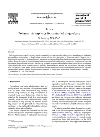
- 1. International Journal of Pharmaceutics 282 (2004) 1–18 Review Polymer microspheres for controlled drug release S. Freiberg, X.X. Zhu∗ Département de chimie, Université de Montréal, C.P. 6128, Succursale Centre-ville, Montréal, Que., Canada H3C 3J7 Received 12 December 2003; received in revised form 21 April 2004; accepted 22 April 2004 Abstract Polymer microspheres can be employed to deliver medication in a rate-controlled and sometimes targeted manner. Medication is released from a microsphere by drug leaching from the polymer or by degradation of the polymer matrix. Since the rate of drug release is controlled by these two factors, it is important to understand the physical and chemical properties of the releasing medium. This review presents the methods used in the preparation of microspheres from monomers or from linear polymers and discusses the physio-chemical properties that affect the formation, structure, and morphology of the spheres. Topics including the effects of molecular weight, blended spheres, crystallinity, drug distribution, porosity, and sphere size are discussed in relation to the characteristics of the release process. Added control over release profiles can be obtained by the employment of core-shell systems and pH-sensitive spheres; the enhancements presented by such systems are discussed through literature examples. © 2004 Elsevier B.V. All rights reserved. Keywords: Polymer microspheres; Microsphere preparation; Drug delivery; Drug release rate 1. Introduction Conventional oral drug administration does not usually provide rate-controlled release or target speci- ficity. In many cases, conventional drug delivery provides sharp increases of drug concentration at potentially toxic levels. Following a relatively short period at the therapeutic level, drug concentration eventually drops off until re-administration. Today new methods of drug delivery are possible: desired drug release can be provided by rate-controlling membranes or by implanted biodegradable polymers containing dispersed medication. Over the past 25 years much research has also been focused on degradable polymer microspheres for drug delivery. Administration of medication via such sys- ∗ Corresponding author. tems is advantageous because microspheres can be ingested or injected; they can be tailored for desired release profiles and in some cases can even provide organ-targeted release. Some reviews covering aspects of microspheres for drug delivery are available (Jalil and Nixon, 1990b; Kawaguchi, 2000; Mueller et al., 2001; Edlund and Albertsson, 2002; Vasir et al., 2003) and this review covers recent works not yet summa- rized and provides information regarding many factors affecting microsphere drug release and the manipula- tion of physical/chemical properties to achieve desired results. The idea of controlled release from polymers dates back to the 1960s through the employment of silicone rubber (Folkman and Long, 1964) and polyethylene (Desai et al., 1965). The lack of degradability in these systems implies the requirement of eventual surgical removal and limits their applicability. In the 1970s biodegradable polymers were suggested as appropriate 0378-5173/$ – see front matter © 2004 Elsevier B.V. All rights reserved. doi:10.1016/j.ijpharm.2004.04.013
- 2. 2 S. Freiberg, X.X. Zhu / International Journal of Pharmaceutics 282 (2004) 1–18 Fig. 1. Chemical structures of chitin, chitosan, amylose poly(lactic Acid) (PLA), and poly(glycolic acid) (PGA). drug delivery materials circumventing the requirement of removal (Jalil and Nixon, 1990b). The idea of poly- mer microcapsules as delivery systems was reported as early as the 1960s (Chang, 1964) and degradation was incorporated by Mason et al. (1976) through the em- ployment of a degradable polymer coating; the topic was reviewed by Marty and Oppenheim (1977). Recent literature shows that suspensions of degrad- able microspheres can be employed for sustained drug release at desirable doses and by implantation without surgical procedures. Biocompatibility can be achieved by the use of natural polymers such as cel- lulose, chitin, and chitosan or by the employment of polymers made from naturally occurring monomers such as lactic and glycolic acids (Fig. 1). Polymers derived from synthetic monomers also show excellent delivery properties. However, their toxicity effects may require evaluation. The factors affecting drug release are controllable; they are attributed to properties such as polymer molecular weight, as well as microsphere size, distri- bution, morphology and make-up. 2. Preparation 2.1. Microspheres prepared by polymerization of monomers Although most microspheres employed for drug de- livery are prepared from linear polymers, the prepa- ration of microspheres from monomers are still of relevance. It involves the polymerization of colloidal monomers dispersed in a liquid with opposite solu- bilities (Kiminta et al., 1996). Spherical droplets are formed by oil-soluble organic monomers dispersed in aqueous media (oil in water, O/W) or by water-soluble monomers dissolved in water dispersed in an organic medium (water in oil, W/O; Candau, 1985). The polymerization of dispersed monomers is achievable by various methods including emulsion, suspension, and dispersion techniques (Piirma, 1985). Emulsions are typically used to form uniform spheres on nanometer scales (10–104(nm). The technique typically involves the dispersion of a hydrocarbon monomer in water with a water-soluble initiator. A surfactant is employed for the formation of uniform micelles; polymerization takes place inside micelles and not inside the dispersed organic monomer droplets since the initiator is not miscible there. The resulting polymer beads can be so uniform on the nano-scale that they may diffract visible light (Weissman et al., 1996). Dispersion polymerization results in particle sizes in the range of 0.5–10(m and all of the reagents in- cluding monomer, initiator, and stabilizer (often an organic polymer consisting of hydrophobic and hy- drophilic parts) are dissolved in an organic medium. Since the initiator is soluble inside the monomer, poly- merization takes place inside the monomer droplets. The polymer beads, insoluble in the organic solvent, precipitate, and the stabilizer prevents bead floccula- tion (Barrett, 1975; Strover and Li, 1996). Suspension polymerizations are typically employed for micron-sized particles (50–500(m). In suspen- sion polymerization the monomer is dispersed in a
- 3. S. Freiberg, X.X. Zhu / International Journal of Pharmaceutics 282 (2004) 1–18 3 water phase with a stabilizer; the initiator is soluble in the monomer phase where polymerization occurs. The size and quantity of the particles is determined by the size and quantity of dispersed monomer droplets and by the speed of mechanical stirring (Piirma, 1985). Recently, Ruckenstein and co-workers have also obtained uniform polymer beads on the millimeter scale by sedimentation polymerization (Ruckenstein and Hong, 1995; Ruckenstein and Sun, 1996) which involves the gravitational ascent of polymerizing aque- ous monomer droplets in hot paraffin oil. At the end of the decent period (7–9(s), the spheres continue to polymerize without coalescing. Amsden (1999) has also developed a similar method for forming beads on the size of millimeters by employing a solvated linear polymer. Droplets of the polymer in organic solution were added to a flowing solution of poly(vinyl alco- hol) (PVA, as stabilizer)/water solution; at the end of the flowing procedure uniform-sized beads were col- lected. Within the last decade there has also been much sig- nificant work on dispersion polymerization in super- critical CO2, which may be beneficial to medical appli- cations since no toxic solvents are involved (Benedetti et al., 1997; Canelas and DeSimone, 1997). Some of the bead forming techniques are listed in Table 1. 2.2. Microspheres prepared from linear polymers Methods involving the preparation of microspheres from linear polymers can be advantageous since a wide range of polymers is available commercially. Synthe- sis, properties and degradation mechanisms of most polymers employed in making such types of micro- spheres have been reviewed by Edlund and Albertsson (2002). This microsphere preparation technique is also useful for polymers that cannot be made by emulsion processes (e.g., biocompatible polylactide (PLA) and polyglycolide (PGA) are usually obtained from an- Table 1 Sizes obtained from various bead-forming techniques Method of preparation Size range Emulsion polymerization 0.01–1(m Dispersion polymerization 0.5–10(m Suspension polymerization 50–500(m Sedimentation polymerization mm sizes ionic polymerization instead of free radical methods) and for naturally occurring polymers such as chitin, chitosan, and cellulose. Some commonly employed microsphere prepara- tion methods are the solvent evaporation technique (or the double emulsion technique) and the spray drying technique. These and other methods are described by Vasir et al. (2003) in a review on bioadhesive micro- spheres (Vasir et al., 2003). The spray drying technique has been described by Masters (1985). Also, Berkland et al. (2001) de- scribed spay drying techniques for the formation of very monodisperse spheres and evaluated their meth- ods for use with poly(lactide-co-glycolide)s (PLGAs; Pavanetto et al., 1993). Witchi and Doelker (1998) also offered a comparison of the properties of micro- spheres prepared by the solvent removal technique or the spray drying technique. 2.3. Preparation by the solvent evaporation method Microspheres can be formed by the evaporation of an organic solvent from dispersed oil droplets contain- ing both polymer and drug (Fig. 2; Jalil and Nixon, 1989, 1990c; Huang et al., 1997; Atkins et al., 1998; Edlund and Albertsson, 1999; Oh et al., 1999; Pistel et al., 1999; Bai et al., 2001). Often, a double emul- sion is employed; first the drug for encapsulation is Fig. 2. Depiction of sphere formation by solvent evaporation. A solvent-polymer droplet disperses inside the continuous phase forming solvent-polymer spheres; the sphere hardens as the organic solvent evaporates.
- 4. 4 S. Freiberg, X.X. Zhu / International Journal of Pharmaceutics 282 (2004) 1–18 dissolved in water; this aqueous phase is dispersed in an organic solvent (usually dichloromethane, DCM), which contains the degradable polymer and the first W/O emulsion is formed. Dispersion of the first emul- sion in a stabilized aqueous medium (usually using poly(vinyl alcohol) as stabilizer) forms the final O/W emulsion; microspheres are formed as the DCM evap- orates and the polymer hardens, trapping the encapsu- lated drug (Bodmeier and McGinity, 1988; Li et al., 1995a,b; Ghaderi et al., 1996). 2.3.1. Encapsulation efficiency Increasing or controlling the encapsulation effi- ciency (EE) is desirable, it can prevent the loss of precious medication and it can help to extend the duration and dosage of treatment. The drug content of the encapsulated microspheres can be described by two quantities. The most common, also used in this paper, is the EE, where EE = D/DT; DT is the total amount of drug employed and D is DT minus the amount of unloaded drug (Gupta and Kumar, 2001). On the other hand, the loading capacity (LC) is de- fined as LC = D/SW, where SW is the weight of the sphere (Gupta and Kumar, 2001). Issues of relevance concerning EE include sphere formation temperature and the nature of the polymer. Yang et al. (2000) have provided a revealing study which correlated EE to sphere preparation tempera- ture. The authors found that the highest EEs occurred at the lowest and highest formation temperatures tested (about 50% at 4 and 38(◦C, and about 19% at 22 and 29(◦C). The non-linear drug loading trend suggested that different mechanisms governed the encapsulation process at different temperatures. At lower tempera- tures, increased immiscibility between the sphere and water resulted in a rapidly forming outer sphere wall, thus trapping the drug early in the evaporation pro- cess (Chung, 1997). At higher temperatures, an in- creased rate of solvent evaporation also resulted in a rapidly hardening sphere wall. In both cases drug trap- ping was enhanced by hardening at the sphere wall, an important point when considering drug encapsula- tion. When considering the relation of the polymer itself to EE, Ghaderi et al. (1996) found that increasing the concentration of polymer in the organic phase increased the EE. An increase in EE from 1 to 25% was observed depending on the concentration of the polymer. Considering the nature of the polymer, LeCorre et al. (1994) obtained different EEs for two very similar polymers, PLA and PLGA, even when the microspheres were prepared under similar con- ditions. Obtained drug loadings for PLAs and PGAs were found to be 21–46%, respectively, this may be attributed to faster precipitation of the PGAs at the sphere interface. 2.3.2. Control of microsphere size Microsphere size can be affected by the polymer concentration in the second emulsion, temperature, viscosity, the stirring rate in the second emulsion step, and the amount of emulsifier employed. Considering the effect of polymer concentration, it has often been reported that increasing the concentration of polymer in the second emulsion increases sphere size (Yan et al., 1994; Ghaderi et al., 1996; McGee et al., 1997; Schlicher et al., 1997; Lin and Vasavada, 2000). In another study, Yang et al. (2000) used scanning electron microscopy (SEM) to show that sphere size was temperature dependent; lower and higher temper- atures produced larger spheres whereas intermediate temperatures produced smaller spheres. Once again, different mechanisms dominated microsphere forma- tion at different temperatures. At lower temperatures, the solution’s higher viscosity resulted in the forma- tion of larger spheres; this has also been confirmed by other researchers (Jeyanthi et al., 1997). Larger spheres were obtained at higher temperatures due to the higher rate of solvent evaporation which resulted in higher solvent flow pressure moving more material from the sphere center outward (Yang et al., 2000). Jalil and Nixon (1990a) studied the variation of sphere size with respect to the stirring rate and the in- fluence of the emulsifier in the second emulsion step. It was shown that microsphere size decreased with in- creasing stirring rate since increased stirring results in the formation of finer emulsions. The authors em- ployed a sorbitan ester as an emulsifier and reported a sharp drop in diameter when the sorbitan ester con- centration was increased from 1 to 2%. Little change in diameter size was reported by increasing emulsi- fier concentration beyond 2%. It is possible that in this particular case, emulsifier packing was optimum at 2% concentration and that no more emulsifier could be adsorbed at the sphere surface above this concen- tration (Jalil and Nixon, 1990a).
- 5. S. Freiberg, X.X. Zhu / International Journal of Pharmaceutics 282 (2004) 1–18 5 2.3.3. Control of sphere porosity Porosity has an important effect on drug release characteristics; a large number of pores may greatly increase the rate of drug expulsion (Yang et al., 2000). The first W/O emulsion can be used to control the microsphere pores; Crotts and Park (1995) have shown that sphere porosity increased with the water content in the first emulsion. When the first water-in-oil sys- tem contained less water, porosity decreased; the for- mation of spheres by skipping the first water-in-oil step resulted in spheres with a non-porous outer skin and a monolithic inner composition. At low water con- tents the inner core contained hollow structures and a non-porous skin. Porosity was observed throughout the particle at higher water content. In a related study, Tuncay et al. (2000) also found that the employment of methanol instead of water in the first emulsion phase may reduce the surface porosity of the microspheres. Li et al. (1995b) determined the effects of varying the amount of water in the second emulsion or con- tinuous phase (CP) on porosity. The CP containing the largest amount of water resulted in faster polymer precipitation and therefore less porous spheres were formed. In another study by Jeyanthi et al. (1996) con- tinuously adding water to the CP of the dispersion up to 1.5 times the initial volume, the authors ob- tained spheres containing a uniform honeycomb struc- ture with no hollow core. Continuous dilution up to 2.5 times the initial volume resulted in similar spheres but with larger pores. The larger pores at increased di- lution were explained by an increased rate of solvent removal with increasing water content (higher solvent flow exit pressures) which occurs in coordination with a faster hardening rate (Ghaderi et al., 1996; Jeyanthi et al., 1996). The rate at which the solvent is removed from the sphere is dependent on temperature, pressure, and the amount of water in the final emulsion phase and can be directly related to sphere porosity (Izumikawa et al., 1991; Jeyanthi et al., 1996; Yang et al., 2000). Yang et al. (2000) have systematically increased sphere preparation temperature; their results suggested that skin porosity tends to decrease with increasing tem- perature until a limiting value. Therefore, a relatively high sphere formation temperature resulted in rapid hardening of the outer wall and low porosity. How- ever, very high temperatures resulted in a highly porous skin and inner core due to the very rapid solvent evaporation. Other researchers also showed that high evaporation rates resulted in more porous spheres (Izumikawa et al., 1991; Chung et al., 2001). Jeyanthi et al. (1996) have investigated sphere porosity at variable temperatures; solvent removal was well correlated to preparation temperature and the solvent was removed more rapidly at higher temperatures. Quick solvent leaching from the soft spheres formed hard spheres with hollow inner cores and thin outer walls, thus showing how the high flow pressure of the evaporating solvent increases porosity. By varying the ramping conditions the authors were able to create spheres with a thicker outer wall and a smaller core whose porosity was controllable by the ramping steps (Jeyanthi et al., 1996). 3. Factors affecting drug release rate Controlled release is an attainable and desirable characteristic for drug delivery systems. The factors affecting the drug release rate revolve around the struc- ture of the matrix where the drug is contained and the chemical properties associated with both the poly- mer and the drug. Conventional oral delivery is not rate controlled. A drug encapsulated in a slowly de- grading matrix provides the opportunity for slower re- lease effects, but polymer degradation is not the only mechanism for the release of a drug. The drug re- lease is also diffusion controlled as the drug can travel through the pores formed during sphere hardening. In some cases, drugs containing nucleophilic groups can cause increased chain scission of the polymer ma- trix, which also increases the rate of drug expulsion. Polymer molecular weight, drug distribution, polymer blending, crystallinity, and other factors are important in manipulating release profiles. The most desirable release profile would show a constant release rate with time. However, in many cases release profiles are more complicated and often contain two main expulsion processes: the first being an initial burst of expelled medication from the sphere surface; the second, a usually more constant stage with release rates dependent on diffusion and degradation (LeCorre et al., 1994; Ghaderi et al., 1996; Mogi et al., 2000). An example showing the initial burst and lin- ear release by Yang et al. (2000) is shown in Fig. 3. Some researchers have been able to achieve a relatively
- 6. 6 S. Freiberg, X.X. Zhu / International Journal of Pharmaceutics 282 (2004) 1–18 Fig. 3. Release profiles of bovine serum albumin from DLPLA and DLPGA microspheres as presented by Yang et al. (2000). Microspheres formed at lower temperatures show a fast burst process followed by slow continued release. Microspheres formed at the highest temperature exhibit the fastest release rate but in a constant fashion due to large uniform pores within the beads. constant release after the initial burst, some have been able to achieve close to zero-order kinetics without a significant burst effect, and others have obtained even more complex but adjustable profiles depending on the desired application (Narayani and Rao, 1996; Makino et al., 2000; Yang et al., 2000; Berkland et al., 2002; Kakish et al., 2002). In the following discussion, the factors responsible for different release profiles will be discussed in terms of physical and chemical prop- erties of the microsphere. 3.1. Polymer molecular weight Degradation of polymer microspheres shows a clear dependence on the molecular weight (MW) of the polymer. In a study by Park (1994), it was found that polymer spheres initially released small molecu- lar weight oligomers by rapid diffusion regardless of MW; following the initial period, low MW degrada- tion products were released. In spheres initially con- taining lower MW chains, the quantity of degradation products increased with time and the polymers making up the microspheres decreased in molecular weight. However, for spheres made from high MW polymers, the quantity of degradation products and the polymer MW remained constant for longer periods of time. Park (1994, 1995) provided evidence suggesting that the varying degradation profiles occur due to the dif- ferences in glass transition temperatures (Tg) and crys- tallinity associated with polymers of different MW. Makino et al. (2000) showed pulsatile drug release in high MW PLGAs as shown in Fig. 4. At lower MWs (19,000), a relatively constant release profile was obtained; increasing the molecular weight to 23,000 (Mogi et al., 2000), 44,000 and 74,000 decreased the linearity of release (Makino et al., 2000). The rate of drug release from particles containing higher MW polymers was initially high, followed by a decrease which was then followed again by an increase. The two-stage release profile suggested the presence of two dominating release mechanisms in high MW poly- mers. Degradation is the main release mechanism for low MW polymers after the initial burst stage(Park, 1994). Spheres containing high MW polymers likely undergo initial slow drug release due to diffusion, fol- lowed by the main drug release due to degradation. Makino et al. (2000) showed this by correlating ob- served drug release with microscopic observation of the microspheres; the drug release was fastest for the degradation of swollen spheres.
- 7. S. Freiberg, X.X. Zhu / International Journal of Pharmaceutics 282 (2004) 1–18 7 Fig. 4. Drug release from poly(lactide-co-glycolide) microspheres fabricated from polymers of different molecular weights; the pulsatile release character increases with molecular weight from Makino et al. (2000) circles, MW = 19000; triangles, MW = 44,000; squares, MW = 74,000. Blending two polymers of different MWs allows the manipulation of the timing associated with the degra- dation release. LeCorre et al. (1994) combined a low molecular weight PLA (MW = 2000) and a higher molecular weight PLA (MW = 9000) to obtain an ex- tra degree of controlled release. Tuncay et al. (2000) and Ravivarapu et al. (2000) obtained similar results using MW blends of PGAs and PLGAs, respectively, where the microspheres containing the lower MW polymer released drug more rapidly. 3.2. Blends of structurally different polymers As already discussed, the physical blending of two polymers can affect the release profiles of polymer spheres. Edlund and Albertsson (2000) have provided an important comparison of a blend system against the corresponding copolymer sys- tem involving poly(l-lactic acid) (PLLA) and poly(1,5-dioxepan-2-one) (PDXO). Spheres made from the linear co-polymers were porous and larger than those made from the blends. During degradation, the molecular weight loss was slower in the blend spheres due to their increased density. The release of a hydrophilic drug was slower for the blend spheres (Fig. 5). Differences in morphology of the two types of spheres made a significant impact on the release profile. It was also observed, by employing varying mixtures of blend composition, that the degradation rate increased with increasing amounts of PDXO. The lower degradation rate of PLLA over PDXO was attributed to the increased crystallinity in PLLA: the crystalline regions degrade more slowly than the amorphous regions (Edlund and Albertsson, 2000). Mi et al. (2002) blended hydrophobic PLGA with hydrophilic chitin and examined the hydration in these systems. It was found that the spheres with a higher content of hydrophilic chitin degraded faster since degradation takes place more rapidly in chitin. Be- ing hydrophilic, chitin degraded by surface erosion, and the more hydrophobic PLGA degraded by bulk erosion, therefore a two-stage degradation profile was observed. In the first part of sphere degradation, fast release occurred due to the readily degradable chitin polymer, which was then followed by a slower release from the PLGA sections.
- 8. 8 S. Freiberg, X.X. Zhu / International Journal of Pharmaceutics 282 (2004) 1–18 Fig. 5. Drug release from a blend system vs. its copolymer system taken from Edlund and Albertsson (2000). The blend system released drug more slowly than its copolymer counterpart due to increased crystallinity in the blend system. Empty symbols represent the blend systems and the solid symbols represent the copolymer systems. The consistency of the trend are shown by the two sets of data; the triangles and the squares represent PLLA–PDXO ratios of a 90:10 and 70:30, respectively. 3.3. Crystallinity Crystallinity in microspheres has been usually in- vestigated by DSC or X-ray diffraction (XRD) stud- ies. DSC can detect phase transitions including the melting of crystalline regions, whereas XRD directly detects the crystallinity properties of a material. LeCorre et al. (1997) observed the crystallinity of a lipophilic drug in polymer microspheres by DSC. Usually, drug has been found to be molecularly dis- persed inside a polymer matrix and crystallinity is not observed (Benita et al., 1984; Benoit et al., 1986; LeCorre et al., 1997; Guyot and Fawaz, 1998). How- ever, in their case, LeCorre et al. (1997) found that the relatively highly loaded drug existed in a particulate dispersion instead of a molecular dispersion, which is possibly due to its lack of solubility in the polymer matrix. Yuksel et al. (1996) used XRD and DSC to in- vestigate crystallinity and drug-polymer interactions. They observed that although a physical mixture of the drug and polymer exhibited crystallinity, the drug was amorphous after dispersion in the microspheres. At- tempts to crystallize the drug inside the microspheres by annealing above the polymer’s Tg, and by heat–cool cycles were unsuccessful showing clear molecular dis- persion of the drug. In the same study it was shown that molecular dispersions may be more favorable than particulate dispersions for drug delivery since the drug was released more readily from a microsphere system than from a particulate form at pH 7.4. The polymer matrix likely disturbs drug crystallinity and initiates rate-controlled delivery with higher drug delivery ef- ficiencies (Yuksel et al., 1996). Considering polymeric crystallinity, Edlund and Albertsson (2000) suggested that degradation occurred first in the amorphous microsphere regions followed by a slower degradation in the crystalline regions. This suggests that the crystallinity in the polymer chains can affect the degradation rate. Furthermore,
- 9. S. Freiberg, X.X. Zhu / International Journal of Pharmaceutics 282 (2004) 1–18 9 at the beginning of sphere degradation, the degree of crystallinity actually increased slightly. This was attributed to the crystallization of partly degraded chains and the preferential degradation of amorphous regions. Izumikawa et al. (1991) studied polymer crys- tallinity and drug crystallinity employing PLA mi- crospheres loaded with progesterone. At low drug loading (5%), XRD and DSC showed that the poly- mer dominates the crystalline properties of the mi- crosphere and no crystallinity arose from the drug; the drug was dispersed in the sphere. At high drug loading (30%), crystallinity was dependent on the organic solvent removal process; at slow solvent re- moval rates sphere crystallinity was observed from both the drug and polymer but fast removal resulted in amorphous spheres. At high concentrations and slow solvent removal, the drug formed a particulate dis- persion resulting in the presence of drug crystallinity. However, the faster solvent removal rate may have resulted in amorphous spheres by not giving the drug and polymer molecules adequate time to crystallize. Release profiles suggest that more amorphous spheres release the drug less rapidly than crystalline spheres. Therefore, the lack of polymer crystallinity suggests better drug dispersion and increased drug–polymer interactions. The drug release rate can be tailored by manipulating the degree of crystallinity; re- duced crystallinity is favorable when slow release is desired. Fig. 6. Tertiary amine containing drugs loaded into a LPLA microsphere matrix as reported by Cha and Pitt (1989). Drugs with less sterically available amines are released more slowly from the matrix. 3.4. Effects of the loaded drug In some cases the drug employed can induce poly- mer chain scission through nucleophilic degradation. Typically this is observed in medications containing amines whose nitrogen atom is nucleophilic, just like the oxygen atom in water. Cha and Pitt (1989) re- ported that sterically available amines increased the rate of polymer degradation. PLLA was loaded with different amine-containing drugs, the polymer MW in spheres containing the most active amine decreased more rapidly and to a greater extent throughout the re- lease process (Fig. 6). In the case of a less active, ster- ically hindered tertiary amine, polymer degradation was not significant nor was drug release, unless it was co-loaded with another drug capable of causing poly- mer chain scission. Other groups have also considered chain scission when reporting their results (Cha and Pitt, 1988; LeCorre et al., 1997; Tuncay et al., 2000). How the drug is distributed in the medium can also vary its release profile (Kakish et al., 2002). Drug re- lease begins at the sphere surface followed by release from the inner layers of the sphere; therefore the dif- fusional distance between the initial drug location in- side the sphere affects the release profile (Lee, 1984, 1985, 1986). Drug uniformly dispersed in the sphere matrix can increase the initial burst effect; Kakish et al. (2002) have successfully modified the drug dis- tribution in microspheres so as to obtain constant drug release. The microspheres were modified by stirring
- 10. 10 S. Freiberg, X.X. Zhu / International Journal of Pharmaceutics 282 (2004) 1–18 dry spheres in an ethanol–water mixture followed by freeze-drying. The resulting distribution is believed to be characterized by an increase in drug concentration toward the center of the microspheres thus resulting in a more constant release rate. Kakish et al. (2002) obtained a system with a relatively constant release rate over 10(h. Their microsphere treatment methods significantly improved controlled release when com- pared to untreated microspheres whose drug release rate decreases with time. 3.5. Porosity The porosity in a system of spheres is determined during microsphere hardening as the organic solvent evaporates during preparation. As already mentioned above, sphere porosity can be controlled by changes in sphere preparation technique and differences in poros- ity do affect release kinetics (Jeyanthi et al., 1996; Yang et al., 2000). This is noticeable in a study by Yang et al. (2000) where a highly porous matrix released a drug at a considerably higher rate than its non-porous counterpart (Fig. 3). Other researchers also reported that sphere porosity affected the release profile in sim- ilar ways (Ghaderi et al., 1996; Yuksel et al., 1996; Chung et al., 2001). Therefore, when preparing mi- crospheres, it should be kept in mind that increasing the number of pores should increase the release rate. Another factor related to sphere porosity is the already mentioned initial burst effect, which corre- sponds to a rapid initial release of drug and is normally followed by relatively-controlled linear release. This is attributed to the leaching which occurs at the outer wall of the sphere as it becomes hydrated (LeCorre et al., 1994; Okada et al., 1994; Ghaderi et al., 1996). This can be minimized by supporting the formation of a non-porous outer sphere skin which can be con- trolled by sphere fabrication temperature (Yang et al., 2000). 3.6. Size distribution The release profiles are also dependent on the size of the microspheres; the rate of drug release was found to decrease with increasing sphere size (Narayani and Rao, 1994, 1995; Akhtar and Lewis, 1997; Sansdrap and Moes, 1997; Bezemer et al., 2000). Therefore, by mixing microspheres of different sizes it is possible Fig. 7. Depiction of a core-shell microsphere containing a drug-loaded microsphere (in some cases only the drug) as the core and another polymer as the outer shell. to obtain another degree of controlling release. More importantly, linear, zero-order kinetics are obtainable by combining the proper formulation of microsphere sizes. Narayani and Rao (1996) have combined micro- spheres of different sizes to obtain linear release pro- files. Employing gelatin microspheres with sizes of 1–35(m, they successfully achieved good zero-order drug release. In a detailed study, Berkland et al. (2001, 2002) have also obtained a zero-order release by mix- ing microspheres of different sizes; sphere size was well controlled by fabricating spheres using the spray dry technique. 3.7. Release from core-shell microspheres Core-shell microspheres (Fig. 7) usually refer to spheres formed by making core units through a nor- mal preparative method, followed by the addition of an outer layer by a dipping procedure, mixing proce- dure, or emulsion procedure (Ermis and Yuksel, 1999; Huang et al., 1999; Jones and Lyon, 2000; Lee et al., 2002; Sparnacci et al., 2002; Zhou et al., 2002). Em- ployment of a shell is usually meant to enhance con- trolled release and possibly reduce the effect of the initial burst. Huang et al. (1999) showed how dipping PLA/poly (ethylene glycol) (PEG) spheres into a gelatin solution can enhance controlled release. The spheres were pre- pared from block copolymers where the PLA block forms a hydrophobic core and the PEG block forms a hydrophilic outer layer after preparation by the sol- vent evaporation method. Spheres made from these polymers are highly porous and have a high burst
- 11. S. Freiberg, X.X. Zhu / International Journal of Pharmaceutics 282 (2004) 1–18 11 effect. However, coating the spheres with gelatin re- sulted in a considerable decrease in the burst effect and offers a release over a longer period of time. This can be useful when the burst effect is too high. In other studies, Pekarek et al. (1994a,b) have pre- pared microspheres containing an inner polyanhydride core and an outer layer of PLA. They found that the more hydrophilic anhydride inner core is degraded into monomers after 2 months which eventually crys- tallized and started to escape the shell after 4 months (Pekarek and Mathiowitz, 1998) without significant degradation of the shell; the spheres also had similar degradation characteristics in vivo or in vitro (Pekarek et al., 1998). This may be useful for the controlled re- lease of a drug inside the core matrix since the release rate is controlled by diffusion through a shell of uni- form thickness (Pekarek et al., 1994a). Along similar lines Yang et al. (2003) observed advanced degrada- tion of the inner core of composite PEO/PLGA mi- crospheres where PEO was the inner core; the same systems are considered as a possible release medium for two drugs simultaneously (Shi et al., 2003). In some cases, researchers have been able to es- tablish targeted release by incorporating pH-sensitive outer shells. The effect is shell degradation at a specific pH values followed by rate-controlled drug release. 3.8. pH controlled release Added control over drug delivery can be achieved by employing pH-triggered release. Therefore by the incorporation of pH-sensitive groups, microspheres can be targeted to various biological environments or to specific organs (Bilia et al., 1996; Cifti et al., 1996; Kumar and Rao, 1997; Mi et al., 1997; Kumar and Rao, 1998; Lorenzo-Lamosa et al., 1998; Bittner et al., 1999; Carelli et al., 1999; Gupta and Kumar, 2001; Jeong et al., 2001; Lynn et al., 2001). Lynn et al. (2001) studied release from micro- spheres at intercellular pH’s. They employed a poly(-amino ester) which was stable in the pH range of 7.0–7.4 but readily solvable below pH 6.5. Very slow release was attained at pH 7.4 but full and im- mediate release occurred at pH 5.1, suggesting a very good material for targeted release. In another study when a chitosan microcore was coated with Eudragit® (poly(methacrylic acid-co- methylmethacrylate)) controlled release occurred only after the pH-sensitive coating was dissolved (Lorenzo-Lamosa et al., 1998). Even though chitosan dissolves rapidly at pH 7.4, no drug was released from the microspheres until the pH of shell degra- dation was attained. Such an outer shell provides usefulness in pH-targeted release; other studies also employed the acrylic Eudragit® for pH-sensitive re- lease (Kawashima et al., 1991; Yuksel et al., 1996; Buonaguidi et al., 1997; Lorenzo-Lamosa et al., 1997; Sriwongjanya and Bodmeier, 1997; Jeong et al., 2001). Researchers also demonstrated that controlled re- lease is possible for acrylamide-based microsphere systems which are both pH- and temperature-sensitive (Kim et al., 1994, 2001; Fang and Kawaguchi, 2002). Temperature sensitivity arises from the lower crit- ical solution temperature (LCST), which may be pH-dependent. Below LCST the polymer is hy- drophilic due to hydrogen bonding with water, and above the LCST the polymer becomes hydrophobic due to the disruption of hydrogen bonds. Since the LCST is pH-dependent, Kim et al. (1994) employed the swelling effect to ensure that a drug was released in the colon at a higher pH instead of the lower stom- ach pH. In the hydrophilic state, the loaded drug was rapidly released and in the hydrophobic state, drug release was very slow. Using similar responsive poly- mers, Kim et al. (2001) also stimulated hypo cortisone release at pH 4 and halted release at pH 7.4 (Fig. 8). 4. Some practical aspects of microspheres for drug delivery 4.1. Applications Micro-scale technologies have long been important and commonplace; nano-scale technologies are also seeing a wider use and have direct implications to the biomedical field (Grainer, 2003). Mueller already provided an interesting review concerning the appli- cability of nanosuspensions for oral and intravenous purposes (Mueller et al., 2001), Vasir also provided a relevant review concerning bioadhesive microspheres (Vasir et al., 2003); therefore only brief comments on the topic will be made here. Microspheres designed for oral treatment target the gastrointestinal (GI) tract, and encapsulation can
- 12. 12 S. Freiberg, X.X. Zhu / International Journal of Pharmaceutics 282 (2004) 1–18 Fig. 8. Hypercortizone release from pH-sensitive acrylamide based microspheres; release is initiated at pH 4 and halted at pH 7 from Kim et al. (2001). enhance GI treatments. Toxic drugs, which can cause side effects when administered in large quantities, or insoluble drugs, which may require large doses to promote absorption, can be administered with a lower frequency and smaller quantity (Davis et al., 1984; Kumar and Rao, 1998). The adhesion properties of biospheres can be exploited so that they stick to the adhesive tissues thus prolonging absorption time at lower doses (Davis et al., 1984; Ch’ng et al., 1985; Kumar and Rao, 1998; Mueller et al., 2001; Vasir et al., 2003). pH-controlled release in these systems is desirable and, as shown in this review, it is very possible (Kim et al., 1994; Lorenzo-Lamosa et al., 1997; Lorenzo-Lamosa et al., 1998; Jeong et al., 2001; Zhou et al., 2002). Intravenous delivery (and needle injection) has implications pertaining to sphere particle size and delivery can be enhanced by pH-controlled release. Particle size is of particular importance as described in reviews by Edlund and Albertsson (2002) and Pouton and Seymour (2001) since particles that are too large, of the order of 6(m, can block blood cap- illaries; in some cases sizes as small as 50–100(nm are required to reach certain organs. Monodis- perse nanospheres with transit capability through nano-sized passages can be readily made by emul- sion or dispersion polymerization (Reese et al., 2000; McPhee et al., 1993; Jones and Lyon, 2000; Sparnacci et al., 2002). In other cases, the drug can also be administered directly to the desired site for local and targeted release (Deurloo et al., 1990; Liu et al., 2001). Targeted sensitivity is also required so that drug is released only upon entry into a desired site. As shown earlier in this review, various systems exist where drug expulsion is retarded in the blood, pH 7.4, but is re- leased at other pH’s (Bezemer et al., 2000; Gupta and Kumar, 2001; Kim et al., 2001; Lynn et al., 2001; Berkland et al., 2002). Kim’s pH- and thermo-sensitive polyacrylamide system may also be realizable in the form of nanospheres instead of microspheres if the spheres are prepared by emulsion polymerization in- stead of solvent evaporation, which may eventually be employable for delivery systems that can transit small passages (Kim et al., 2001). 4.2. In vivo administration of microspheres The use of microspheres in mammals is not a the- oretical issue but an applied reality. Sandstrap et al. (1999) showed how it was possible to obtain reason- able accordance between in vitro and in vivo results using of PGLA microsphere systems in rats for the release of nifedipine.
- 13. S. Freiberg, X.X. Zhu / International Journal of Pharmaceutics 282 (2004) 1–18 13 In examples representative of animal testing, Khan et al. (2000) and Chen et al. (1997) have sepa- rately injected rats with antisense oligonucleotides and anti-cancer agents, respectively, with promising results. Khan et al. (2000) employed PGLA micro- spheres and showed that the oligonucleotides are re- leased in a controlled manner and their system causes improved oligonucleotide distribution in the brain. Chen et al. (1997) delivered anti-cancer medication (carboplatin) to rats in the brain and showed that the microspheres evoked a local inflammatory reaction that was well tolerated by the rats. Other studies also show significant release advantages in the use of mi- crospheres when treating brain tumors (Eroglu et al., 2001) and the article of Fournier et al. (2003) pro- vided some important references on the topic. Many in vivo studies employ PLA or PLGA microspheres (Sandstrap et al., 1999; Khan et al., 2000; Tuncay et al., 2000; Eroglu et al., 2001; Woo et al., 2002) but poly(methilidine malonate) (Fournier et al., 2003) and Eudragit® (Jeong et al., 2001) microspheres have also been used. Recently, Wang et al. (2004) made microspheres from poly(ortho-ester) for the delivery of DNA vac- cines and tested them in mice. The polymer can pre- vent the DNA from degradation and the release takes place inside the cell where the weakly acidic environ- ment degrades the polymer without compromising the biological activity of the DNA. The diameter of the spheres was adjusted to about 5(m, a size believed to be taken up preferentially by the cells. Microsphere testing is also seen in humans; Katz et al. (2003) recently obtained promising results in a study of an orally delivered vaccine with the employ- ment of PLGA microspheres. In another study, Paque- tte et al. also obtained promising results when using microsphere encapsulated medication in the treatment of gum disease. Patents mentioning polymer microsphere systems are also seen which shows their potential commercial importance (Carrasquillo and Adamis, 2003; Hanes et al., 2003; Lee et al., 2003). 4.3. Commercialization of microspheres The use of microspheres for drug delivery is not limited to any specific illness, rather they can be widely applied to many situations where continuous and controlled drug administration is essential. How- ever, some of references noted in this article were performed for the release of specific medications including: antibiotics (Atkins et al., 1998), peptides (Li et al., 1995b; Jeyanthi et al., 1997), proteins (Yan et al., 1994; Bezemer et al., 2000; Yang et al., 2000), anaesthetics (LeCorre et al., 1994), anti-virals (Schlicher et al., 1997), hypertension drugs (Yuksel et al., 1996; Guyot and Fawaz, 1998; Sansdrap et al., 1999), anti-HIV drugs(Akhtar and Lewis, 1997), and anti-cancer drugs (Deurloo et al., 1990; Narayani and Rao, 1996; Liu et al., 2001). Drug delivery is a primary topic in the biophar- maceuticals industry and microspheres do overcome many of the shortcomings of conventional drug de- livery routes. To date a limited number of companies provide commercially available microspheres and/or have active product development programs in the field. For market applications, microsphere systems are ex- pected to undergo phase and clinical testing just as non-encapsulated drug systems. However, the use of materials already approved for in vivo degradation, such as PLGs may increase their likelihood and speed of acceptance. A typical application cited by companies man- ufacturing and developing microspheres is for use in the treatment of cancers. Currently, an injectable polyphosphoester microsphere is undergoing de- velopment which boasts the potential for localized chemotherapy thus reducing adverse effects to the pa- tient. However, many companies don’t suggest a spe- cific medical ailment for which their products should be used, rather they indicate that treatment doses and duration times can be modulated for the desired task. In some cases, customizable release profiles are even offered. 5. Conclusions The controlled release of medications from poly- mer microspheres is achievable by manipulating the physical and chemical properties of the polymer as well as those of the microsphere. Issues such as poly- mer molecular weight, blend composition, polymer and drug crystallinity, drug distribution, sphere poros- ity, and sphere size all influence the release profile and can be tailored to fit a desired release. Extra control
- 14. 14 S. Freiberg, X.X. Zhu / International Journal of Pharmaceutics 282 (2004) 1–18 over microsphere release can be obtained by the ad- dition of a pH-sensitive outer core, and/or by the em- ployment of a pH-sensitive inner shell. Microspheres provide sustained release in localized areas and can be employed to reduce medication doses and its fre- quency of use. The utility and potential of microsphere drug deliv- ery systems have been demonstrated and it has been shown that tailored delivery is possible. Site specific applications would normally imply site injection. Oral delivery is also desirable for medications that are ef- fective upon intestinal absorption and can be admin- istered with microspheres that are unaffected by the stomach followed by adherence and degradation at the colon wall. Many chemical and engineering questions to these designed systems have been addressed. Furthermore, in vivo testing of specific drug/microsphere systems has been successfully accomplished by various re- search groups. Broad scope application of microsphere systems requires testing on case-by-case studies and it may not always be clear how systems will perform during in vivo tests as compared to their controlled laboratory counterpart environments. In particular, the variability of degradation environments in biological systems may in some cases require the necessity of new innovative release triggers. Bead functionaliza- tion with chemical groups of specific reactivities may enhance targeted specificity. References Akhtar, S., Lewis, K., 1997. Antisense oligonucleotide delivery to cultured macrophages is improved by incorporation into sustained-release biodegradable polymer microspheres. Int. J. Pharm. 151, 57–67. Amsden, B., 1999. The production of uniformly sized polymer microspheres. Pharm. Res. 16, 1140–1143. Atkins, T.W., Peacock, S.J., Yates, D.J., 1998. Incorporation and release of vancomycin from poly(d,l-lactide-co-glycocide) microspheres. J. Microencapsul. 15, 31–44. Bai, X.-L., Yang, Y.-Y., Chung, T.-S., Ng, S., Heller, J., 2001. Effect of polymer compositions on the fabrication of poly(ortho-ester) microspheres for controlled release of protein. J. Appl. Polym. Sci. 80, 1630–1642. Barrett, K.E.J., 1975. Dispersion Polymerization in Organic Media. Wiley, London. Benedetti, L., Bertucco, A., Pallado, P., 1997. Production of microparticles of a biocompatible polymer using supercritical carbon dioxide. Biotechnol. Bioeng. 53, 232–237. Benita, S., Benoit, J.P., Puisieux, F., Thies, C., 1984. Charac- terization of drug-loaded poly(d,l-lactide) microspheres. J. Pharm. Sci. 73, 1721–1724. Benoit, J.P., Courteille, F., Theis, C., 1986. A phsyiochemical study of the morphology of progestrerone-loaded poly(d,l-lactide) microspheres. Int. J. Pharm. 29, 95–102. Berkland, C., Kim, K., Pack, D.W., 2001. Fabrication of PLG microspheres with precisely controlled and monodisperse size distributions. J. Control Release 73, 59–74. Berkland, C., King, M., Cox, A., Kim, K., Pack, D.W., 2002. Precise control of PLG microsphere size provides enhanced control of drug release rate. J. Control Release 82, 137–147. Bezemer, J.M., Radersma, R., Grijpma, D.W., Dijkstra, P.J., Blitterswijk, C.A.V., Feijen, J., 2000. Microspheres for protein delivery prepared from amphiphilic multiblock copolymers 2. Modulation of release rate. J. Control Release 67, 249–260. Bilia, A., Carelli, V., Colo, G.D., Nannipieri, E., 1996. In vitro evaluation of a pH sensitive hydrogel for control of GI drug delivery from silicone-based matrices. Int. J. Pharm. 130, 83– 92. Bittner, B., Witt, C., Mader, K., Kissel, T., 1999. Degradation and protein release properties of microspheres prepared from biodegradable poly(lactide-co-glycolide) and ABA triblock copolymers: influence of buffer media on polymer erosion and bovine serum albumin release. J. Control Release 60, 297–309. Bodmeier, R., McGinity, J.W., 1988. Solvent selection in preparation of poly(d,l-lactide) microspheres prepeared by the solvent evaporation method. Int. J. Pharm. 43, 179–186. Buonaguidi, M., Carelli, V., Colo, G.D., Nannipieri, E., Serafini, M.F., 1997. Evaluation of a pH-sensitive semi-interpenetrating polymer network for control of GI drug delivery. Int. J. Pharm. 147, 1–10. Candau, F., 1985. Microemulsion polymerization. In: Mark, H.F., Bikales, N.M., Overberger, C.G., Menges, G., Kroschwitz, J.I. (Eds.), Encyclopedia of Polymer Science and Engineering, 2nd ed. John Wiley and Sons, New York, pp. 719–723. Canelas, D.A., DeSimone, J.M., 1997. Polymerizations in liquid and supercritical carbon dioxide. Adv. Polym. Sci. 133, 103– 140. Carelli, V., Coltelli, S., Colo, G.D., Nannipieri, E., Serafini, M.F., 1999. Silicone microspheres from pH-controlled gastrointestinal drug delivery. Int. J. Pharm. 179, 73–78. Carrasquillo, K.G., Adamis, A., 2003. Ocular Drug Delivery Systems for Aptamers PCT. International Applied Massachusetts Eye and Ear Infirmary, USA. Cha, Y., Pitt, C.G., 1988. A one-week subdermal delivery system for l-methadone based on biodegradable microcapsules. J. Control Release 7, 69–78. Cha, Y., Pitt, C.G., 1989. The acceleration of degradation- controlled drug delivery from polyester microspheres. J. Control Release 8, 259–265. Chang, T.M.S., 1964. Semipermeable microcapsules. Science 146, 524–525. Chen, W., He, J., Olson, J.J., Lu, D.R., 1997. Carboplatin-loaded PLGA microspheres for intracerebral implantation: in vivo characterization. Drug Deliv. 4, 301–311.
- 15. S. Freiberg, X.X. Zhu / International Journal of Pharmaceutics 282 (2004) 1–18 15 Ch’ng, H.S., Park, H., Kelly, P., Robinson, J.R., 1985. Bioahesive polymers as platforms for oral controlled drug delivery II: synthesis and evaluation of some swelling, water-insoluble bioadhesive polymers. J. Pharm. Sci. 74, 399. Chung, T.S., 1997. The limitiations of using Flory-Huggins equation for the states of the solutions during asymmetric hollow fiber membrane formation. J. Membr. Sci. 126, 19. Chung, T.-W., Huang, Y.-Y., Liu, Y.-Z., 2001. Effects of the rate of solvent evaporation on the characteristics of drug loaded PLLA and PDLLA microspheres. Int. J. Pharm. 212, 161–169. Cifti, K., Kas, H.S., Hincal, A.A., Ercan, T.M., Guven, O., Ruacan, S., 1996. In vitro and in vivo evaluation of PLAGA (50/50) microspheres containing 5-fluorouracil prepared by a solvent evaporation method. Int. J. Pharm. 131, 73–82. Crotts, G., Park, T.G., 1995. Preparation of porous and nonporous biodegradable polymeric hollow spheres. J. Control Release 35, 91–105. Davis, S.S., Hardy, J.G., Taylor, M.J., Whalley, D.R., Willson, C.G., 1984. A comparative study of gastrointestinal transit of a pellet and tablet formation. Int. J. Pharm. 21, 167–177. Desai, S.J., Siminelli, A.P., Higuchi, W.I., 1965. Investigation of factors influencing release of solid drug dispersed in inert matrixes. J. Pharm. Sci. 54, 1459–1464. Deurloo, M.J.M., Bohlken, S., Kop, W., Lerk, C.F., Hennink, W., Bartelink, H., Begg, A.C., 1990. Intratumoural administration of cisplatin in slow-release devices. Cancer Chemother. Phamacol. 27, 135–140. Edlund, U., Albertsson, A.-C., 1999. Novel drug delivery microspheres from poly(1,5-dioxepan-2-one-co-l-lactide). J. Polym. Sci.: Part A: Pol. Chem. 37, 1877–1884. Edlund, U., Albertsson, A.-C., 2000. Morphology engineering of a novel poly(l-lactide)/poly(1,5-dioxepane-2-one) microsphere system for controlled drug delivery. J. Polym. Sci.: Part A: Pol. Chem. 38, 786–796. Edlund, U., Albertsson, A.-C., 2002. Degradable polymer microspheres for controlled drug delivery. Adv. Polym. Sci. 157, 67–112. Ermis, D., Yuksel, A., 1999. Preparation of spray-dried microspheres of indomethacin and examination of the effects of coating on dissolution rates. J. Microencapsul. 16, 315–324. Eroglu, H., Kas, H.S., Oner, L., Turkoglu, O.F., Akalan, N., Sargon, M.F., Ozer, N., 2001. The in-vitro and in-vivo characterization of PGLA: l-PLA microspheres containing dexamethasone sodium phosphate. J. Microencapsul. 18, 603–612. Fang, S.-J., Kawaguchi, H., 2002. A thermosensitive amphoteric microsphere and its potential application as a biological carrier. Colloid Polym. Sci. 280, 984–989. Folkman, J., Long, D.M., 1964. The use of silicone rubber as a carrier for prolonged drug therapy. J. Surg. Res. 4, 139–142. Fournier, E., Passirani, C., Montero-Menei, C., Colin, N., Breton, P., Sagodira, S., Menei, P., Benoit, J.-P., 2003. Therapeutic effectiveness of novel 5-fluorouracil-loaded poly(methylidene malonate 2.1.2)-based microspheres on F98 glioma-bearing rats. Cancer 97, 2822–2829. Ghaderi, R., Struesson, C., Carlfors, J., 1996. Effect of preparative parameters on the characteristics of poly(d,l-lactide- co-glocolide) microspheres made by the double emulsion method. Int. J. Pharm. 141, 205–216. Grainer, D.W., 2003. Biomedical micro- and nanotechnology. Adv. Drug. Deliv. Rev. 55, 311–313. Gupta, K.C., Kumar, M.N.V.R., 2001. pH dependent hydrolysis and drug release behavior of chitosan/poly(ethylene glycol) polymer network microspheres. J. Mater. Sci. Mater. Med. 12, 753–759. Guyot, M., Fawaz, F., 1998. Nifedipine loaded polymeric microspheres: preparation and physical characteristics. Int. J. Pharm. 175, 61–74. Hanes, J., Fu, J., Fiegel, J., 2003. Biodegradable Polymer, Compositions, and Pharmaceutical Uses. PCT International Applied Johns Hopkins University School of Medicine, USA. Huang, Y.-Y., Chung, T.-W., Tzeng, T.-W., 1997. Drug release from PLA/PEG microparticulates. Int. J. Pharm. 156, 9–15. Huang, Y.-Y., Chung, T.-W., Tzeng, T.-W., 1999. A method using biodegradable polylactides/polyethylene glycol for drug release with reduced initial burst. Int. J. Pharm. 182, 93–100. Izumikawa, S., Yoshioka, S., Aso, Y., Takeda, Y., 1991. Preparation of poly(l-lactide) microspheres of different crystalline morphology on drug release rate. J. Control Release 15, 133–140. Jalil, R., Nixon, J.R., 1989. Microencapsulation using poly(l-lactic acid) I: microcapsul properties affected by the preparative technique. J. Microencapsul. 6, 473–484. Jalil, R., Nixon, J.R., 1990a. Microencapsulation using poly(l-lactic acid) II: preparative variables affecting microcapsule properties. J. Microencapsul. 7, 25–39. Jalil, R., Nixon, J.R., 1990b. Biodegradable poly(lactic acid) and poly(lactide-co-glycocide) microcapsules: problems associated with preparative techniques and release properties. J. Microencapsul. 7, 297–325. Jalil, R., Nixon, J.R., 1990c. Microencapsulation using poly(l-lactic acid) IV: Release properties of microcapsules containing phenobarbitone. J. Microencapsul. 7, 53–66. Jeong, Y.-I., Prasad, Yv.R., Ohno, T., Yoshikawa, Y., Shibata, N., Kato, S., Takeuchi, K., Takada, K., 2001. Application of Eudragit P-4135F for the delivery of ellagic acid to the rat lower small intestine. J. Pharm. Pharmacol. 53, 1079–1085. Jeyanthi, R., Thanoo, B.C., Metha, R.C., DeLuca, P.P., 1996. Effect of solvent removal technique on the matrix characteristics of polylactide/glcolide microspheres for peptide delivery. J. Control Release 38, 235–244. Jeyanthi, R., Mehta, R.C., Thanoo, B.C., DeLuca, P.P., 1997. Effect of processing parameters on the properties of peptide-containing PLGA microspheres. J. Microencapsul. 14, 163–174. Jones, C.D., Lyon, L.A., 2000. Synthesis and characterization of multiresponsive core-shell microgels. Macromolecules 33, 8301–8306. Kakish, H.F., Tashtoush, B., Ibrahim, H.G., Najib, N.M., 2002. A novel approach for the preparation of highly loaded polymeric controlled release dosage forms of diltiazem HCl and diclofenac sodium. Eur. J. Pharm. Biopharm. 54, 75–81. Katz, D.E., DeLorimier, A.J., Wolf, M.K., Hall, E.R., Cassels, F.J., van Hamont, J.E., Newcomer, R.L., Davachi, M.A., Taylor, D.N., McQueen, C.E., 2003. Oral immunization of adult volunteers with microencapsulated enterotoxigenic Escherichia coli (ETEC) CS6 antigen. Vaccine 21, 341–346.
- 16. 16 S. Freiberg, X.X. Zhu / International Journal of Pharmaceutics 282 (2004) 1–18 Kawaguchi, H., 2000. Functional polymer microspheres. Prog. Polym. Sci. 25, 1171–1210. Kawashima, Y., Iwamoto, T., Niwa, T., Takeuchi, H., Itoh, Y., 1991. Preparation and characterization of a new controlled release ibuprofen suspension for improving suspendability. Int. J. Pharm. 75, 25–36. Khan, A., Sommer, W., Fuxe, K., Akhtar, S., 2000. Site- specific administration of antisense oligonucleotides using biodegradable polymer microspheres provides sustained delivery and improved subcellular biodistribution in the neostriatum of the rat brain. J. Drug Target 8, 319. Kim, Y.H., Bae, Y.H., Kim, S.W., 1994. pH/temperature sensitive polymers DOE macromolecular drug loading and release. J. Control Release 28, 143–152. Kim, E.J., Cho, S.H., Yuk, S.H., 2001. Polymeric microspheres composed of pH/temperature-sensitive polymer complex. Biomaterials 22, 2495–2499. Kiminta, D.M.O., Braithwaite, G., Luckham, P.F., 1996. Colloidal dispersions, nanogels. In: Salamone, J.C. (Ed.), Polymer Materials Encyclopedia. CRC Press, Boca Raton, pp. 1298–1309. Kumar, A.B.M., Rao, K.P., 1997. Poly(palmitoyl-l-hydroproline ester) microspheres as potential oral controlled drug delivery system. Int. J. Pharm. 149, 107–114. Kumar, A.B.M., Rao, K.P., 1998. Preparation and characterization of pH-sensitive proteinoid microspheres for the oral delivery of methotrexate. Biomaterials 19, 725–732. LeCorre, P., LeGuevello, P., Gajan, V., Chevanne, F., LeVerge, R., 1994. Preparation and characterization of bupivacaine-loaded polylactide and polylactide-co-glycolide microspheres. Int. J. Pharm. 107, 41–49. LeCorre, P., Rytting, J.H., Gajan, V., Chevanne, F., Verge, R.L., 1997. In vitro controlled release kinetics of local anasthetics from PDLLA and PLGA microspheres. J. Microencapsul. 14, 243–255. Lee, P.I., 1984. Novel approach to zero-order delivery via immobilized nonuniform drug distribution in glassy hydrogels. J. Pharm. Sci. 73, 1344–1347. Lee, P.I., 1985. Kinetics of drug release from hydrogel matrices. J. Control Release 2, 277–288. Lee, P.I., 1986. Initial concentration distribution as a mechanism for regulating drug release from diffusion controlled and surface errosion controlled matrix systems. J. Control Release 4, 1–7. Lee, T.H., Wang, J., Wang, C.-H., 2002. Double-walled microspheres for the sustained release of a highly water soluble drug: characterization and irradiation studies. J. Control Release 83, 437–452. Lee, H.-y., Lee, H.-s., Kim, J.-s., Kim, S.-b., Lee, J.-s., Choi, H.-i., Chang, S.-g., 2003. Polymer-based injectable sustained release pharmaceutical compositions for peptide and protein drugs. PCT International Applied Peptron, Inc., South Korea. Li, W.-I., Anderson, K.W., DeLuca, P.P., 1995a. Kinetic and thermodynamic modeling of the formation of polymeric microspheres using solvent extraction/evaporation method. J. Control Release 37, 187–198. Li, W.-I., Anderson, K.W., Mehta, R.C., DeLuca, P.P., 1995b. Prediction of solvent removal profile and effect on properties for peptide loaded PLGA microspheres prepared by solvent extraction/evaporation method. J. Control Release 37, 199–214. Lin, Y.-H.E., Vasavada, R.C., 2000. Studies on microencapsulation of 5-fluorouracil with poly(ortho ester) polymers. J. Microencapsul. 17, 1–11. Liu, Z., Bendayan, R., Wu, X.Y., 2001. Triton-X-100-modified polymer and microspheres for reversal of multidrug resistance. J. Pharm. Pharmacol. 53, 779–787. Lorenzo-Lamosa, M.L., Remunan-Lopez, C., Vila-Jato, J.L., Alonso, M.J., 1998. Design for microencapsulated chitosan microspheres for colonic drug delivery. J. Control Release 52, 109–118. Lorenzo-Lamosa, M.L., Cuna, M., Vila-Jato, J.L., Torres, D., Alonso, M.J., 1997. Development of a microencapsulated form of cefuroxime axetil using pH-sensitive acrylic polymers. J. Microencapsul. 14, 607–616. Lynn, D., Amiji, M., Langer, R., 2001. pH-responsive polymer microspheres: rapid release of encapsulated material within the range of intracellular pH. Angev. Chem. Int. Ed. 40, 1707– 1710. Makino, K., Mogi, T., Ohtake, N., Yoshida, M., Ando, S., Nakajima, T., Ohshima, H., 2000. Pulsatile drug release from poly(lactide-co-glycolide) microspheres: how does the composition of the polymer matrixes affect the time interval between the initial burst and the pulsatile release of drugs? Colloid. Surf. B: Biointerfaces 19, 173–179. Marty, J.J., Oppenheim, R.C., 1977. Colloidal systems for drug delivery. Aust. J. Pharm. 6, 65–76. Mason, N., Thies, C., Cicero, T.J., 1976. In-vivo and in vitro evaluation of a microencapsulated narcotic anagonist. J. Pharm. Sci. 65, 847–850. Masters, K., 1985. Spray Drying Handbook, 4th ed. George Goodwin, Ltd., London. McGee, J.P., Singh, M., Li, X.M., Qui, H., O’Hagen, D.T., 1997. The encapsulation of a model protein in poly(d,l-lactide- co-glycocide) microparticles of various sizes: an evaluation of process reproduciblity. J. Microencapsul. 14, 197–210. McPhee, W., Tam, K.C., Pelton, R., 1993. Poly(N- isopropylacrylamide) lactices prepared with sodium dodecyl sulfate. J. Colloid Interface Sci. 156, 24–30. Mi, F.-L., Chen, C.-T., Tseng, Y.-C., Kuan, C.-Y., Shyu, S.-S., 1997. Iron(III)-carboxymethylchitin microsphere for the pH-sensitive release of 6-mercaptopurine. J. Control Release 44, 19–32. Mi, F.-L., Lin, Y.-M., Wu, Y.-B., Shyu, S.-S., Tsai, Y.-H., 2002. Chitin/PLGA blend microspheres as a biodegradable drug-delivery system: phase-separation. Biomaterials 23, 3257– 3267. Mogi, T., Ohtake, N., Yoshida, M., Chimura, R., Kamaga, Y., Ando, S., Tsukamoto, T., Nakajima, T., Uenodan, H., Otsuka, M., Matsuda, Y., Ohshima, K., Makino, K., 2000. Sustained release of 17-estradiol from poly(lactide-co-glycocide) microspheres in vitro and in vivo. Colloid Surface B. 17, 153–165. Mueller, R.H., Jacobs, C., Kayser, O., 2001. Nanosuspensions as particulate drug formulations in therapy rationale for development and what we can expect for the future. Adv. Drug. Deliv. Rev. 47, 3–19.
- 17. S. Freiberg, X.X. Zhu / International Journal of Pharmaceutics 282 (2004) 1–18 17 Narayani, R., Rao, K.P., 1994. Controlled release of anticancer drug methotrexate from biodegradable gelatin microspheres. J. Microencapsul. 11, 69–77. Narayani, R., Rao, K.P., 1995. pH responsive gelatin microspheres for the oral delivery of anticancer drug methotrexate. J. Appl. Polym. Sci. 58, 1761–1769. Narayani, R., Rao, K.P., 1996. Gelatin microsphere cocktails of different sizes for the controlled release of anticancer drugs. Int. J. Pharm. 143, 255–258. Oh, J.E., Nam, Y.S., Lee, K.H., Park, T.G., 1999. Conjugation of drug poly(d,l-lactic-co-glycolic acid) for controlled release from biodegradable microspheres. J. Control Release 57, 269– 280. Okada, H., Yamomoto, M., Heya, T., Inoue, Y., Kamei, S., Ogawa, Y., Toguchi, H., 1994. Drug delivery using biodegradable microspheres. J. Control Release 28, 121–129. Park, T.G., 1994. Degradation of poly(d,l-lactic acid) microspheres: effect of molecular weight. J. Control Release 30, 161–173. Park, T.G., 1995. Degradation of poly(lactic-co-glcocic acid) microspheres: effect of copolymer composition. Biomaterials 16, 1123–1130. Pavanetto, F., Genta, I., Giunchedi, P., Conti, B., 1993. Evaluation of spray drying as a method for polyhlactide and polylactide-co-glycocide microsphere preparation. J. Microencapsul. 10, 487–497. Pekarek, K.J., Mathiowitz, E., 1998. Degradation of double-walled polymer microspheres of PLLA and P(CPP:SA) 20:80. I. In vitro degradation. Biomaterials 19, 1973–1980. Pekarek, K.J., Jacob, J.S., Mathiowitz, E., 1994a. Double walled polymer microspheres for controlled drug release. Nature 367, 258–260. Pekarek, K.J., Jacob, J.S., Mathiowitz, E., 1994b. One-step preoaration of double-walled microspheres. Adv. Mater. 6, 684–687. Pekarek, K.J., Takahashi, S., Mathowitz, E., 1998. Degradation of double-walled polymer microspheres of PLLA and P(CPP: SA) 20:80. II. In vivo degradation. Biomaterials 19, 1981–1988. Piirma, I., 1985. Colloids. In: Mark, H.F., Bikales, N.M., Overberger, C.G., Menges, G., Kroschwitz, J.I. (Eds.), Encyclopedia of Polymer Science and Engineering, 2nd ed. John Wiley and Sons, New York, pp. 125–130. Pistel, K.F., Bittner, B., Koll, H., Winter, G., Kissel, T., 1999. Biodegradable recombinant human erythropoietin loaded microspheres prepared from linear and start-branched block copolymers: influence of encapsulation technique and polymer composition on particle characteristics. J. Control Release 59, 309–325. Pouton, C.W., Seymour, L.W., 2001. Key issues in non-viral gene delivery. Adv. Drug. Deliv. Rev. 46, 187–203. Ravivarapu, H.B., Burton, K., DeLuca, P.P., 2000. Polymer and microsphere blending to alter the release of a peptide from PLGA microspheres. Eur. J. Pharm. Biopharm. 50, 263–270. Reese, C.E., Guerrero, C.C., Weissman, J.M., Lee, K., Asher, S.A., 2000. Synthesis of highly charged, monodisperse polystyrene colloidal particles for the fabrication of photonic crystals. J. Colloid Interface Sci. 232, 76–80. Ruckenstein, E., Hong, L., 1995. Sedimentation polymerization. Polymer 36, 2857–2860. Ruckenstein, E., Sun, Y., 1996. Preparation and characteristics of polymer-based large adsorbent particles. J. Appl. Polym. Sci. 61, 1949–1956. Sandstrap, P., Fontaine, J., Moes, A.J., 1999. Nifedipine-loaded PLGA microspheres: in vitro/in vivo comparison of drug release and polymer degradation. S.T.P. Pharm. 9, 443–446. Sansdrap, P., Moes, A.J., 1997. In vitro evaluation of the hydrolytic degradation of dispersed and aggregated poly(d,l-lactide-co-glycolide) microspheres. J. Control Release 43, 47–58. Sansdrap, P., Fontaine, J., Moes, A.J., 1999. Nifedipine-loaded PLGA microspheres: in vitro/in vivo comparison of drug release and polymer degradation. S.T.P. Pharm. Sci. 9, 443. Schlicher, E.J.A.M., Postma, N.S., Zuidema, J., Talsma, H., Hennink, W.E., 1997. Preparation and characterisation of poly(d,l-lactic-co-glycolic acid) microspheres containing desferrioxamine. Int. J. Pharm. 153, 235–245. Shi, M., Yang, Y.-Y., Chaw, C.-S., Goh, S.-H., Moochhala, S.M., Ng, S., Heller, J., 2003. Double walled POE/PLGA microspheres: encapsulation of water-soluble and water- insoluble proteins and their release properties. J. Control Release 89, 167–177. Sparnacci, K., Laus, M., Tondelli, L., Magnani, L., Bernardi, C., 2002. Core-shell microspheres by dispersion polymerization as drug delivery systems. Macromol. Chem. Phys. 203, 1364– 1369. Sriwongjanya, M., Bodmeier, R., 1997. Entrapment of drug-loaded ion exchange particles within polymeric microparticles. Int. J. Pharm. 158, 28–38. Strover, H.D.H., Li, K., 1996. Dispersion polymerization. In: Polymer Materials Encyclopedia. CRC Press, Boca Raton, pp. 1900–1905. Tuncay, M., Calis, S., Kas, H.S., Ercan, M.T., Peksoy, I., Hincal, A.A., 2000. Diclofenac sodium incorporated PLGA (50:50) microspheres: formulation considerations and in vitro/in vivo evaluation. Int. J. Pharm. 195, 179–188. Vasir, J.K., Tambwekar, K., Garg, S., 2003. Bioadhesive microspheres as a controlled drug delivery system. Int. J. Pharm. 255, 13–32. Wang, C., Ge, Q., Ting, D., Nguyen, D., Shen, H.R., Chen, J.Z., Eisen, H.N., Heller, J., Langer, R., Putnam, D., 2004. Molecularly engineered poly(ortho ester) microspheres for enhanced delivery of DNA vaccines. Nat. Mater. 3, 190–196. Weissman, J.M., Sunkara, H.B., Tee, A.S., Asher, S.A., 1996. Thermally switchable periodicities and diffraction from mesoscopically ordered materials. Science 274, 959–965. Witchi, C., Doelker, E., 1998. Influence of the microencapsulation method and peptide loading on poly(lactic acid) and poly(lactic-co-glycolic acid) degradation during in vitro testing. J. Control Release 51, 327–341. Woo, B.H., Na, K.-H., Dani, B.A., Jiang, G., Thanoo, B.C., DeLuca, P.P., 2002. In vitro characterization and in vivo testosterone suppression of 6-month release poly(d,l-lactide) microspheres. Pharm. Res. 19, 546–550. Yan, C., Resau, J.H., Hewetson, J., West, M., Rill, W.L., Kende, M., 1994. Characterization and morphological analysis of protein
- 18. 18 S. Freiberg, X.X. Zhu / International Journal of Pharmaceutics 282 (2004) 1–18 loaded poly(lactide-co-glycolide) microparticles prepared by water-in-oil–in-water emulsion technique. J. Control Release 32, 231–241. Yang, Y.-Y., Chung, T.-S., Bai, X.-L., Chan, W.-K., 2000. Effect of preparation conditions on morphology and release profiles of biodegradable polymeric microspheres containing protein fabricated by double-emulsion method. Chem. Eng. Sci. 55, 2223–2236. Yang, Y.-Y., Shi, M., Goh, S.-H., Moochhala, S.M., Ng, S., Heller, J., 2003. POE/PLGA composite microspheres: formation and in vitro behavior of double walled microspheres. J. Control Release 88, 201–213. Yuksel, N., Tincer, T., Baykara, T., 1996. Interaction between nicardipine hydrochloride and polymeric microspheres for a controlled release system. Int. J. Pharm. 140, 145– 154. Zhou, S., Deng, X., Yuan, M., Li, X., 2002. Investigation on preparation and protein release of biodegradable polymer microspheres as drug-delivery system. J. Appl. Polym. Sci. 84, 778–784.