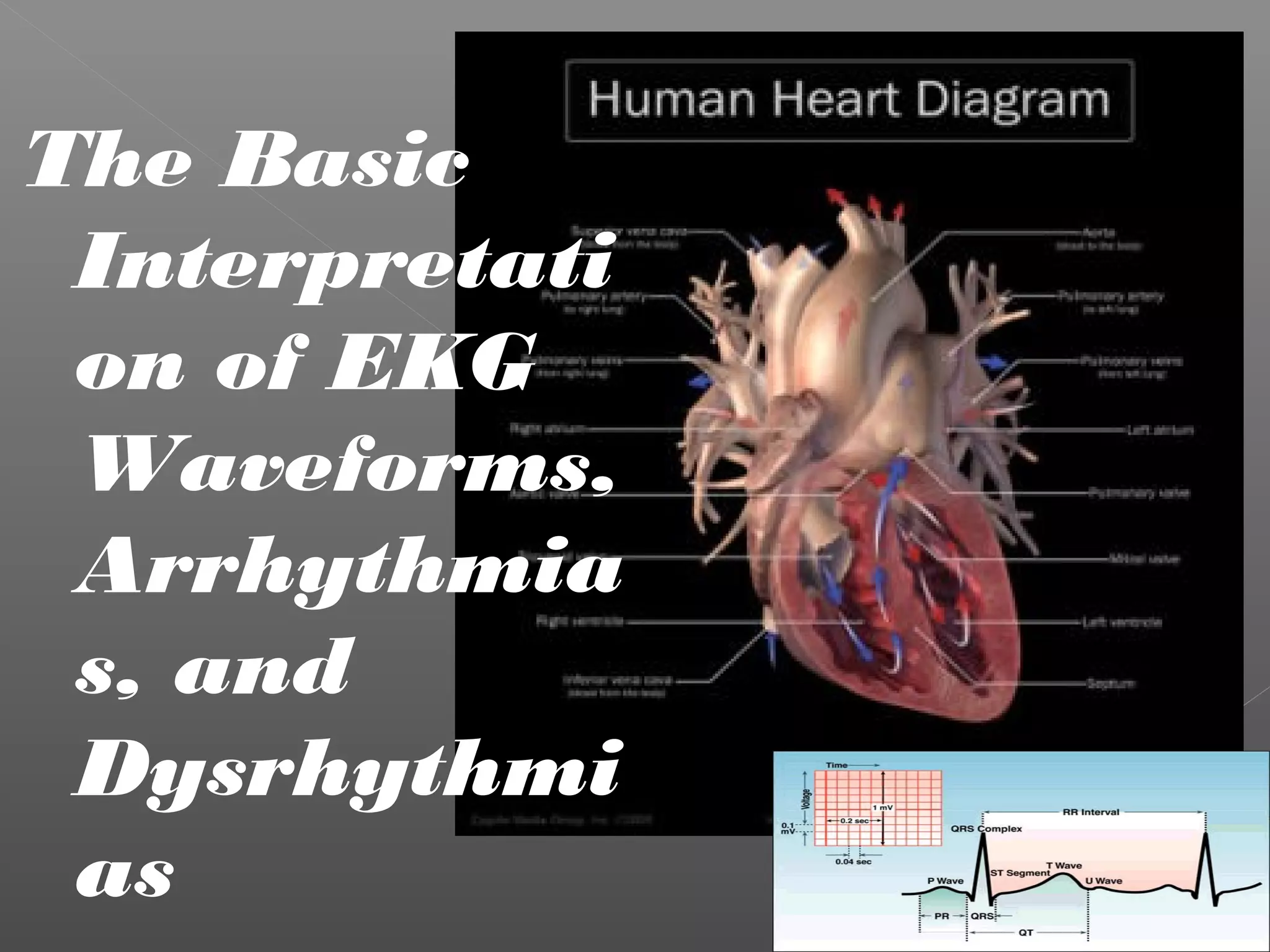
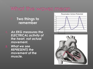
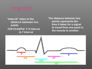
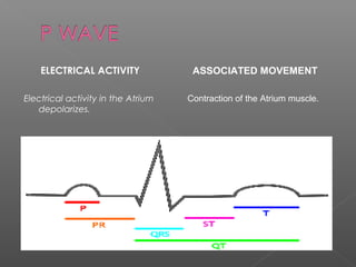
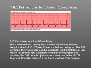
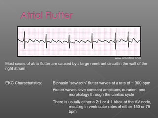
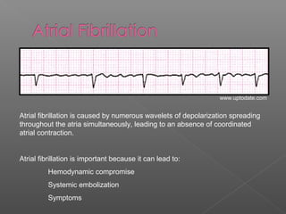
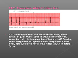
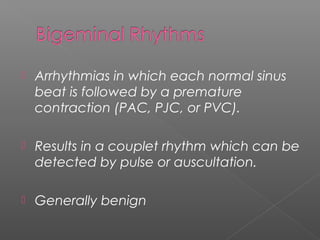
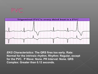
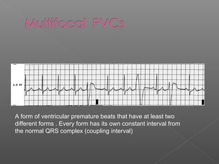
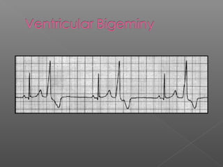
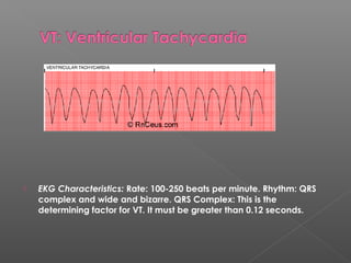
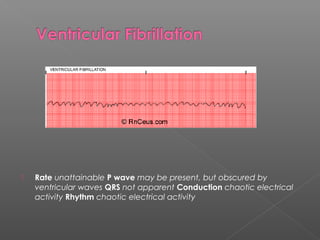
The Basic
Interpretati
on of EKG
Waveforms,
Arrhythmia
s, and
Dysrhythmi
as
 Two things to
remember
1. An EKG measures the
ELECTRICAL activity of
the heart, not actual
movement.
2. What we see
REPRESENTS the
movement of the
muscle.
“Interval” refers to the
distance between two
points.
FOR EXAMPLE: P-R interval,
Q-T Interval
The distance between two
points represents the
time it takes for a signal
to travel from one point in
the muscle to another.
ELECTRICAL ACTIVITY
Electrical activity in the Atrium
depolarizes.
ASSOCIATED MOVEMENT
Contraction of the Atrium muscle.
ELECTRICAL ACTIVITY
Electrical activity in the
Ventricle depolarizes.
ASSOIATED MOVEMENT
Contraction of the Ventricle
muscles.
ELECTRICAL ACTIVITY
Electrical activity in the
Ventricle repolarizes.
ASSOIATED MOVEMENT
No movement.
1. Calculate the Rate
2. Measure the Regularity (Rhythm) of the R-
Waves
3. Examine the P-Waves
4. Examine the P-R Interval
5. Examine the QRS Complex
1. The Rate is normal.
2. The Regularity
(Rhythm) of the R-
Waves is
abnormal.
3. The P-Waves are
normal.
4. The P-R Intervals
are abnormal.
5. The QRS Complex
are normal.
These findings will lead you to a type of
Heart Block. Comparing of the different
types of Heart Blocks to the EKG we
are looking at, we can see the type of
block we have here is a 2nd
Degree
Type 1
Now, by comparing
the answers from
those 5 steps to a
list of EKG rhythms
and their
descriptions, you
can assume the
type of beat or
rhythm you are
seeing. The
example to the left
is Sinus Arrhythmia!
Common Examples
 AV Block (relatively common)
› 1st
degree AV block
› Type 1 2nd
degree AV block
› Type 2 2nd
degree AV block
› 3rd
degree AV block
 SA Block (relatively rare)
EKG Characteristics: Prolongation of the PR interval, which is constant
All P waves are conducted
The Alan E. Lindsay ECG Learning Center ; http://medstat.med.utah.edu/kw/ecg/
Type 1 (Wenckebach)
EKG Characteristics: Progressive prolongation of the PR interval until a P
wave is not conducted.
As the PR interval prolongs, the RR interval actually
shortens
EKG Characteristics: Constant PR interval with intermittent failure to conduct
Type 2
EKG Characteristics: No relationship between P waves and QRS complexes
Relatively constant PP intervals and RR intervals
Greater number of P waves than QRS complexes
www.uptodate.com
EKG Characteristics: Presence of sinus P waves
Variation of the PP interval which cannot be q
attributed to either SA nodal block or PACs
When the variations in PP interval occur in phase with respiration, this is
considered to be a normal variant. When they are unrelated to respiration,
they may be caused by the same etiologies leading to sinus bradycardia.
Braunwald's Heart Disease: A Textbook of Cardiovascular Medicine, 7th ed., 2005.
Sinus bradycardia (rate of ~43 bpm) with a sinus pause
 PJC: Premature Junctional Complexes 
 EKG Characteristics: Usually 60-100 beats per minute. Rhythm:
Irregular, due to PJC. P Wave: Can occur before, during, or after QRS
complex. The P wave also can be inverted in lead II. PR Interval: Less
than 0.12 seconds. QRS Complex: Normal in configuration and
duration. The QRS complex just occurs sooner due to the PJC. ST
Segment: Could be distorted if the P wave follows QTS complex. 
Most cases of atrial flutter are caused by a large reentrant circuit in the wall of the
right atrium
EKG Characteristics: Biphasic “sawtooth” flutter waves at a rate of ~ 300 bpm
Flutter waves have constant amplitude, duration, and
morphology through the cardiac cycle
There is usually either a 2:1 or 4:1 block at the AV node,
resulting in ventricular rates of either 150 or 75
bpm
www.uptodate.com
Atrial fibrillation is caused by numerous wavelets of depolarization spreading
throughout the atria simultaneously, leading to an absence of coordinated
atrial contraction.
Atrial fibrillation is important because it can lead to:
Hemodynamic compromise
Systemic embolization
Symptoms
www.uptodate.com
 EKG Characteristics. Rate: Atrial and ventricular usually normal.
Rhythm: Irregular. P Wave: Ectopic P Wave. PR Interval: Usually
normal, but could also be greater than 020 seconds. QRS Complex:
normal configuration. ST Segment: Normal configuration. T Wave:
Usually normal, but could have P Wave hidden in it, which distorts T
Waves. 
 Arrhythmias in which each normal sinus
beat is followed by a premature
contraction (PAC, PJC, or PVC).
 Results in a couplet rhythm which can be
detected by pulse or auscultation.
 Generally benign
EKG Characteristics: The QRS fires too early. Rate:
Normal for the intrinsic rhythm. Rhythm: Regular, except
for the PVC. P Wave: None. PR Interval: None. QRS
Complex: Greater than 0.12 seconds.
A form of ventricular premature beats that have at least two
different forms . Every form has its own constant interval from
the normal QRS complex (coupling interval)
 EKG Characteristics: Rate: 100-250 beats per minute. Rhythm: QRS
complex and wide and bizarre. QRS Complex: This is the
determining factor for VT. It must be greater than 0.12 seconds.
 Rate unattainable P wave may be present, but obscured by
ventricular waves QRS not apparent Conduction chaotic electrical
activity Rhythm chaotic electrical activity
For more examples and information, check
out:
http://www.skillstat.com/ECG_Sim_demo.html
http://www.rnceus.com/course_frame.asp?exam_id=16&directory=ekg
The End

Ekg presentation

  • 1.
    The Basic Interpretati on ofEKG Waveforms, Arrhythmia s, and Dysrhythmi as
  • 2.
     Two thingsto remember 1. An EKG measures the ELECTRICAL activity of the heart, not actual movement. 2. What we see REPRESENTS the movement of the muscle.
  • 3.
    “Interval” refers tothe distance between two points. FOR EXAMPLE: P-R interval, Q-T Interval The distance between two points represents the time it takes for a signal to travel from one point in the muscle to another.
  • 4.
    ELECTRICAL ACTIVITY Electrical activityin the Atrium depolarizes. ASSOCIATED MOVEMENT Contraction of the Atrium muscle.
  • 5.
    ELECTRICAL ACTIVITY Electrical activityin the Ventricle depolarizes. ASSOIATED MOVEMENT Contraction of the Ventricle muscles.
  • 6.
    ELECTRICAL ACTIVITY Electrical activityin the Ventricle repolarizes. ASSOIATED MOVEMENT No movement.
  • 7.
    1. Calculate theRate 2. Measure the Regularity (Rhythm) of the R- Waves 3. Examine the P-Waves 4. Examine the P-R Interval 5. Examine the QRS Complex
  • 8.
    1. The Rateis normal. 2. The Regularity (Rhythm) of the R- Waves is abnormal. 3. The P-Waves are normal. 4. The P-R Intervals are abnormal. 5. The QRS Complex are normal. These findings will lead you to a type of Heart Block. Comparing of the different types of Heart Blocks to the EKG we are looking at, we can see the type of block we have here is a 2nd Degree Type 1
  • 9.
    Now, by comparing theanswers from those 5 steps to a list of EKG rhythms and their descriptions, you can assume the type of beat or rhythm you are seeing. The example to the left is Sinus Arrhythmia!
  • 10.
  • 11.
     AV Block(relatively common) › 1st degree AV block › Type 1 2nd degree AV block › Type 2 2nd degree AV block › 3rd degree AV block  SA Block (relatively rare)
  • 12.
    EKG Characteristics: Prolongationof the PR interval, which is constant All P waves are conducted The Alan E. Lindsay ECG Learning Center ; http://medstat.med.utah.edu/kw/ecg/
  • 13.
    Type 1 (Wenckebach) EKGCharacteristics: Progressive prolongation of the PR interval until a P wave is not conducted. As the PR interval prolongs, the RR interval actually shortens EKG Characteristics: Constant PR interval with intermittent failure to conduct Type 2
  • 14.
    EKG Characteristics: Norelationship between P waves and QRS complexes Relatively constant PP intervals and RR intervals Greater number of P waves than QRS complexes www.uptodate.com
  • 15.
    EKG Characteristics: Presenceof sinus P waves Variation of the PP interval which cannot be q attributed to either SA nodal block or PACs When the variations in PP interval occur in phase with respiration, this is considered to be a normal variant. When they are unrelated to respiration, they may be caused by the same etiologies leading to sinus bradycardia. Braunwald's Heart Disease: A Textbook of Cardiovascular Medicine, 7th ed., 2005.
  • 16.
    Sinus bradycardia (rateof ~43 bpm) with a sinus pause
  • 17.
     PJC: PrematureJunctional Complexes   EKG Characteristics: Usually 60-100 beats per minute. Rhythm: Irregular, due to PJC. P Wave: Can occur before, during, or after QRS complex. The P wave also can be inverted in lead II. PR Interval: Less than 0.12 seconds. QRS Complex: Normal in configuration and duration. The QRS complex just occurs sooner due to the PJC. ST Segment: Could be distorted if the P wave follows QTS complex. 
  • 18.
    Most cases ofatrial flutter are caused by a large reentrant circuit in the wall of the right atrium EKG Characteristics: Biphasic “sawtooth” flutter waves at a rate of ~ 300 bpm Flutter waves have constant amplitude, duration, and morphology through the cardiac cycle There is usually either a 2:1 or 4:1 block at the AV node, resulting in ventricular rates of either 150 or 75 bpm www.uptodate.com
  • 19.
    Atrial fibrillation iscaused by numerous wavelets of depolarization spreading throughout the atria simultaneously, leading to an absence of coordinated atrial contraction. Atrial fibrillation is important because it can lead to: Hemodynamic compromise Systemic embolization Symptoms www.uptodate.com
  • 20.
     EKG Characteristics.Rate: Atrial and ventricular usually normal. Rhythm: Irregular. P Wave: Ectopic P Wave. PR Interval: Usually normal, but could also be greater than 020 seconds. QRS Complex: normal configuration. ST Segment: Normal configuration. T Wave: Usually normal, but could have P Wave hidden in it, which distorts T Waves. 
  • 21.
     Arrhythmias inwhich each normal sinus beat is followed by a premature contraction (PAC, PJC, or PVC).  Results in a couplet rhythm which can be detected by pulse or auscultation.  Generally benign
  • 23.
    EKG Characteristics: TheQRS fires too early. Rate: Normal for the intrinsic rhythm. Rhythm: Regular, except for the PVC. P Wave: None. PR Interval: None. QRS Complex: Greater than 0.12 seconds.
  • 24.
    A form ofventricular premature beats that have at least two different forms . Every form has its own constant interval from the normal QRS complex (coupling interval)
  • 26.
     EKG Characteristics:Rate: 100-250 beats per minute. Rhythm: QRS complex and wide and bizarre. QRS Complex: This is the determining factor for VT. It must be greater than 0.12 seconds.
  • 27.
     Rate unattainableP wave may be present, but obscured by ventricular waves QRS not apparent Conduction chaotic electrical activity Rhythm chaotic electrical activity
  • 28.
    For more examplesand information, check out: http://www.skillstat.com/ECG_Sim_demo.html http://www.rnceus.com/course_frame.asp?exam_id=16&directory=ekg
  • 29.