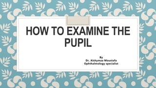
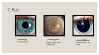
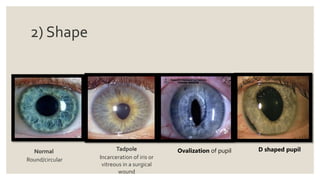
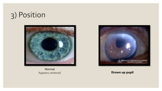
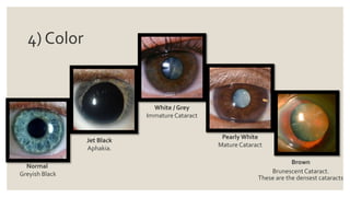
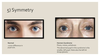
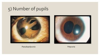
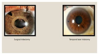
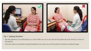
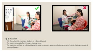
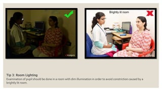
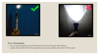
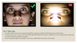
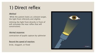
HOW TO EXAMINE THE
PUPIL
By
Dr. Alshymaa Moustafa
Ophthalmology specialist
WHAT TO EXAMINE?
1) Size
Normal Pupil
2mm-8mm Size .
Abnormally Small
Opiods, miotic agents
(pilocarpine)
Abnormally Large
Dilating drops, CN3
palsy
2) Shape
Tadpole
Incarceration of iris or
vitreous in a surgical
wound
Normal
Round/circular
Ovalization of pupil D shaped pupil
Festooned
Posterior synechiae.
Irregular
Adies sectoral dilation. This is
subtle.
Note how the iris is slightly
more dilated at 2-3 o’clock.
Iris coloboma
3) Position
Normal
Appears centered Drown up pupil
4) Color
Normal
Greyish Black
Jet Black
Aphakia.
White / Grey
Immature Cataract
Pearly White
Mature Cataract
Brown
Brunescent Cataract.
These are the densest cataracts
5) Symmetry
Normal
<1mm difference in
pupil size.
Horners Syndrome
Ptosis, miosis, anhidrosis
The abnormal pupil in this syndrome is the
smaller, left pupil. Note also the left lid
droop (ptosis).
Cranial Nerve 3 Palsy
Note the right pupil is enlarged and the right eye is
outwards and downwards.
5) Number of pupils
Pseudopolycoria Polycoria
Surgical iridectomy Temporal laser iridotomy
TIPS FOR
EXAMINATION
Tip 1: Seating Position:
◦ Ask the patient to sit comfortably on a chair while you sit on one side of the patient & never directly in front of
the patient.
◦ Sit to the side to avoid obstructing the patient’s view when you ask the patient to fixate at a distant target.
Tip 2: Fixation
• Ask the patient to maintain fixation on a distant target.
• The pupils constrict when the eyes focus at near.
• The patient must look at a distant target in order to prevent accommodation associated miosis that can confound
your pupil exam.
Tip 3: Room Lighting
Examination of pupil should be done in a room with dim illumination in order to avoid constriction caused by a
brightly lit room.
Tip 4: Illumination
• Use an evenly bright source of illumination for examining the light reflexes.
• Lights with uneven illumination patterns may cause variable constriction of the pupils.
Tip 5: Dark Irises
• It is often difficult to view a consensual pupillary response in patients with dark irises in dark rooms.
• In these cases, you can use a dim secondary light held below the pupils in addition to your bright primary light that
you use to elicit the pupillary response.
• If your secondary light is too bright it will cause undesirable constriction of the pupil.
HOW TO EXAMINE
PUPIL REFLEXES?
1) Direct reflex
Method:
While the patient looks at a distant target,
the light from inferiorly and slightly
(shining the light from directly in front of
will stimulate the near reflex that will
constriction).
Normal response:
contraction of pupils capture by sphincter
Record the speed of reaction:
brisk, sluggish, or fixed
2) Consensual reflex
Method:
While the patient is looking at a distant
the light on one pupil and notice the
other eye.
Normal response:
contraction of the contralateral pupil when
shone in one eye which should be
equal in velocity and extent to the direct
response.
Record the speed of reaction:
brisk, sluggish, or fixed
3) Near reflex
Method:
Ask patient to focus on a distant object in a moderately lit room.
Hold a Snellen chart about 30 cm in front of the patient’s eyes.
Instruct the patient to look at the Snellen chart.
Observe
pupillary constriction, which may take up to 10 seconds in certain situations.
Note:
There is actually a triad of things occurring – convergence of gaze, accommodation of the lens,
constriction.
4) Test for relative afferent pupillary
defect (RAPD)/Marcus Gunn Pupil
Method:
◦ Use a bright handheld light in a dim room.
◦ Shine the light in one of the patient’s eyes.
◦ After ~3 seconds, rapidly swing the light to the opposite pupil.
◦ After ~3 seconds, swing back to the first eye.
Observe:
a reaction for each step.
What is a “positive RAPD”?
◦ A positive RAPD is noted when the pupil dilates when you move the light from one side to
◦ Sometimes, the pupil will bounce a little when you swing the flashlight over. However, if you
shining on the pupil, it will return to the normal constricted size. This is physiologic and
Relative Afferent Pupillary Defect (RAPD)/Marcus Gunn Pupil:
Usually occurs as a result of optic nerve pathology or severe retinal disease. A
is whether or not a dense white cataract can cause an RAPD – it won’t!
Adie’s Pupil:
One or both eyes are abnormally dilated due to loss of parasympathetic innervation to the
iris sphincter and ciliary muscle.
Argyll Robertson Pupil:
Colloquially known as “prostitute’s pupils”. These are pupils that do not constrict to
bilaterally accomodate with the near response.
Thanks

Pupil examination

  • 1.
    HOW TO EXAMINETHE PUPIL By Dr. Alshymaa Moustafa Ophthalmology specialist
  • 2.
  • 3.
    1) Size Normal Pupil 2mm-8mmSize . Abnormally Small Opiods, miotic agents (pilocarpine) Abnormally Large Dilating drops, CN3 palsy
  • 4.
    2) Shape Tadpole Incarceration ofiris or vitreous in a surgical wound Normal Round/circular Ovalization of pupil D shaped pupil
  • 5.
    Festooned Posterior synechiae. Irregular Adies sectoraldilation. This is subtle. Note how the iris is slightly more dilated at 2-3 o’clock. Iris coloboma
  • 6.
  • 7.
    4) Color Normal Greyish Black JetBlack Aphakia. White / Grey Immature Cataract Pearly White Mature Cataract Brown Brunescent Cataract. These are the densest cataracts
  • 8.
    5) Symmetry Normal <1mm differencein pupil size. Horners Syndrome Ptosis, miosis, anhidrosis The abnormal pupil in this syndrome is the smaller, left pupil. Note also the left lid droop (ptosis).
  • 9.
    Cranial Nerve 3Palsy Note the right pupil is enlarged and the right eye is outwards and downwards.
  • 10.
    5) Number ofpupils Pseudopolycoria Polycoria
  • 11.
  • 12.
  • 13.
    Tip 1: SeatingPosition: ◦ Ask the patient to sit comfortably on a chair while you sit on one side of the patient & never directly in front of the patient. ◦ Sit to the side to avoid obstructing the patient’s view when you ask the patient to fixate at a distant target.
  • 14.
    Tip 2: Fixation •Ask the patient to maintain fixation on a distant target. • The pupils constrict when the eyes focus at near. • The patient must look at a distant target in order to prevent accommodation associated miosis that can confound your pupil exam.
  • 15.
    Tip 3: RoomLighting Examination of pupil should be done in a room with dim illumination in order to avoid constriction caused by a brightly lit room.
  • 16.
    Tip 4: Illumination •Use an evenly bright source of illumination for examining the light reflexes. • Lights with uneven illumination patterns may cause variable constriction of the pupils.
  • 17.
    Tip 5: DarkIrises • It is often difficult to view a consensual pupillary response in patients with dark irises in dark rooms. • In these cases, you can use a dim secondary light held below the pupils in addition to your bright primary light that you use to elicit the pupillary response. • If your secondary light is too bright it will cause undesirable constriction of the pupil.
  • 18.
  • 19.
    1) Direct reflex Method: Whilethe patient looks at a distant target, the light from inferiorly and slightly (shining the light from directly in front of will stimulate the near reflex that will constriction). Normal response: contraction of pupils capture by sphincter Record the speed of reaction: brisk, sluggish, or fixed
  • 20.
    2) Consensual reflex Method: Whilethe patient is looking at a distant the light on one pupil and notice the other eye. Normal response: contraction of the contralateral pupil when shone in one eye which should be equal in velocity and extent to the direct response. Record the speed of reaction: brisk, sluggish, or fixed
  • 21.
    3) Near reflex Method: Askpatient to focus on a distant object in a moderately lit room. Hold a Snellen chart about 30 cm in front of the patient’s eyes. Instruct the patient to look at the Snellen chart. Observe pupillary constriction, which may take up to 10 seconds in certain situations. Note: There is actually a triad of things occurring – convergence of gaze, accommodation of the lens, constriction.
  • 22.
    4) Test forrelative afferent pupillary defect (RAPD)/Marcus Gunn Pupil Method: ◦ Use a bright handheld light in a dim room. ◦ Shine the light in one of the patient’s eyes. ◦ After ~3 seconds, rapidly swing the light to the opposite pupil. ◦ After ~3 seconds, swing back to the first eye. Observe: a reaction for each step. What is a “positive RAPD”? ◦ A positive RAPD is noted when the pupil dilates when you move the light from one side to ◦ Sometimes, the pupil will bounce a little when you swing the flashlight over. However, if you shining on the pupil, it will return to the normal constricted size. This is physiologic and
  • 24.
    Relative Afferent PupillaryDefect (RAPD)/Marcus Gunn Pupil: Usually occurs as a result of optic nerve pathology or severe retinal disease. A is whether or not a dense white cataract can cause an RAPD – it won’t! Adie’s Pupil: One or both eyes are abnormally dilated due to loss of parasympathetic innervation to the iris sphincter and ciliary muscle. Argyll Robertson Pupil: Colloquially known as “prostitute’s pupils”. These are pupils that do not constrict to bilaterally accomodate with the near response.
  • 25.