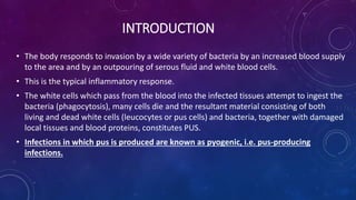
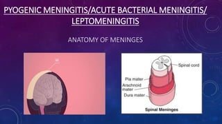
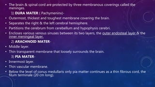
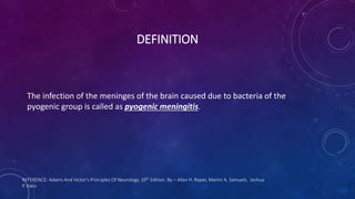
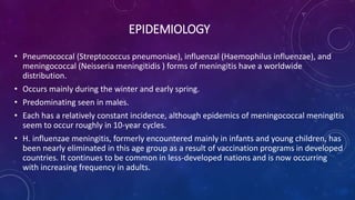
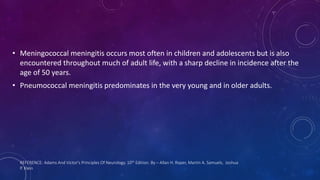
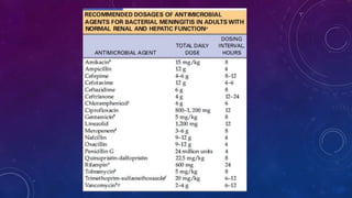
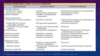
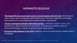
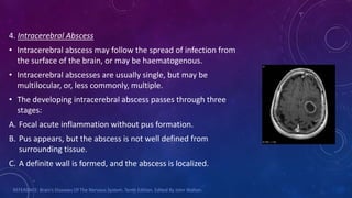
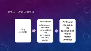
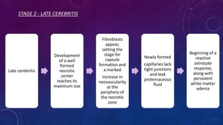
PYOGENIC
INFECTION
SAYALI GUJJEWAR
MPT II YEAR
QUESTIONS
Paper VI
• Describe the aetiology, clinical manifestations, laboratory investigations and
medical management of acute bacterial meningitis. (20 m)
• Signs of meningeal irritation. (8 m)
• Extradural abscess. (8 m)
• Changes in CSF in meningitis. (8 m)
• Neurosyphilis. (8 m)
• Laboratory investigation findings in bacterial meningitis. (8 m)
• Meningitis sequelae. ( 8 m)
Paper VIII
• Enumerate the guidelines for the physiotherapeutic management of
meningitis. (20 m)
• Treatment principles of neurosyphilis. (8 m)
INTRODUCTION
• The body responds to invasion by a wide variety of bacteria by an increased blood supply
to the area and by an outpouring of serous fluid and white blood cells.
• This is the typical inflammatory response.
• The white cells which pass from the blood into the infected tissues attempt to ingest the
bacteria (phagocytosis), many cells die and the resultant material consisting of both
living and dead white cells (leucocytes or pus cells) and bacteria, together with damaged
local tissues and blood proteins, constitutes PUS.
• Infections in which pus is produced are known as pyogenic, i.e. pus-producing
infections.
REFERENCE: An Introduction To Microbiology For Nurses (Third Edition), Chapter Nine - The Pyogenic Infections.
Https://Www.Sciencedirect.Com/Science/Article/Pii/B9780433303015500138
• Pus may be present as a localised collection deep in the tissues—an ABSCESS, it may be
produced on a surface, e.g. the mucosa of the pharynx, the mucosa of the bladder, the
méninges, indeed any body surface, it is then known as a PURULENT EXUDATE.
• Alternatively infection may spread evenly through the tissues causing a diffuse
inflammation CELLULITIS.
• The type of pus production will depend on the organism causing the infection, on the
tissue in which the infective process is taking place, and also on the body resistance to
the infection.
• Although the pyogenic infections have very similar appearances whatever the causative
organism, different sites of the body have a tendency to be infected with particular
species of bacteria.
PYOGENIC MENINGITIS/ACUTE BACTERIAL MENINGITIS/
LEPTOMENINGITIS
ANATOMY OF MENINGES
• The brain & spinal cord are protected by three membranous coverings called the
meninges.
1) DURA MATER ( Pachymeninx)-
• Outermost, thickest and toughest membrane covering the brain.
• Separates the right & the left cerebral hemisphere.
• Partitions the cerebrum from cerebellum and hypophysis cerebri.
• Encloses various venous sinuses between its two layers, the outer endosteal layer & the
inner meningeal layer.
2) ARACHNOID MATER-
• Middle layer.
• Thin transparent membrane that loosely surrounds the brain.
3) PIA MATER-
• Innermost layer.
• Thin vascular membrane.
• Below the level of conus medullaris only pia matter continues as a thin fibrous cord, the
filum terminale (20 cm long).
REFERENCE: B D Chaurasia’s Human Anatomy, Volume 3, Fifth Edition.
• The arachnoid mater & the pia mater are together called as leptomeninges.
• The extradural or epidural space is the space between the inner aspect of
skull bone and the endosteal layer of dura mater.
• The subdural space is the space between the dura and arachnoid maters.
• The subarachnoid space is the space between the arachnoid and the pia
maters.
• The dura mater & the arachnoid mater along with subacrachnoid space
containing CSF extend up to second sacral vertebra.
• Between the lower border of L1 & S2 vertebrae, the subarachnoid space
contains spinal nerve roots which constitute the cauda equina.
DEFINITION
The infection of the meninges of the brain caused due to bacteria of the
pyogenic group is called as pyogenic meningitis.
REFERENCE: Adams And Victor's Principles Of Neurology. 10th Edition. By – Allan H. Roper, Martin A. Samuels, Joshua
P. Klein
EPIDEMIOLOGY
• Pneumococcal (Streptococcus pneumoniae), influenzal (Haemophilus influenzae), and
meningococcal (Neisseria meningitidis ) forms of meningitis have a worldwide
distribution.
• Occurs mainly during the winter and early spring.
• Predominating seen in males.
• Each has a relatively constant incidence, although epidemics of meningococcal meningitis
seem to occur roughly in 10-year cycles.
• H. influenzae meningitis, formerly encountered mainly in infants and young children, has
been nearly eliminated in this age group as a result of vaccination programs in developed
countries. It continues to be common in less-developed nations and is now occurring
with increasing frequency in adults.
REFERENCE: Adams And Victor's Principles Of Neurology. 10th Edition. By – Allan H. Roper, Martin A. Samuels, Joshua
P. Klein
• Meningococcal meningitis occurs most often in children and adolescents but is also
encountered throughout much of adult life, with a sharp decline in incidence after the
age of 50 years.
• Pneumococcal meningitis predominates in the very young and in older adults.
CAUSES
• There are three principal ways in which meninges may become infected:
1. Infection through the bloodstream - Sometimes meningitis may be secondary to a focal
infection elsewhere in the body or secondary to a blood-borne infection that first settles in
the brain.
2. Extension to the meninges of a pre-existing pyogenic infection of one of the nasal sinuses,
the middle ear, or mastoid.
3. After fracture of the skull - In the case of a penetrating injury of the cranial vault,
organisms may be carried in from the scalp. When the base of the skull is fractured, they
may spread to the meninges from the nasopharynx, middle ear, or mastoid.
REFERENCE: Brain & Bannister’s Clinical Neurology, Seventh Edition
TYPES
1. H. influenza, N. meningitides, S. pneumoniae are most common, which account for
approximately 75 percent of sporadic cases.
2. L. monocytogenes is now the fourth most common type of nonsurgical bacterial meningitis
in adults.
3. Staphylococcus aureus and group A (Streptococcus pyogenes) and group D streptococci,
usually in association with brain abscess, epidural abscess, head trauma, neurosurgical
procedures, or cranial thrombophlebitis; are less frequent causes.
4. E. coli and group B streptococci in newborns.
5. Pseudomonas and the Enterobacteriaceae, such as Klebsiella, Proteus, which are usually a
consequence of lumbar puncture, spinal anesthesia, or shunting procedures to relieve
hydrocephalus.
REFERENCE: Adams And Victor's Principles Of Neurology. 10th Edition. By – Allan H. Roper, Martin A. Samuels, Joshua
P. Klein
• Less-common meningeal pathogens include Salmonella, Shigella, Clostridium,
Neisseria gonorrhoeae, and Acinetobacter calcoaceticus.
PATHOPHYSIOLOGY
CLINICAL FEATURES
• All forms of acute meningitis, whatever their cause, have certain symtoms in common.
• The onset may be fulminating, acute, or, less commonly, insidious.
1. Headache:
• Usually the first symptom.
• Increases in severity.
• “Bursting” character.
• Diffuse or mainly frontal, and usually radiates down the neck and into the back, sometimes
being associated with pain in the spine radiating to the limbs, especially the lower.
2. Fever:
• The temperature is usually between 37.8°C and 38°C though hyperpyrexia may occur,
especially terminally.
3. Pulse Rate:
• Sometimes slow in the early stages, for example between 50 and 60, but always rises as
the illness progresses and at the end is usually very rapid and often irregular.
4. Respiratory Rate:
• Usually slightly increased , Cheyne-Stokes breathing may occur.
5. Vomiting:
• May occur, especially in early stages.
6. Convulsions:
• Common in children, especially in influenzal meningitis but rare in adults.
7. The patient tends to lie in an attitude of general flexion, curled up under the bedclothes
and resenting interference.
8. There may be a high pitched “meningeal” cry in infants.
9. Signs of Meningeal Irritation
A. Cervical Rigidity (Neck Stiffness):
• Present at an early stage in most cases but may be minimal or absent in the very old, or
the very young patient.
• It is elicited by the observer placing his hand beneath the patient’s occiput and
endeavouring to flex the head so as to bring the chin towards the chest. In a normal
patient individual this is accomplished with ease and without pain. In meningitis there is
resistance due to spasm of the extensor muscles of the neck, and an attempt to
overcome this causes pain.
• Cervical rigidity is usually associated with some rigidity of the lower spine.
B. Head Retraction:
• Head retraction is an extreme degree of cervical rigidity brought about by spasm of
extensor muscles.
• Flexion of the neck causes a rise in the CSF pressure in the cisterna magna.
• When the meninges are inflamed this is painful; neck stiffness and head retraction
represent reflex protective spasm.
C. Kernig’s and Brudzinski’s Signs:
• To elicit Kernig’s sign one knee is extended with the hip fully flexed; when positive there is
pain and spasm of the hamstrings.
• Brudzinski’s sign consists first of spontaneous flexion of the knees and hips on attempted
neck flexion and secondly spontaneous flexion of one leg when the other is flexed
passively.
• These signs result from the presence of inflammatory exudate around the roots in the
lumbar theca.
10. Other Signs:
A. The mental state of the patient varies according to the stage and progress of the
disease.
B. Delirium is common in the early stages, but often, as the disease progresses, gives
place to drowsiness and stupor, which is followed by coma.
C. Photophobia
D. The fundi may be normal or may show venous congestion or, sometimes,
papilloedema.
E. The pupils are often unequal and may react sluggishly; they may be dilated and
fixed.
F. Ptosis is common as are squint and diplopia.
G. Any of the ocular muscles may be paralysed, most frequently one or both lateral
recti.
I. Facial paresis
J. Dysphagia may also occur in the later stages.
K. Muscular power in the limbs is usually preserved, though slight incoordination
and tremor are common and there is often marked hypotonia.
L. Diffuse flaccid paralysis is a terminal event.
M. The tendon reflexes are usually sluggish and often soon lost; the abdominal
reflexes also disappear early; the plantar reflexes are usually flexor at first,
though later one or both may become extensor.
N. Sensory loss is rare.
O. True paralysis of sphincter control occurs only late, but stupor or coma may
lead to retention or incontinence of urine early in illness.
11. Late Complications:
• Meningitis localized for a time to one hemisphere may cause jacksonian
convulsions, hemiparesis, and even hemianopia.
• Dementia, amnesia, epilepsy, paresis of the limbs, ataxia, blindness, deafness,
paraplegia, cerebral herniation.
REFERENCE: Brain’s Diseases Of The Nervous System. Tenth Edition. Edited By John Walton.
DIAGNOSTIC PROCEDURE
CSF EXAMINATION
• The CSF is under increased pressure.
• A pleocytosis in the spinal fluid is diagnostic.
• The number of leukocytes ranges from 250 to 1,00,000/mm3, but the usual number is from
1,000 to 10,000.
• Occasionally, in pneumococcal and influenzal meningitis, the CSF may contain a large
number of bacteria but few, if any, neutrophils for the first few hours.
• Neutrophils predominate (85 to 95 percent of the total), but an increasing proportion of
mononuclear cells is found as the infection continues for days, and especially in partially
treated meningitis.
• The protein content is higher than 45 mg/dL in more than 90 percent of the cases; in most
cases, it falls in the range of 100 to 500 mg/dL.
• The glucose content is diminished, usually to a concentration below 40 mg/dL, or less than
40 percent of the blood glucose concentration (measured concomitantly or within the
previous hour), provided that the latter is less than 250 mg/dL.
• The Gram stain of the spinal fluid sediment permits identification of the causative agent in
most cases of bacterial meningitis; pneumococci and H. influenzae are identified more
readily than meningococci.
• Cultures of the spinal fluid proves to be positive in 70 to 90 percent of cases of bacterial
meningitis.
• Chloride concentrations in the CSF are usually found to be low, possibly reflecting
dehydration and low serum chloride levels.
• A rise in total CSF lactate dehydrogenase (LDH) activity is consistently observed in patients
with bacterial meningitis.
• Levels of lactic acid in the CSF are also elevated in both bacterial and fungal meningitides
(greater than 35 mg/dL) and may be helpful in distinguishing these disorders from viral
meningitides, in which lactic acid levels remain normal.
OTHER LABORATORY FINDINGS
• Blood cultures are positive in 40 to 60 percent of patients with H. influenzae,
meningococcal, and pneumococcal meningitis, and may provide the only
definite clue as to the causative agent.
• The leukocyte count in the blood is generally elevated, and immature forms
are usually present.
• Severe hyponatremia may be seen as a result of inappropriate secretion of
antidiuretic hormone (ADH).
IMAGING STUDIES
• In patients with bacterial meningitis, chest films are essential because they may disclose an
area of pneumonia or abscess.
• Sinus and skull films may provide clues to the presence of cranial osteomyelitis, paranasal
sinusitis, mastoiditis, or cranial osteomyelitis, but these structures are better visualized on
CT scans, which have supplanted conventional films in most cases.
• The CT scan is particularly useful in detecting lesions that erode the skull or spine and
provide a route for bacterial invasion, such as tumors or sinus wall defects, as well as
demonstrating a brain abscess or subdural empyema.
• MRI with gadolinium enhancement may display the meningeal exudate and cortical
reaction, and both types of imaging, with appropriate techniques, will demonstrate venous
occlusions and adjacent infarctions.
REFERENCE: Adams and Victor's Principles of Neurology. 10th edition. By – Allan H. Roper, Martin A. Samuels, Joshua P. Klein
DIFFERENTIAL DIAGNOSIS
Pyogenic meningitis must be distinguished from:
1. General infection with toxaemia, especially when headache is a prominent
symptom
2. Meningism
3. Acute cerebral infection, including encephalitis and intracranial absess
4. Subarachnoid haemorrhage
5. Other forms of meningitis & meningeal irritation
REFERENCE: Brain’s Diseases Of The Nervous System. Tenth Edition. Edited By John Walton.
PROGNOSIS
• The prognosis of pyogenic meningitis depends upon the nature of the
invading organism, the number of organisms present in the CSF, the possibility
of removing the source of infection and the effectiveness of treatment.
• Early & effective treatment should lead to recovery without residual
symptoms.
• In any form of pyogenic meningitis treated late or inadequately, permanent
damage may lead to dementia, epilepsy, deafness, blindness or spastic
weakness.
REFERENCE: Brain’s Diseases Of The Nervous System. Tenth Edition. Edited By John Walton.
TREATMENT
REFERENCE: Adams and Victor's Principles of Neurology. 10th edition. By – Allan H. Roper, Martin A. Samuels, Joshua P. Klein
MENINGITIS SEQUELAE
• Meningeal fibrosis around optic nerves or around spinal cord and roots: blindness and
optic atrophy, spastic paraparesis with sensory loss in the lower segments of the body
(opticochlasmatic arachnoiditis and meningomyelitis, respectively).
• Chronic meningoencephalitis with hydrocephalus: dementia, stupor or coma, and
paralysis (e.g., general paralysis of the insane). If lumbosacral posterior roots are
chronically damaged, a tabetic syndrome results. Deep infarcts.
• Persistent hydrocephalus in the child: blindness, arrest of mental activity, bilateral spastic
hemiplegia.
REFERENCE: Adams and Victor's Principles of Neurology. 10th edition. By – Allan H. Roper, Martin A. Samuels, Joshua P. Klein
BRAIN ABSCESS
DEFINITION
• Brain abscess is a focal suppurative process within the brain parenchyma.
• It is an abscess caused by inflammation and collection of infected material,
coming from local (ear infection, dental abscess, infection of paranasal
sinuses, infection of the mastoid air cells of the temporal bone, epidural
abscess) or remote (lung, heart, kidney etc.) infectious sources, within
the brain tissue.
REFERENCE:
1. R. K. Gupta et al. (eds.), MR Imaging and Spectroscopy of Central Nervous System Infection © Kluwer Academic
/ Plenum Publishers 2001
2. https://en.wikipedia.org/wiki/Brain_abscess
EPIDEMIOLOGY
• The incidence of brain abscess varies from 0.18 to 1.3% in the developed
world, but remains a significant problem in the developing world.
• Paradoxically, the mortality and morbidity from a brain abscess remained high
until relatively recently despite potent, specific antimicrobial therapy and
advances in neurosurgical technique.
• Three times more common in males than females.
• 29% of abscesses are temporal, 25% frontal, 10% parietal, 6% cerebellar, 3%
occipital, 7% subdural or thalamic & 20% multiple.
REFERENCE: 1. Brain’s Diseases Of The Nervous System. Tenth Edition. Edited By John Walton.
2. R. K. Gupta et al. (eds.), MR Imaging and Spectroscopy of Central Nervous System Infection © Kluwer Academic / Plenum Publishers
2001
CAUSES
1. Infection of the middle ear or nasal sinuses
• Infection of the middle ear is 4 - 9 times as common a cause as is sinus infection; the frontal
sinus is most often involved, the sphenoid sinus next.
2. Pyaemia or bacteraemia
3. Metastasis from intrathoracic suppuration
• When brain abscess is secondary to infection elsewhere, the thorax is often the source, and
rarely the primary abscess is elsewhere, as in the liver.
4. Head injury
• Fracture of the skull is liable to cause abscess when an injury leads to free
communication between the body surface and the brain, especially when fragments of
bone, clothing or a missile penetrate the latter.
• Pencil tip injuries penetrating the orbital roof or temporal bone may have this effect in
children whereas in adults compound depressed skull fracture with a dural tear is the
commonest cause.
5. There is a clear association between brain abscess and cyanotic congenital heart
disease.
6. Single or multiple abscesses may also develop in the immunosuppresed patient, as
after cardiac transplantation.
7. Most common causative organisms are Staphylococcus aureus, streptococci,
pneumococci and E. coli.
REFERENCE: 1. Brain’s Diseases Of The Nervous System. Tenth Edition. Edited By John Walton.
TYPES
1. Extradural Abscess:
• Secondary to osteitis of a cranial
bone.
• The infection passes through the
bone, but as its further advance is
arrested by the dura , the
accumulating pus strips off dura.
2. Subdural Abscess
• The pus lies between dura &
arachnoid.
3. Subarachnoid Abscess
• A rare form in which the pus is
confined to the subarachnoid space
& spreads along the brain surface.
4. Intracerebral Abscess
• Intracerebral abscess may follow the spread of infection from
the surface of the brain, or may be haematogenous.
• Intracerebral abscesses are usually single, but may be
multilocular, or, less commonly, multiple.
• The developing intracerebral abscess passes through three
stages:
A. Focal acute inflammation without pus formation.
B. Pus appears, but the abscess is not well defined from
surrounding tissue.
C. A definite wall is formed, and the abscess is localized.
REFERENCE: Brain’s Diseases Of The Nervous System. Tenth Edition. Edited By John Walton.
PATHOPHYSIOLOGY
The evaluation of an abscess includes four histopathologic stages:
• First Stage - Early Cerebritis (days 1-3).
• Second Stage - Late Cerebritis (days 4- 9).
• Third Stage - Early Capsule Formation (days 10-13).
• Fourth Stage - Late Capsule Formation (days 14 and later).
STAGE 1 – EARLY CEREBRITIS
Early
cerebritis
Perivascular
inflammatory
response
surrounding
the
developing
necrotic
center
Profound
edema in
the
surrounding
white
matter
develops
STAGE 2 - LATE CEREBRITIS
Late cerebritis
Development
of a well
formed
necrotic
center
reaches its
maximum size
Fibroblasts
appear,
setting the
stage for
capsule
formation and
a marked
increase in
neovascularity
at the
periphery of
the necrotic
zone
Newly formed
capillaries lack
tight junctions
and leak
proteinaceous
fluid
Beginning of a
reactive
astrocyte
response,
along with
persistent
white matter
edema
STAGE 3 - EARLY CAPSULE FORMATION
Early capsule
formation
slight
decrease in
the
size of the
necrotic
center
Well-
developed
layer of
fibroblasts,
with
significantly
more reticulin
deposition on
the cortical
side of the
lesion than
on the
ventricular
side
Outside this
developing
capsule is a
region of
persistent
cerebritis and
neovascularity,
with a further
increase in
reactive
astrocytes
These
processes
continue in
the fourth and
final stage
STAGE 4 - LATE CAPSULE FORMATION
late capsule formation
The capsule continues
to thicken, with an
abundance of collagen
present by the third
week
REFERENCE: R. K. Gupta et al. (eds.), MR Imaging and Spectroscopy of Central Nervous System Infection
© Kluwer Academic / Plenum Publishers 2001
CLINICAL FEATURES
The symptoms of brain abscess can be divided into:
1. General symptoms of infection
2. Symptoms of increased intracranial pressure
3. Focal symptoms
4. Changes in the CSF
1. General Symptoms Of Infection
• In acute cases – irregular pyrexia.
• In chronic cases the temperature may be intermittently raised but not
invariably.
• In both there may be a neutrophil leucocytosis in the blood.
2. Symptoms Of Increased Intracranial Pressure
Headache:
• In chronic abscess it is often paroxysmal, being increased by stooping & exertion, and presenting the other
features of headache due to increased intracranial pressure.
• In more acute cases it is persistent and severe.
Papilloedema:
• It is a late sign & is often absent or slight.
• When present it is sometimes more marked on the side of the lesion.
Bradycardia:
• Common, but is not constant, and when it occurs usually indicates a rapid increase in the severity of the
condition
In severe cases delirium, somnolence, stupor and coma develop.
Exceptionally, signs of increased intracranial pressure are slight or lacking, even when a large abscess is present.
Epilepsy may be the presenting feature especially in the neonate.
3. Focal Symptoms
Extradural Abscess:
• Difficult to diagnose because, unless the abscess is large, focal symptoms are absent,
except for headache radiating from the ear or from the infected sinus towards the vertex.
• Increasing local headache with tenderness and/or oedema of the scalp in the region of
the infected ear or sinus when surgical drainage seems adequate are suspicious signs.
Subdural Abscess:
• High fever
• Fits (either focal or generalized)
• Rapidly developing hemiplegia, with aphasia if the major hemisphere is involved.
Intracerebral Abscess:
1. Temporal Lobe Abscess:
• If situated on the left side in a right-handed individual, can cause aphasia, often of the
nominal or amnestic type.
• A patient who can name familiar objects accurately often hesitates or misnames less
familiar articles.
• Visual-field defect – usually a homonymous upper quadrantic defect on the opposite
side due to involvement of the lower fibres of the optic radiation.
• Damage to the corticospinal tract is usually slight, and weakness is most marked in the
face and tongue.
• The opposite plantar reflex may be extensor.
• Oculomotor paralyses may result from pressure upon the third or sixth cranial nerves.
2. Cerebellar Abscess:
• Headache is often predominantly suboccipital. May radiate down the neck and be
associated with neck stiffness.
• The head may be flexed to the side of the lesion or retracted.
• Signs of cerebellar dysfunction vary in severity and may be slight. The most important
are:
Phasic nystagmus, most marked on looking to the side of lesion.
Hypotonia and incoordination in the limbs on the affected side, with an inability to carry
out rapid alternating movements with the upper limb on the affected side as well as on
the normal side.
• Pressure upon the brainstem may occur, leading to compression of cranial nerves,
especially the sixth & seventh, on the side of the abscess, and slight signs of corticospinal
defect on the opposite side.
• Pass-pointing outwards, with the affected hand and a tendency to deviate or fall to the
side of the lesion when walking are seen in some cases.
3. Frontal Abscess:
• Headache
• Drowsiness
• Apathy
• Impairments of memory and attention
• A large abscess with oedema may cause aphasia or hemiparesis.
• Unilateral anosmia and slight exophthalmos may be present.
4. Abscesses in other situations:
• The focal symptoms depend upon the position of the abscess, and usually resemble
those of tumour in the same situation.
Subarachnoid Abscess:
• This rare condition may be suspected when the signs suggest abscess of otitic
origin.
• Convulsions may occur with a superficial abscess over a cerebral hemisphere.
• When the signs suggest involvement of the cerebellum but no abscess is
evident in the cerebellum itself, a superficial abscess in the cerebellopontine
angle may be present.
REFERENCE: Brain’s Diseases Of The Nervous System. Tenth Edition. Edited By John Walton.
DIAGNOSTIC PROCEDURE
• Computerized Tomography:
Allows determination of the site and size of the abscess, whether it is single or multiple.
Obviates the need for doing lumbar puncture which may be hazardous in this condition.
Allows visualization of the middle-ear cavities, mastoid air cells, frontal and sphenoid
sinuses.
• Radionuclide scanning is also a sensitive method of localizing abscesses.
• Angiography: if associated thrombosis is suspected.
• Electroencephalography may help to distinguish a single focal lesion from a more
generalized encephalitis.
• Lumbar puncture:
Opening pressure is increased and there is an excess of cells, usually <100 per mm3 , and
mainly lymphocytes.
The protein is moderately elevated up to 2.0 g/l
Organisms are generally absent unless the abscess has ruptured into the CSF.
• There may be a neutrophil leucocytosis in the peripheral blood.
• Depending on the aetiology, blood culture may yield an organism.
REFERENCE: Brain’s Diseases Of The Nervous System. Tenth Edition. Edited By John Walton.
DIFFERENTIAL DIAGNOSIS
• Viral encephalitis
• Bacterial meningitis
• Infective embolic infarction from endocarditis
• Sinus thrombosis secondary to superficial infection
• Cerebellar infarction
• Intracranial tumour
• Cyanotic heart disease
REFERENCE: Brain’s Diseases Of The Nervous System. Tenth Edition. Edited By John Walton.
PROGNOSIS
• Almost uniformly fatal in the absence of antibiotic and surgical treatment.
• Recovery by spontaneous drainage through the ear or nose may occur.
• Spreading encephalitis, rupture of the abscess into the ventricular system,
meningitis and sinus thrombosis are the usual terminations.
• Epilepsy is an important sequel, occurring in between 50 and 72 % of cases,
usually within 12 months.
REFERENCE: Brain’s Diseases Of The Nervous System. Tenth Edition. Edited By John Walton.
TREATMENT
• Antibiotics:
Broad-spectrum combination like a third-generation cephalosporin with good brain
penetration, such as ceftriaxone, which will cover aerobic Gram-positive and Gram-
negative organisms, and metronidazole to cover anaerobic organisms.
Penicillin in high dosage, amoxicillin, chloramphenicol, and gentamicin are also used as
alternatives.
• Aspiration of the abscess
• Excision of the abscess
REFERENCE: Brain’s Diseases Of The Nervous System. Tenth Edition. Edited By John Walton.
Pyogenic infection

Pyogenic infection

  • 1.
  • 2.
    QUESTIONS Paper VI • Describethe aetiology, clinical manifestations, laboratory investigations and medical management of acute bacterial meningitis. (20 m) • Signs of meningeal irritation. (8 m) • Extradural abscess. (8 m) • Changes in CSF in meningitis. (8 m) • Neurosyphilis. (8 m) • Laboratory investigation findings in bacterial meningitis. (8 m) • Meningitis sequelae. ( 8 m)
  • 3.
    Paper VIII • Enumeratethe guidelines for the physiotherapeutic management of meningitis. (20 m) • Treatment principles of neurosyphilis. (8 m)
  • 4.
    INTRODUCTION • The bodyresponds to invasion by a wide variety of bacteria by an increased blood supply to the area and by an outpouring of serous fluid and white blood cells. • This is the typical inflammatory response. • The white cells which pass from the blood into the infected tissues attempt to ingest the bacteria (phagocytosis), many cells die and the resultant material consisting of both living and dead white cells (leucocytes or pus cells) and bacteria, together with damaged local tissues and blood proteins, constitutes PUS. • Infections in which pus is produced are known as pyogenic, i.e. pus-producing infections.
  • 5.
    REFERENCE: An IntroductionTo Microbiology For Nurses (Third Edition), Chapter Nine - The Pyogenic Infections. Https://Www.Sciencedirect.Com/Science/Article/Pii/B9780433303015500138 • Pus may be present as a localised collection deep in the tissues—an ABSCESS, it may be produced on a surface, e.g. the mucosa of the pharynx, the mucosa of the bladder, the méninges, indeed any body surface, it is then known as a PURULENT EXUDATE. • Alternatively infection may spread evenly through the tissues causing a diffuse inflammation CELLULITIS. • The type of pus production will depend on the organism causing the infection, on the tissue in which the infective process is taking place, and also on the body resistance to the infection. • Although the pyogenic infections have very similar appearances whatever the causative organism, different sites of the body have a tendency to be infected with particular species of bacteria.
  • 6.
    PYOGENIC MENINGITIS/ACUTE BACTERIALMENINGITIS/ LEPTOMENINGITIS ANATOMY OF MENINGES
  • 7.
    • The brain& spinal cord are protected by three membranous coverings called the meninges. 1) DURA MATER ( Pachymeninx)- • Outermost, thickest and toughest membrane covering the brain. • Separates the right & the left cerebral hemisphere. • Partitions the cerebrum from cerebellum and hypophysis cerebri. • Encloses various venous sinuses between its two layers, the outer endosteal layer & the inner meningeal layer. 2) ARACHNOID MATER- • Middle layer. • Thin transparent membrane that loosely surrounds the brain. 3) PIA MATER- • Innermost layer. • Thin vascular membrane. • Below the level of conus medullaris only pia matter continues as a thin fibrous cord, the filum terminale (20 cm long).
  • 8.
    REFERENCE: B DChaurasia’s Human Anatomy, Volume 3, Fifth Edition. • The arachnoid mater & the pia mater are together called as leptomeninges. • The extradural or epidural space is the space between the inner aspect of skull bone and the endosteal layer of dura mater. • The subdural space is the space between the dura and arachnoid maters. • The subarachnoid space is the space between the arachnoid and the pia maters. • The dura mater & the arachnoid mater along with subacrachnoid space containing CSF extend up to second sacral vertebra. • Between the lower border of L1 & S2 vertebrae, the subarachnoid space contains spinal nerve roots which constitute the cauda equina.
  • 9.
    DEFINITION The infection ofthe meninges of the brain caused due to bacteria of the pyogenic group is called as pyogenic meningitis. REFERENCE: Adams And Victor's Principles Of Neurology. 10th Edition. By – Allan H. Roper, Martin A. Samuels, Joshua P. Klein
  • 10.
    EPIDEMIOLOGY • Pneumococcal (Streptococcuspneumoniae), influenzal (Haemophilus influenzae), and meningococcal (Neisseria meningitidis ) forms of meningitis have a worldwide distribution. • Occurs mainly during the winter and early spring. • Predominating seen in males. • Each has a relatively constant incidence, although epidemics of meningococcal meningitis seem to occur roughly in 10-year cycles. • H. influenzae meningitis, formerly encountered mainly in infants and young children, has been nearly eliminated in this age group as a result of vaccination programs in developed countries. It continues to be common in less-developed nations and is now occurring with increasing frequency in adults.
  • 11.
    REFERENCE: Adams AndVictor's Principles Of Neurology. 10th Edition. By – Allan H. Roper, Martin A. Samuels, Joshua P. Klein • Meningococcal meningitis occurs most often in children and adolescents but is also encountered throughout much of adult life, with a sharp decline in incidence after the age of 50 years. • Pneumococcal meningitis predominates in the very young and in older adults.
  • 12.
    CAUSES • There arethree principal ways in which meninges may become infected: 1. Infection through the bloodstream - Sometimes meningitis may be secondary to a focal infection elsewhere in the body or secondary to a blood-borne infection that first settles in the brain. 2. Extension to the meninges of a pre-existing pyogenic infection of one of the nasal sinuses, the middle ear, or mastoid. 3. After fracture of the skull - In the case of a penetrating injury of the cranial vault, organisms may be carried in from the scalp. When the base of the skull is fractured, they may spread to the meninges from the nasopharynx, middle ear, or mastoid. REFERENCE: Brain & Bannister’s Clinical Neurology, Seventh Edition
  • 13.
    TYPES 1. H. influenza,N. meningitides, S. pneumoniae are most common, which account for approximately 75 percent of sporadic cases. 2. L. monocytogenes is now the fourth most common type of nonsurgical bacterial meningitis in adults. 3. Staphylococcus aureus and group A (Streptococcus pyogenes) and group D streptococci, usually in association with brain abscess, epidural abscess, head trauma, neurosurgical procedures, or cranial thrombophlebitis; are less frequent causes. 4. E. coli and group B streptococci in newborns. 5. Pseudomonas and the Enterobacteriaceae, such as Klebsiella, Proteus, which are usually a consequence of lumbar puncture, spinal anesthesia, or shunting procedures to relieve hydrocephalus.
  • 14.
    REFERENCE: Adams AndVictor's Principles Of Neurology. 10th Edition. By – Allan H. Roper, Martin A. Samuels, Joshua P. Klein • Less-common meningeal pathogens include Salmonella, Shigella, Clostridium, Neisseria gonorrhoeae, and Acinetobacter calcoaceticus.
  • 15.
  • 16.
    CLINICAL FEATURES • Allforms of acute meningitis, whatever their cause, have certain symtoms in common. • The onset may be fulminating, acute, or, less commonly, insidious. 1. Headache: • Usually the first symptom. • Increases in severity. • “Bursting” character. • Diffuse or mainly frontal, and usually radiates down the neck and into the back, sometimes being associated with pain in the spine radiating to the limbs, especially the lower.
  • 17.
    2. Fever: • Thetemperature is usually between 37.8°C and 38°C though hyperpyrexia may occur, especially terminally. 3. Pulse Rate: • Sometimes slow in the early stages, for example between 50 and 60, but always rises as the illness progresses and at the end is usually very rapid and often irregular. 4. Respiratory Rate: • Usually slightly increased , Cheyne-Stokes breathing may occur. 5. Vomiting: • May occur, especially in early stages. 6. Convulsions: • Common in children, especially in influenzal meningitis but rare in adults.
  • 18.
    7. The patienttends to lie in an attitude of general flexion, curled up under the bedclothes and resenting interference. 8. There may be a high pitched “meningeal” cry in infants. 9. Signs of Meningeal Irritation A. Cervical Rigidity (Neck Stiffness): • Present at an early stage in most cases but may be minimal or absent in the very old, or the very young patient. • It is elicited by the observer placing his hand beneath the patient’s occiput and endeavouring to flex the head so as to bring the chin towards the chest. In a normal patient individual this is accomplished with ease and without pain. In meningitis there is resistance due to spasm of the extensor muscles of the neck, and an attempt to overcome this causes pain. • Cervical rigidity is usually associated with some rigidity of the lower spine.
  • 19.
    B. Head Retraction: •Head retraction is an extreme degree of cervical rigidity brought about by spasm of extensor muscles. • Flexion of the neck causes a rise in the CSF pressure in the cisterna magna. • When the meninges are inflamed this is painful; neck stiffness and head retraction represent reflex protective spasm. C. Kernig’s and Brudzinski’s Signs: • To elicit Kernig’s sign one knee is extended with the hip fully flexed; when positive there is pain and spasm of the hamstrings. • Brudzinski’s sign consists first of spontaneous flexion of the knees and hips on attempted neck flexion and secondly spontaneous flexion of one leg when the other is flexed passively. • These signs result from the presence of inflammatory exudate around the roots in the lumbar theca.
  • 20.
    10. Other Signs: A.The mental state of the patient varies according to the stage and progress of the disease. B. Delirium is common in the early stages, but often, as the disease progresses, gives place to drowsiness and stupor, which is followed by coma. C. Photophobia D. The fundi may be normal or may show venous congestion or, sometimes, papilloedema. E. The pupils are often unequal and may react sluggishly; they may be dilated and fixed. F. Ptosis is common as are squint and diplopia. G. Any of the ocular muscles may be paralysed, most frequently one or both lateral recti. I. Facial paresis
  • 21.
    J. Dysphagia mayalso occur in the later stages. K. Muscular power in the limbs is usually preserved, though slight incoordination and tremor are common and there is often marked hypotonia. L. Diffuse flaccid paralysis is a terminal event. M. The tendon reflexes are usually sluggish and often soon lost; the abdominal reflexes also disappear early; the plantar reflexes are usually flexor at first, though later one or both may become extensor. N. Sensory loss is rare. O. True paralysis of sphincter control occurs only late, but stupor or coma may lead to retention or incontinence of urine early in illness.
  • 22.
    11. Late Complications: •Meningitis localized for a time to one hemisphere may cause jacksonian convulsions, hemiparesis, and even hemianopia. • Dementia, amnesia, epilepsy, paresis of the limbs, ataxia, blindness, deafness, paraplegia, cerebral herniation. REFERENCE: Brain’s Diseases Of The Nervous System. Tenth Edition. Edited By John Walton.
  • 23.
    DIAGNOSTIC PROCEDURE CSF EXAMINATION •The CSF is under increased pressure. • A pleocytosis in the spinal fluid is diagnostic. • The number of leukocytes ranges from 250 to 1,00,000/mm3, but the usual number is from 1,000 to 10,000. • Occasionally, in pneumococcal and influenzal meningitis, the CSF may contain a large number of bacteria but few, if any, neutrophils for the first few hours. • Neutrophils predominate (85 to 95 percent of the total), but an increasing proportion of mononuclear cells is found as the infection continues for days, and especially in partially treated meningitis.
  • 24.
    • The proteincontent is higher than 45 mg/dL in more than 90 percent of the cases; in most cases, it falls in the range of 100 to 500 mg/dL. • The glucose content is diminished, usually to a concentration below 40 mg/dL, or less than 40 percent of the blood glucose concentration (measured concomitantly or within the previous hour), provided that the latter is less than 250 mg/dL. • The Gram stain of the spinal fluid sediment permits identification of the causative agent in most cases of bacterial meningitis; pneumococci and H. influenzae are identified more readily than meningococci. • Cultures of the spinal fluid proves to be positive in 70 to 90 percent of cases of bacterial meningitis.
  • 25.
    • Chloride concentrationsin the CSF are usually found to be low, possibly reflecting dehydration and low serum chloride levels. • A rise in total CSF lactate dehydrogenase (LDH) activity is consistently observed in patients with bacterial meningitis. • Levels of lactic acid in the CSF are also elevated in both bacterial and fungal meningitides (greater than 35 mg/dL) and may be helpful in distinguishing these disorders from viral meningitides, in which lactic acid levels remain normal.
  • 26.
    OTHER LABORATORY FINDINGS •Blood cultures are positive in 40 to 60 percent of patients with H. influenzae, meningococcal, and pneumococcal meningitis, and may provide the only definite clue as to the causative agent. • The leukocyte count in the blood is generally elevated, and immature forms are usually present. • Severe hyponatremia may be seen as a result of inappropriate secretion of antidiuretic hormone (ADH).
  • 27.
    IMAGING STUDIES • Inpatients with bacterial meningitis, chest films are essential because they may disclose an area of pneumonia or abscess. • Sinus and skull films may provide clues to the presence of cranial osteomyelitis, paranasal sinusitis, mastoiditis, or cranial osteomyelitis, but these structures are better visualized on CT scans, which have supplanted conventional films in most cases. • The CT scan is particularly useful in detecting lesions that erode the skull or spine and provide a route for bacterial invasion, such as tumors or sinus wall defects, as well as demonstrating a brain abscess or subdural empyema. • MRI with gadolinium enhancement may display the meningeal exudate and cortical reaction, and both types of imaging, with appropriate techniques, will demonstrate venous occlusions and adjacent infarctions. REFERENCE: Adams and Victor's Principles of Neurology. 10th edition. By – Allan H. Roper, Martin A. Samuels, Joshua P. Klein
  • 28.
    DIFFERENTIAL DIAGNOSIS Pyogenic meningitismust be distinguished from: 1. General infection with toxaemia, especially when headache is a prominent symptom 2. Meningism 3. Acute cerebral infection, including encephalitis and intracranial absess 4. Subarachnoid haemorrhage 5. Other forms of meningitis & meningeal irritation REFERENCE: Brain’s Diseases Of The Nervous System. Tenth Edition. Edited By John Walton.
  • 29.
    PROGNOSIS • The prognosisof pyogenic meningitis depends upon the nature of the invading organism, the number of organisms present in the CSF, the possibility of removing the source of infection and the effectiveness of treatment. • Early & effective treatment should lead to recovery without residual symptoms. • In any form of pyogenic meningitis treated late or inadequately, permanent damage may lead to dementia, epilepsy, deafness, blindness or spastic weakness. REFERENCE: Brain’s Diseases Of The Nervous System. Tenth Edition. Edited By John Walton.
  • 30.
  • 32.
    REFERENCE: Adams andVictor's Principles of Neurology. 10th edition. By – Allan H. Roper, Martin A. Samuels, Joshua P. Klein
  • 33.
    MENINGITIS SEQUELAE • Meningealfibrosis around optic nerves or around spinal cord and roots: blindness and optic atrophy, spastic paraparesis with sensory loss in the lower segments of the body (opticochlasmatic arachnoiditis and meningomyelitis, respectively). • Chronic meningoencephalitis with hydrocephalus: dementia, stupor or coma, and paralysis (e.g., general paralysis of the insane). If lumbosacral posterior roots are chronically damaged, a tabetic syndrome results. Deep infarcts. • Persistent hydrocephalus in the child: blindness, arrest of mental activity, bilateral spastic hemiplegia. REFERENCE: Adams and Victor's Principles of Neurology. 10th edition. By – Allan H. Roper, Martin A. Samuels, Joshua P. Klein
  • 34.
    BRAIN ABSCESS DEFINITION • Brainabscess is a focal suppurative process within the brain parenchyma. • It is an abscess caused by inflammation and collection of infected material, coming from local (ear infection, dental abscess, infection of paranasal sinuses, infection of the mastoid air cells of the temporal bone, epidural abscess) or remote (lung, heart, kidney etc.) infectious sources, within the brain tissue. REFERENCE: 1. R. K. Gupta et al. (eds.), MR Imaging and Spectroscopy of Central Nervous System Infection © Kluwer Academic / Plenum Publishers 2001 2. https://en.wikipedia.org/wiki/Brain_abscess
  • 35.
    EPIDEMIOLOGY • The incidenceof brain abscess varies from 0.18 to 1.3% in the developed world, but remains a significant problem in the developing world. • Paradoxically, the mortality and morbidity from a brain abscess remained high until relatively recently despite potent, specific antimicrobial therapy and advances in neurosurgical technique. • Three times more common in males than females. • 29% of abscesses are temporal, 25% frontal, 10% parietal, 6% cerebellar, 3% occipital, 7% subdural or thalamic & 20% multiple. REFERENCE: 1. Brain’s Diseases Of The Nervous System. Tenth Edition. Edited By John Walton. 2. R. K. Gupta et al. (eds.), MR Imaging and Spectroscopy of Central Nervous System Infection © Kluwer Academic / Plenum Publishers 2001
  • 36.
    CAUSES 1. Infection ofthe middle ear or nasal sinuses • Infection of the middle ear is 4 - 9 times as common a cause as is sinus infection; the frontal sinus is most often involved, the sphenoid sinus next. 2. Pyaemia or bacteraemia 3. Metastasis from intrathoracic suppuration • When brain abscess is secondary to infection elsewhere, the thorax is often the source, and rarely the primary abscess is elsewhere, as in the liver.
  • 37.
    4. Head injury •Fracture of the skull is liable to cause abscess when an injury leads to free communication between the body surface and the brain, especially when fragments of bone, clothing or a missile penetrate the latter. • Pencil tip injuries penetrating the orbital roof or temporal bone may have this effect in children whereas in adults compound depressed skull fracture with a dural tear is the commonest cause. 5. There is a clear association between brain abscess and cyanotic congenital heart disease. 6. Single or multiple abscesses may also develop in the immunosuppresed patient, as after cardiac transplantation. 7. Most common causative organisms are Staphylococcus aureus, streptococci, pneumococci and E. coli. REFERENCE: 1. Brain’s Diseases Of The Nervous System. Tenth Edition. Edited By John Walton.
  • 38.
    TYPES 1. Extradural Abscess: •Secondary to osteitis of a cranial bone. • The infection passes through the bone, but as its further advance is arrested by the dura , the accumulating pus strips off dura.
  • 39.
    2. Subdural Abscess •The pus lies between dura & arachnoid. 3. Subarachnoid Abscess • A rare form in which the pus is confined to the subarachnoid space & spreads along the brain surface.
  • 40.
    4. Intracerebral Abscess •Intracerebral abscess may follow the spread of infection from the surface of the brain, or may be haematogenous. • Intracerebral abscesses are usually single, but may be multilocular, or, less commonly, multiple. • The developing intracerebral abscess passes through three stages: A. Focal acute inflammation without pus formation. B. Pus appears, but the abscess is not well defined from surrounding tissue. C. A definite wall is formed, and the abscess is localized. REFERENCE: Brain’s Diseases Of The Nervous System. Tenth Edition. Edited By John Walton.
  • 41.
    PATHOPHYSIOLOGY The evaluation ofan abscess includes four histopathologic stages: • First Stage - Early Cerebritis (days 1-3). • Second Stage - Late Cerebritis (days 4- 9). • Third Stage - Early Capsule Formation (days 10-13). • Fourth Stage - Late Capsule Formation (days 14 and later).
  • 42.
    STAGE 1 –EARLY CEREBRITIS Early cerebritis Perivascular inflammatory response surrounding the developing necrotic center Profound edema in the surrounding white matter develops
  • 43.
    STAGE 2 -LATE CEREBRITIS Late cerebritis Development of a well formed necrotic center reaches its maximum size Fibroblasts appear, setting the stage for capsule formation and a marked increase in neovascularity at the periphery of the necrotic zone Newly formed capillaries lack tight junctions and leak proteinaceous fluid Beginning of a reactive astrocyte response, along with persistent white matter edema
  • 44.
    STAGE 3 -EARLY CAPSULE FORMATION Early capsule formation slight decrease in the size of the necrotic center Well- developed layer of fibroblasts, with significantly more reticulin deposition on the cortical side of the lesion than on the ventricular side Outside this developing capsule is a region of persistent cerebritis and neovascularity, with a further increase in reactive astrocytes These processes continue in the fourth and final stage
  • 45.
    STAGE 4 -LATE CAPSULE FORMATION late capsule formation The capsule continues to thicken, with an abundance of collagen present by the third week REFERENCE: R. K. Gupta et al. (eds.), MR Imaging and Spectroscopy of Central Nervous System Infection © Kluwer Academic / Plenum Publishers 2001
  • 46.
    CLINICAL FEATURES The symptomsof brain abscess can be divided into: 1. General symptoms of infection 2. Symptoms of increased intracranial pressure 3. Focal symptoms 4. Changes in the CSF
  • 47.
    1. General SymptomsOf Infection • In acute cases – irregular pyrexia. • In chronic cases the temperature may be intermittently raised but not invariably. • In both there may be a neutrophil leucocytosis in the blood.
  • 48.
    2. Symptoms OfIncreased Intracranial Pressure Headache: • In chronic abscess it is often paroxysmal, being increased by stooping & exertion, and presenting the other features of headache due to increased intracranial pressure. • In more acute cases it is persistent and severe. Papilloedema: • It is a late sign & is often absent or slight. • When present it is sometimes more marked on the side of the lesion. Bradycardia: • Common, but is not constant, and when it occurs usually indicates a rapid increase in the severity of the condition In severe cases delirium, somnolence, stupor and coma develop. Exceptionally, signs of increased intracranial pressure are slight or lacking, even when a large abscess is present. Epilepsy may be the presenting feature especially in the neonate.
  • 49.
    3. Focal Symptoms ExtraduralAbscess: • Difficult to diagnose because, unless the abscess is large, focal symptoms are absent, except for headache radiating from the ear or from the infected sinus towards the vertex. • Increasing local headache with tenderness and/or oedema of the scalp in the region of the infected ear or sinus when surgical drainage seems adequate are suspicious signs. Subdural Abscess: • High fever • Fits (either focal or generalized) • Rapidly developing hemiplegia, with aphasia if the major hemisphere is involved.
  • 50.
    Intracerebral Abscess: 1. TemporalLobe Abscess: • If situated on the left side in a right-handed individual, can cause aphasia, often of the nominal or amnestic type. • A patient who can name familiar objects accurately often hesitates or misnames less familiar articles. • Visual-field defect – usually a homonymous upper quadrantic defect on the opposite side due to involvement of the lower fibres of the optic radiation. • Damage to the corticospinal tract is usually slight, and weakness is most marked in the face and tongue. • The opposite plantar reflex may be extensor. • Oculomotor paralyses may result from pressure upon the third or sixth cranial nerves.
  • 51.
    2. Cerebellar Abscess: •Headache is often predominantly suboccipital. May radiate down the neck and be associated with neck stiffness. • The head may be flexed to the side of the lesion or retracted. • Signs of cerebellar dysfunction vary in severity and may be slight. The most important are: Phasic nystagmus, most marked on looking to the side of lesion. Hypotonia and incoordination in the limbs on the affected side, with an inability to carry out rapid alternating movements with the upper limb on the affected side as well as on the normal side. • Pressure upon the brainstem may occur, leading to compression of cranial nerves, especially the sixth & seventh, on the side of the abscess, and slight signs of corticospinal defect on the opposite side. • Pass-pointing outwards, with the affected hand and a tendency to deviate or fall to the side of the lesion when walking are seen in some cases.
  • 52.
    3. Frontal Abscess: •Headache • Drowsiness • Apathy • Impairments of memory and attention • A large abscess with oedema may cause aphasia or hemiparesis. • Unilateral anosmia and slight exophthalmos may be present. 4. Abscesses in other situations: • The focal symptoms depend upon the position of the abscess, and usually resemble those of tumour in the same situation.
  • 53.
    Subarachnoid Abscess: • Thisrare condition may be suspected when the signs suggest abscess of otitic origin. • Convulsions may occur with a superficial abscess over a cerebral hemisphere. • When the signs suggest involvement of the cerebellum but no abscess is evident in the cerebellum itself, a superficial abscess in the cerebellopontine angle may be present. REFERENCE: Brain’s Diseases Of The Nervous System. Tenth Edition. Edited By John Walton.
  • 54.
    DIAGNOSTIC PROCEDURE • ComputerizedTomography: Allows determination of the site and size of the abscess, whether it is single or multiple. Obviates the need for doing lumbar puncture which may be hazardous in this condition. Allows visualization of the middle-ear cavities, mastoid air cells, frontal and sphenoid sinuses. • Radionuclide scanning is also a sensitive method of localizing abscesses. • Angiography: if associated thrombosis is suspected. • Electroencephalography may help to distinguish a single focal lesion from a more generalized encephalitis.
  • 55.
    • Lumbar puncture: Openingpressure is increased and there is an excess of cells, usually <100 per mm3 , and mainly lymphocytes. The protein is moderately elevated up to 2.0 g/l Organisms are generally absent unless the abscess has ruptured into the CSF. • There may be a neutrophil leucocytosis in the peripheral blood. • Depending on the aetiology, blood culture may yield an organism. REFERENCE: Brain’s Diseases Of The Nervous System. Tenth Edition. Edited By John Walton.
  • 56.
    DIFFERENTIAL DIAGNOSIS • Viralencephalitis • Bacterial meningitis • Infective embolic infarction from endocarditis • Sinus thrombosis secondary to superficial infection • Cerebellar infarction • Intracranial tumour • Cyanotic heart disease REFERENCE: Brain’s Diseases Of The Nervous System. Tenth Edition. Edited By John Walton.
  • 57.
    PROGNOSIS • Almost uniformlyfatal in the absence of antibiotic and surgical treatment. • Recovery by spontaneous drainage through the ear or nose may occur. • Spreading encephalitis, rupture of the abscess into the ventricular system, meningitis and sinus thrombosis are the usual terminations. • Epilepsy is an important sequel, occurring in between 50 and 72 % of cases, usually within 12 months. REFERENCE: Brain’s Diseases Of The Nervous System. Tenth Edition. Edited By John Walton.
  • 58.
    TREATMENT • Antibiotics: Broad-spectrum combinationlike a third-generation cephalosporin with good brain penetration, such as ceftriaxone, which will cover aerobic Gram-positive and Gram- negative organisms, and metronidazole to cover anaerobic organisms. Penicillin in high dosage, amoxicillin, chloramphenicol, and gentamicin are also used as alternatives. • Aspiration of the abscess • Excision of the abscess REFERENCE: Brain’s Diseases Of The Nervous System. Tenth Edition. Edited By John Walton.