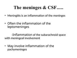
The meninges &CSF……
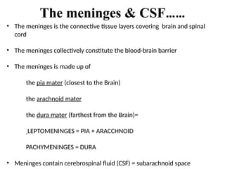
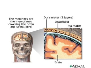
• The meninges is the connective tissue layers covering brain and spinal
cord
• The meninges collectively constitute the blood-brain barrier
• The meninges is made up of
the pia mater (closest to the Brain)
the arachnoid mater
the dura mater (farthest from the Brain)=
LEPTOMENINGES = PIA + ARACCHNOID
PACHYMENINGES = DURA
• Meninges contain cerebrospinal fluid (CSF) = subarachnoid space
5.
The meninges &CSF……
• Meningitis is an inflammation of the meninges
• Often the inflammation of the
leptomeninges
-Inflammation of the subarachnoid space
with meningeal involvement
• May involve inflammation of the
pachymeninges
6.
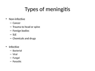
Types of meningitis
•Non-infective
– Cancer
– Trauma to head or spine
– Foreign bodies
– SLE
– Chemicals and drugs
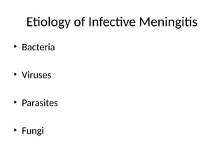
• Infective
– Bacterial
– Viral
– Fungal
– Parasitic
7.
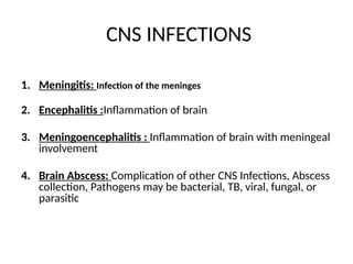
CNS INFECTIONS
1. Meningitis:Infection of the meninges
2. Encephalitis :Inflammation of brain
3. Meningoencephalitis : Inflammation of brain with meningeal
involvement
4. Brain Abscess: Complication of other CNS Infections, Abscess
collection, Pathogens may be bacterial, TB, viral, fungal, or
parasitic
8.
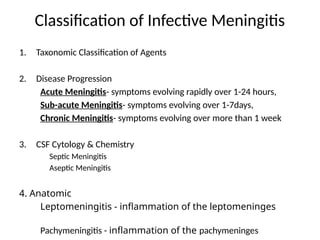
Classification of InfectiveMeningitis
1. Taxonomic Classification of Agents
2. Disease Progression
Acute Meningitis- symptoms evolving rapidly over 1-24 hours,
Sub-acute Meningitis- symptoms evolving over 1-7days,
Chronic Meningitis- symptoms evolving over more than 1 week
3. CSF Cytology & Chemistry
Septic Meningitis
Aseptic Meningitis
4. Anatomic
Leptomeningitis - inflammation of the leptomeninges
Pachymeningitis - inflammation of the pachymeninges
9.
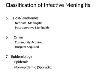
Classification of InfectiveMeningitis
5. Host/Syndromes
Neonatal Meningitis
Post-operative Meningitis
6. Origin
Community Acquired
Hospital Acquired
7. Epidemiology
Epidemic
Non-epidemic (Sporadic)
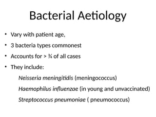
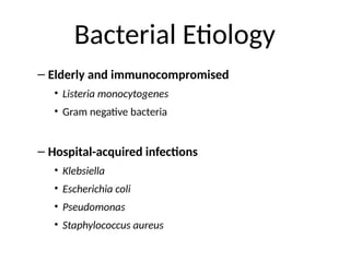
Bacterial Aetiology
• Varywith patient age,
• 3 bacteria types commonest
• Accounts for ˃ ¾ of all cases
• They include:
Neisseria meningitidis (meningococcus)
Haemophilus influenzae (in young and unvaccinated)
Streptococcus pneumoniae ( pneumococcus)
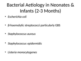
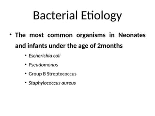
Bacterial Etiology
• Themost common organisms in Neonates
and infants under the age of 2months
• Escherichia coli
• Pseudomonas
• Group B Streptococcus
• Staphylococcus aureus
15.
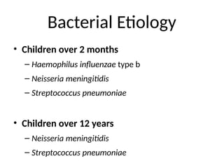
Bacterial Etiology
• Childrenover 2 months
– Haemophilus influenzae type b
– Neisseria meningitidis
– Streptococcus pneumoniae
• Children over 12 years
– Neisseria meningitidis
– Streptococcus pneumoniae
16.
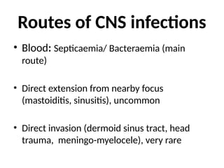
Routes of CNSinfections
• Blood: Septicaemia/ Bacteraemia (main
route)
• Direct extension from nearby focus
(mastoiditis, sinusitis), uncommon
• Direct invasion (dermoid sinus tract, head
trauma, meningo-myelocele), very rare
17.
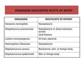
ORGANISM ROUTE/SITE OFENTERY
Neisseria meningitidis Nasopharynx
Streptococcus pneumoniae Nasopharynx or direct extension
across
skull fracture
Listeria monocytogenes GI tract, placenta
Haemophilus influenzae Nasopharynx
Staphylococcus aureus Bacteremia, skin, or foreign body
Staphylococcus epidermidis Skin or foreign body
ORGANISM ASSOCIATED ROUTE OF ENTRY
18.
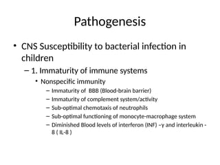
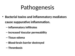
Pathogenesis
• CNS Susceptibilityto bacterial infection in
children
– 1. Immaturity of immune systems
• Nonspecific immunity
– Immaturity of BBB (Blood-brain barrier)
– Immaturity of complement system/activity
– Sub-optimal chemotaxis of neutrophils
– Sub-optimal functioning of monocyte-macrophage system
– Diminished Blood levels of interferon (INF) –γ and interleukin -
8 ( IL-8 )
19.
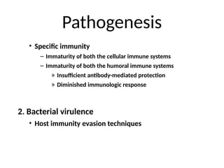
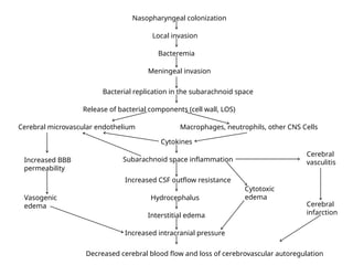
Pathogenesis
• Specific immunity
–Immaturity of both the cellular immune systems
– Immaturity of both the humoral immune systems
» Insufficient antibody-mediated protection
» Diminished immunologic response
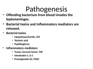
2. Bacterial virulence
• Host immunity evasion techniques
Nasopharyngeal colonization
Local invasion
Bacteremia
Meningealinvasion
Bacterial replication in the subarachnoid space
Release of bacterial components (cell wall, LOS)
Cerebral microvascular endothelium Macrophages, neutrophils, other CNS Cells
Cytokines
Subarachnoid space inflammation
Cerebral
vasculitis
Increased CSF outflow resistance
Hydrocephalus
Interstitial edema
Increased intracranial pressure
Decreased cerebral blood flow and loss of cerebrovascular autoregulation
Cytotoxic
edema
Cerebral
infarction
Increased BBB
permeability
Vasogenic
edema
23.
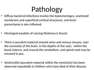
Pathology
• Diffuse bacterialinfections involve the leptomeninges, arachnoid
membrane and superficial cortical structures, and brain
parenchyma is also inflamed.
• Meningeal exudate of varying thickness is found.
• There is purulent material around veins and venous sinuses, over
the convexity of the brain, in the depths of the sulci, within the
basal cisterns, and around the cerebellum, and spinal cord may be
encased in pus.
• Ventriculitis (purulent material within the ventricles) has been
observed repeatedly in children who have died of their disease.
24.
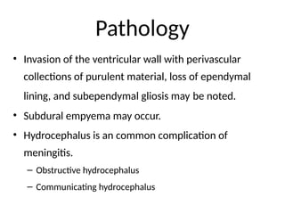
Pathology
• Invasion ofthe ventricular wall with perivascular
collections of purulent material, loss of ependymal
lining, and subependymal gliosis may be noted.
• Subdural empyema may occur.
• Hydrocephalus is an common complication of
meningitis.
– Obstructive hydrocephalus
– Communicating hydrocephalus
25.
Pathology
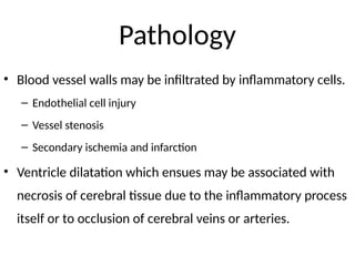
• Blood vesselwalls may be infiltrated by inflammatory cells.
– Endothelial cell injury
– Vessel stenosis
– Secondary ischemia and infarction
• Ventricle dilatation which ensues may be associated with
necrosis of cerebral tissue due to the inflammatory process
itself or to occlusion of cerebral veins or arteries.
26.
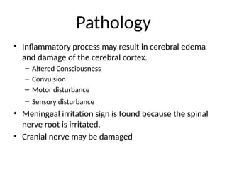
Pathology
• Inflammatory processmay result in cerebral edema
and damage of the cerebral cortex.
– Altered Consciousness
– Convulsion
– Motor disturbance
– Sensory disturbance
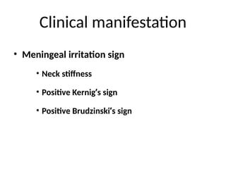
• Meningeal irritation sign is found because the spinal
nerve root is irritated.
• Cranial nerve may be damaged
27.
Meningitis and Septicaemia
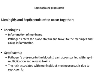
Meningitisand Septicaemia often occur together:
• Meningitis
– Inflammation of meninges
– Pathogen enters the blood stream and travel to the meninges and
cause inflammation.
• Septicaemia
– Pathogen’s presence in the blood stream accompanied with rapid
multiplication and release toxins.
– The rash associated with meningitis of meningococcus is due to
septicaemia
28.
Meningitis and Septicemia
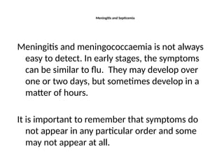
Meningitisand meningococcaemia is not always
easy to detect. In early stages, the symptoms
can be similar to flu. They may develop over
one or two days, but sometimes develop in a
matter of hours.
It is important to remember that symptoms do
not appear in any particular order and some
may not appear at all.
29.
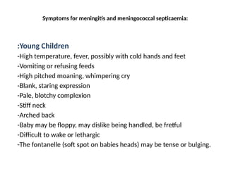
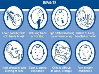
Symptoms for meningitisand meningococcal septicaemia:
:Young Children
-High temperature, fever, possibly with cold hands and feet
-Vomiting or refusing feeds
-High pitched moaning, whimpering cry
-Blank, staring expression
-Pale, blotchy complexion
-Stiff neck
-Arched back
-Baby may be floppy, may dislike being handled, be fretful
-Difficult to wake or lethargic
-The fontanelle (soft spot on babies heads) may be tense or bulging.
31.
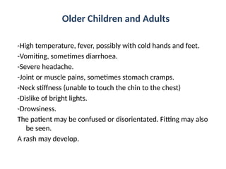
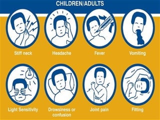
Older Children andAdults
-High temperature, fever, possibly with cold hands and feet.
-Vomiting, sometimes diarrhoea.
-Severe headache.
-Joint or muscle pains, sometimes stomach cramps.
-Neck stiffness (unable to touch the chin to the chest)
-Dislike of bright lights.
-Drowsiness.
The patient may be confused or disorientated. Fitting may also
be seen.
A rash may develop.
33.
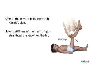
One of thephysically demonstrable sign of meningitis is
Kernig's sign.
Severe stiffness of the hamstrings causes an inability to
straighten the leg when the hip is flexed to 90 degrees.
34.
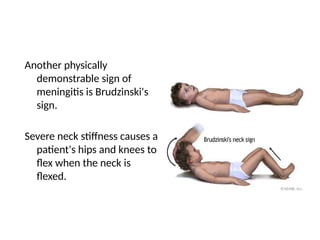
Another physically
demonstrable signof
meningitis is Brudzinski's
sign.
Severe neck stiffness causes a
patient's hips and knees to
flex when the neck is
flexed.
35.
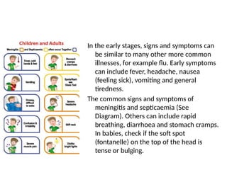
In the earlystages, signs and symptoms can
be similar to many other more common
illnesses, for example flu. Early symptoms
can include fever, headache, nausea
(feeling sick), vomiting and general
tiredness.
The common signs and symptoms of
meningitis and septicaemia (See
Diagram). Others can include rapid
breathing, diarrhoea and stomach cramps.
In babies, check if the soft spot
(fontanelle) on the top of the head is
tense or bulging.
36.
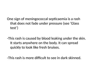
One sign ofmeningococcal septicaemia is a rash
that does not fade under pressure (see ‘Glass
test’)
-This rash is caused by blood leaking under the skin.
It starts anywhere on the body. It can spread
quickly to look like fresh bruises.
-This rash is more difficult to see in dark skinned.
37.
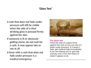
‘Glass Test’
A rashthat does not fade under
pressure will still be visible
when the side of a clear
drinking glass is pressed firmly
against the skin.
If someone is ill or obviously
getting worse, do not wait for
a rash. It may appear late or
not at all.
A fever with a rash that does not
fade under pressure is a
medical emergency.
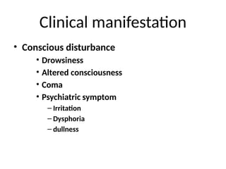
Clinical manifestation
• Seizures
•Seizures occur in about 20%-30% of children with
bacterial meningitis.
• Seizures is often found in Haemophilus influenzae
and pneumococal infection.
• Seizures is correlative with the inflammation of brain
parenchyma, cerebral infarction and electrolyte
disturbances.
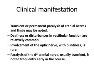
Clinical manifestation
– Transientor permanent paralysis of cranial nerves
and limbs may be noted.
– Deafness or disturbances in vestibular function are
relatively common.
– Involvement of the optic nerve, with blindness, is
rare.
– Paralysis of the 6th
cranial nerve, usually transient, is
noted frequently early in the course.
44.
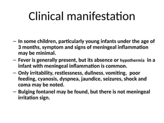
Clinical manifestation
– Insome children, particularly young infants under the age of
3 months, symptom and signs of meningeal inflammation
may be minimal.
– Fever is generally present, but its absence or hypothermia in a
infant with meningeal inflammation is common.
– Only irritability, restlessness, dullness, vomiting, poor
feeding, cyanosis, dyspnea, jaundice, seizures, shock and
coma may be noted.
– Bulging fontanel may be found, but there is not meningeal
irritation sign.
45.
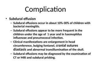
Complication
• Subdural effusion
–Subdural effusions occur in about 10%-30% of children with
bacterial meningitis.
– Subdural effusions appear to be more frequent in the
children under the age of 1 year and in haemophilus
influenzae and pneumococal infection.
– Clinical manifestations are enlargement in head
circumference, bulging fontanel, cranial sutures
diastasis and abnormal transillumination of the skull.
– Subdural effusions may be diagnosed by the examination of
CT or MRI and subdural pricking.
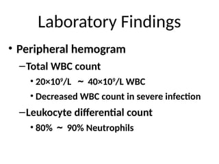
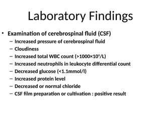
Laboratory Findings
• Examinationof cerebrospinal fluid (CSF)
– Increased pressure of cerebrospinal fluid
– Cloudiness
– Increased total WBC count (>1000×109
/L)
– Increased neutrophils in leukocyte differential count
– Decreased glucose (<1.1mmol/l)
– Increased protein level
– Decreased or normal chloride
– CSF film preparation or cultivation : positive result
51.
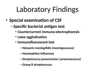
Laboratory Findings
• Specialexamination of CSF
–Specific bacterial antigen test
• Countercurrent immuno-electrophoresis
• Latex agglutination
• Immunofluorescent test
– Neisseria meningitidis (meningococcus)
– Haemophilus influenzae
– Streptococcus pneumoniae ( pneumococcus)
– Group B streptococcus
52.
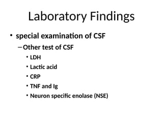
Laboratory Findings
• specialexamination of CSF
–Other test of CSF
• LDH
• Lactic acid
• CRP
• TNF and Ig
• Neuron specific enolase (NSE)
53.
Laboratory Findings
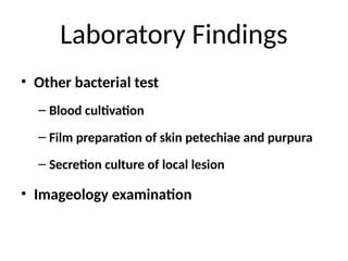
• Otherbacterial test
– Blood cultivation
– Film preparation of skin petechiae and purpura
– Secretion culture of local lesion
• Imageology examination
54.
Diagnosis
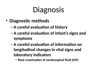
• Diagnostic methods
–Acareful evaluation of history
–A careful evaluation of infant’s signs and
symptoms
–A careful evaluation of information on
longitudinal changes in vital signs and
laboratory indicators
• Rout examination of cerebrospinal fluid (CSF)
55.
Differential diagnosis
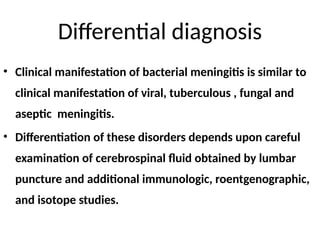
• Clinicalmanifestation of bacterial meningitis is similar to
clinical manifestation of viral, tuberculous , fungal and
aseptic meningitis.
• Differentiation of these disorders depends upon careful
examination of cerebrospinal fluid obtained by lumbar
puncture and additional immunologic, roentgenographic,
and isotope studies.
56.
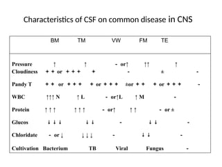
Characteristics of CSFon common disease in CNS
BM TM VW FM TE
Pressure ↑ ↑ - or↑ ↑↑ ↑
Cloudiness ++ or +++ + - ± -
Pandy T ++ or +++ + or +++ ±or ++ + or +++ -
WBC ↑↑↑ N ↑ L - or↑L ↑ M -
Protein ↑ ↑ ↑ ↑ ↑ ↑ - or↑ ↑ ↑ - or ±
Glucos ↓ ↓ ↓ ↓ ↓ - ↓ ↓ -
Chloridate - or ↓ ↓ ↓ ↓ - ↓ ↓ -
Cultivation Bacterium TB Viral Fungus -
57.
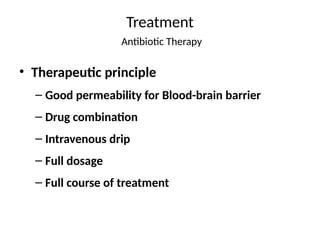
Treatment
Antibiotic Therapy
• Therapeuticprinciple
– Good permeability for Blood-brain barrier
– Drug combination
– Intravenous drip
– Full dosage
– Full course of treatment
58.
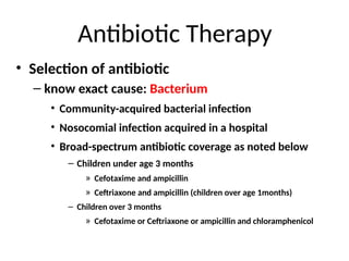
Antibiotic Therapy
• Selectionof antibiotic
– know exact cause: Bacterium
• Community-acquired bacterial infection
• Nosocomial infection acquired in a hospital
• Broad-spectrum antibiotic coverage as noted below
– Children under age 3 months
» Cefotaxime and ampicillin
» Ceftriaxone and ampicillin (children over age 1months)
– Children over 3 months
» Cefotaxime or Ceftriaxone or ampicillin and chloramphenicol
59.
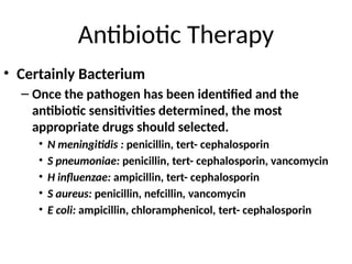
Antibiotic Therapy
• CertainlyBacterium
– Once the pathogen has been identified and the
antibiotic sensitivities determined, the most
appropriate drugs should selected.
• N meningitidis : penicillin, tert- cephalosporin
• S pneumoniae: penicillin, tert- cephalosporin, vancomycin
• H influenzae: ampicillin, tert- cephalosporin
• S aureus: penicillin, nefcillin, vancomycin
• E coli: ampicillin, chloramphenicol, tert- cephalosporin
60.
Antibiotic Therapy
• Courseof treatment
– 7 days for meningococcal infection
– 10 ~ 14 days for H influenzae or S pneumoniae
infection
– More than 21 days for S aureus or E coli infection
– 14 ~ 21 days for other organisms
61.
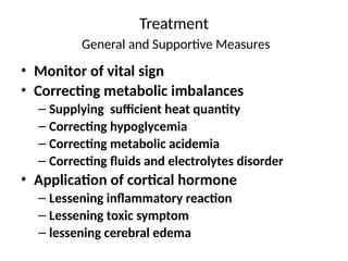
Treatment
General and SupportiveMeasures
• Monitor of vital sign
• Correcting metabolic imbalances
– Supplying sufficient heat quantity
– Correcting hypoglycemia
– Correcting metabolic acidemia
– Correcting fluids and electrolytes disorder
• Application of cortical hormone
– Lessening inflammatory reaction
– Lessening toxic symptom
– lessening cerebral edema
62.
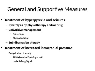
General and SupportiveMeasures
• Treatment of hyperpyrexia and seizures
– Pyretolysis by physiotherapy and/or drug
– Convulsive management
• Diazepam
• Phenobarbital
– Subhibernation therapy
• Treatment of increased intracranial pressure
– Dehydration therapy
• 20%Mannitol 5ml/kg vi q6h
• Lasix 1-2mg/kg vi
63.
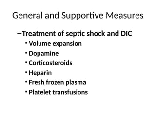
General and SupportiveMeasures
–Treatment of septic shock and DIC
• Volume expansion
• Dopamine
• Corticosteroids
• Heparin
• Fresh frozen plasma
• Platelet transfusions
64.
Treatment
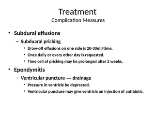
Complication Measures
• Subduraleffusions
– Subduaral pricking
• Draw-off effusions on one side is 20-30ml/time.
• Once daily or every other day is requested.
• Time cell of pricking may be prolonged after 2 weeks.
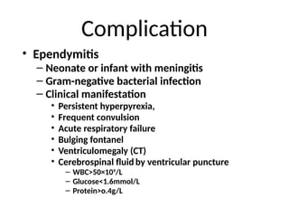
• Ependymitis
– Ventricular puncture — drainage
• Pressure in ventricle be depressed.
• Ventricular puncture may give ventricle an injection of antibiotic.
65.
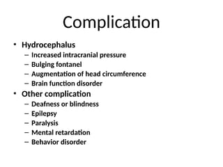
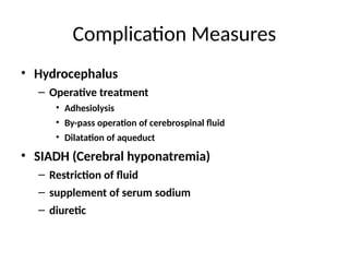
Complication Measures
• Hydrocephalus
–Operative treatment
• Adhesiolysis
• By-pass operation of cerebrospinal fluid
• Dilatation of aqueduct
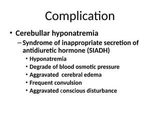
• SIADH (Cerebral hyponatremia)
– Restriction of fluid
– supplement of serum sodium
– diuretic
66.
Prognosis
• Appropriate antibiotictherapy reduces the
mortality rate for bacterial meningitis in
children, but mortality remain high.
• Overall mortality in the developed countries
ranges between 5% and 30%.
• 50 percent of the survivors have some sequelae
of the disease.
67.
Prognosis
• Prognosis dependsupon many factors:
– Age
– Causative organism
– Number of organisms and bacterial virulence
– Duration of illness prior to effective antibiotic therapy
– Presence of disorders that may compromise host
response to infection
68.
Aseptic Meningitis
Definition: Asyndrome characterized by acute onset of meningeal symptoms,
fever, and cerebrospinal fluid pleocytosis, with bacteriologically sterile
cultures.
Laboratory criteria for diagnosis:
CSF showing ≥ 5 WBC/cu mm
No evidence of bacterial or fungal meningitis.
Case classification
Confirmed: a clinically compatible illness diagnosed by a physician as aseptic
meningitis, with no laboratory evidence of bacterial or fungal meningitis
Comment
Aseptic meningitis is a syndrome of multiple etiologies, but most cases are
caused by a viral agent
69.
Viral Meningitis
Etiological Agents:
Enteroviruses(Coxsackie's and echovirus): most common.
-Adenovirus
-Arbovirus
-Measles virus
-Herpes Simplex Virus
-Varicella
Reservoirs:
-Humans for Enteroviruses, Adenovirus, Measles, Herpes Simplex, and Varicella
-Natural reservoir for arbovirus birds, rodents etc.
Modes of transmission:
-Primarily person to person and arthopod vectors for Arboviruses
Incubation Period:
-Variable. For enteroviruses 3-6 days, for arboviruses 2-15 days
Treatment: No specific treatment available.
Most patients recover completely on their own.