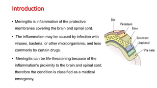
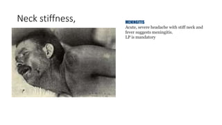
2. Introduction
• Meningitis is inflammation of the protective
membranes covering the brain and spinal cord.
• The inflammation may be caused by infection with
viruses, bacteria, or other microorganisms, and less
commonly by certain drugs.
• Meningitis can be life-threatening because of the
inflammation's proximity to the brain and spinal cord;
therefore the condition is classified as a medical
emergency.
3. Causes
• Meningitis is usually caused by infection from viruses or
microorganisms.
• Most cases are due to infection with viruses, with bacteria, fungi, and
parasites being the next most common causes.
• It may also result from various non-infectious causes.
4. Bacterial cause
The types of bacteria that cause bacterial meningitis vary by age
group.
• In premature babies and newborns up to three months old,
common causes are group B streptococci a
• Escherichia coli
• Listeria monocytogenes .
Older children are more commonly affected by
• Neisseria meningitidis (meningococcus),
• Streptococcus pneumoniae
• Haemophilus influenzae type B
5. Bacterial cont’d
In adults,
• N. meningitidis and
• S. pneumoniae
Since the pneumococcal vaccine was introduced, however,
rates of pneumococcal meningitis have declined in children and
adults.
• Recent trauma to the skull gives bacteria in the nasal cavity the
potential to enter the meningeal space
6. Bacterial cont’d
• Individuals with a cerebral shunt or related device are increased
the risk of infection through those devices. In these cases,
infections with staphylococci are more likely.
• In a small proportion of people, an infection in the head and
neck area, such as otitis media or mastoiditis, can lead to
meningitis
• Recipients of cochlear implants for hearing loss are at an
increased risk of pneumococcal meningitis.
• Tuberculous meningitis, meningitis due to infection with
Mycobacterium tuberculosis, is more common in those from
countries where tuberculosis is common, but is also
encountered in those with immune problems, such as AIDS.
7. Viral & Parasitic
Viruses that can cause meningitis include
• enteroviruses
• herpes simplex virus type 2 (and less commonly type 1),
• varicella zoster virus (known for causing chickenpox and shingles),
• mumps virus
• HIV
• Parasitic
• A parasitic cause is often assumed when there is a predominance of eosinophils
(a type of white blood cell) in the CSF.
• The most common parasites implicated are
• -Angiostrongylus cantonensis, - Gnathostoma spinigerum,
• -Schistosoma,
8. Non-infectious
• Meningitis may occur as the result of several non-infectious causes:
• spread of cancer to the meninges (malignant or neoplastic meningitis)
• certain drugs (mainly non-steroidal anti-inflammatory drugs, antibiotics and
intravenous immunoglobulins).
o It may also be caused by several inflammatory conditions such as
sarcoidosis (which is then called neurosarcoidosis),
oconnective tissue disorders such as systemic lupus erythematosus
o certain forms of vasculitis (inflammatory conditions of the blood vessel
wall) such as Behçet's disease.
• Epidermoid cysts and dermoid cysts may cause meningitis by releasing
irritant matter into the subarachnoid space.
• Rarely, migraine may cause meningitis.
9. Mechanism
The meninges comprise three membranes that, together with the cerebrospinal
fluid, enclose and protect the brain and spinal cord (the central nervous
system).
• The pia mater is a very delicate impermeable membrane that firmly adheres
to the surface of the brain, following all the minor contours.
• The arachnoid matter (so named because of its spider-web-like appearance) is
a loosely fitting sac on top of the pia mater.
• The subarachnoid space separates the arachnoid and pia mater membranes,
and is filled with cerebrospinal fluid.
• The outermost membrane, the dura mater, is a thick durable membrane,
which is attached to both the arachnoid membrane and the skull.
10. Mechanism cont’d
• In bacterial meningitis, bacteria reach the meninges by one of two main routes: through the
bloodstream or through direct contact between the meninges and either the nasal cavity or
the skin.
• In most cases, meningitis follows invasion of the bloodstream by organisms that live upon
mucous surfaces such as the nasal cavity.
• This is often in turn preceded by viral infections, which break down the normal barrier
provided by the mucous surfaces.
• Once bacteria have entered the bloodstream, they enter the subarachnoid space in places
where the blood-brain barrier is vulnerable—such as the choroid plexus.
• Meningitis occurs in 25% of newborns with bloodstream infections due to group B
streptococci; this phenomenon is less common in adults.
• Direct contamination of the cerebrospinal fluid may arise from indwelling devices, skull
fractures, or infections of the nasopharynx or the nasal sinuses that have formed a tract with
the subarachnoid space ; (occasionally, congenital defects of the dura mater can be
identified.)
11. Mechanism cont’d
• The large-scale inflammation that occurs in the subarachnoid
space during meningitis is not a direct result of bacterial
infection but can rather largely be attributed to the response of
the immune system to the entrance of bacteria into the central
nervous system.
• When components of the bacterial cell membrane are identified
by the immune cells of the brain (astrocytes and microglia), they
respond by releasing large amounts of cytokines, hormone-like
mediators that recruit other immune cells and stimulate other
tissues to participate in an immune response.
• The blood-brain barrier becomes more permeable, leading to
"vasogenic" cerebral edema (swelling of the brain due to fluid
12. Mechanism cont’d
• Large numbers of white blood cells enter the CSF, causing
inflammation of the meninges, and leading to "interstitial"
edema (swelling due to fluid between the cells).
• In addition, the walls of the blood vessels themselves become
inflamed (cerebral vasculitis), which leads to a decreased blood
flow and a third type of edema, "cytotoxic" edema.
• The three forms of cerebral edema all lead to an increased
intracranial pressure; together with the lowered blood pressure
often encountered in acute infection, this means that it is harder
for blood to enter the brain, and brain cells are deprived of
oxygen and undergo apoptosis (automated cell death).
13. Signs and symptoms
Clinical features
In adults,
• A severe headache is the most common symptom of meningitis,
• Nuchal rigidity (inability to flex the neck forward passively due to
increased neck muscle tone and stiffness).
• The classic triad of diagnostic signs consists of nuchal rigidity, sudden
high fever, and altered mental status; however, all three features are
present in only 44–46% of all cases of bacterial meningitis.
If none of the three signs is present, meningitis is extremely unlikely.
Other signs commonly associated with meningitis include:
• Photophobia (intolerance to bright light) and
• Phonophobia (intolerance to loud noises).
15. Signs and symptoms cont’d
• Small children often do not exhibit the aforementioned symptoms, and may only
be irritable and look unwell.
• In infants up to 6 months of age, bulging of the fontanelle (the soft spot on top of
a baby's head) may be present.
• Other features that might distinguish meningitis from less severe illnesses in
young children are leg pain, cold extremities, and an abnormal skin color
16. Signs and symptoms cont’d
• Nuchal rigidity occurs in 70% of adult cases of bacterial meningitis.
• Other signs of meningism include the presence of positive Kernig's sign or Brudzinski's sign.
• Kernig's sign is assessed with the patient lying supine, with the hip and knee flexed to 90 degrees.
In a patient with a positive Kernig's sign, pain limits passive extension of the knee.
• A positive Brudzinski's sign occurs when flexion of the neck causes involuntary flexion of the knee
and hip.
• Although Kernig's sign and Brudzinski’s neck sign are both commonly used to screen for meningitis,
the sensitivity of these tests is limited.
• They have very good specificity for meningitis: the signs rarely occur in other diseases.
• Another test, known as the "jolt accentuation maneuver" helps determine whether meningitis is
present in patients reporting fever and headache. The patient is told to rapidly rotate his or her head
horizontally; if this make the headache worse, it shows meningitis.
19. Signs and symptoms cont’d
• Meningitis caused by the bacterium Neisseria meningitidis
(meningococcal meningitis) can be differentiated from
meningitis with other causes by a rapidly spreading
petechial rash which may precede other symptoms.
• The rash consists of numerous small, irregular purple or
red spots on the trunk, lower extremities, mucous
membranes, conjuctiva, and (occasionally) the palms of the
hands or soles of the feet.
• The rash is typically non-blanching: the redness does not
disappear when pressed with a finger.
20. Signs and symptoms cont’d
• The inflammation of the meninges may lead to abnormalities of
the cranial nerves, a group of nerves arising from the brain stem
that supply the head and neck area and control eye movement,
facial muscles and hearing, among other functions.
• Visual symptoms and hearing loss may persist after an episode of
meningitis .
• Inflammation of the brain (encephalitis) or its blood vessels
(cerebral vasculitis), as well as the formation of blood clots in the
veins (cerebral venous thrombosis), may all lead to weakness, loss
of sensation, or abnormal movement or function of the part of the
body supplied by the affected area in the brain.
21. Diagnosis
Type of meningitis Glucose Protein Cells
Acute bacterial Low High PMNs, often > 300/mm³
Acute viral Normal
Normal or
high
mononuclear, < 300/mm³
Tuberculous Low High
mononuclear and
PMNs, < 300/mm³
Fungal Low High < 300/mm³
Malignant Low High Usually mononuclear
Analysis of Cerebrospinal Fluid (CSF) Assessment of CSF is critical
for patients with suspected meningitis or encephalitis
CSF findings in different forms of meningitis
22. Treatment
Initial treatment
• Meningitis is potentially life-threatening and has a high mortality
rate if untreated.
• delay in treatment has been associated with a poorer outcome.
• Thus treatment with wide-spectrum antibiotics should not be
delayed while confirmatory tests are being conducted.
• If meningococcal disease is suspected in primary care, guidelines
recommend that benzylpenicillin be administered before transfer to
hospital.
• Intravenous fluids should be administered if hypotension (low blood
pressure) or shock are present.
• Given that meningitis can cause a number of early severe
complications, regular medical review is recommended to identify
these complications early as well as admission to an intensive care
unit if necessary.
23. Treatment cont’d
• Mechanical ventilation may be needed if the level of
consciousness is very low, or if there is evidence of respiratory
failure.
• If there are signs of raised intracranial pressure, measures to
monitor the pressure may be taken; this would allow the
optimization of the cerebral perfusion pressure and various
treatments to decrease the intracranial pressure with medication
(e.g. mannitol). Seizures are treated with anticonvulsants.
• Hydrocephalus (obstructed flow of CSF) may require insertion of
a temporary or long-term drainage device, such as a cerebral
shunt.
24. Treatment cont’d
• One of the third-generation cefalosporin antibiotics recommended for
the initial treatment of bacterial meningitis in young children and those
over 50 years of age, as well as those who are immunocompromised,
addition of ampicillin is recommended to cover Listeria monocytogenes
• Tuberculous meningitis requires prolonged treatment with antibiotics
• Steroids: Adjuvant treatment with corticosteroids (usually
dexamethasone) has been shown in some studies to reduce rates of
mortality
• Viral meningitis typically requires supportive therapy only; most viruses
responsible for causing meningitis are not amenable to specific
treatment.
• Fungal meningitis, such as cryptococcal meningitis, is treated with long
courses of highly dosed antifungals, such as amphotericin B and
flucytosineb
25. Physiotherapy management
Can be divided into two phase.
•Acute stage
•Recovery stage
Physiotherapy treatment may also vary weather
patient is :
•Adult
•child
26. Aim of physiotherapy management in acute
stage (in ICU)
• Maintain a clear airway
• Prevent accumulation of secretion
• To maintain proper ventilation
• To maintain proper oxygenation of blood
• To maintain full range of motion in all joint and prevent any
deformity
• To prevent DVT
• To facilitate tone and contracture of muscle
27. Aim of physiotherapy management in
after discharge from ICU
• To give psychological support, To reduce pain
• Continuation of chest care
• Maintain and improve joint range of motion
• Strengthening program, Balance training
• Re-education of patient
• Ambulation, Orthotic management
• To improve endurance
• Functional independence