
Download to read offline
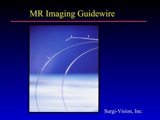
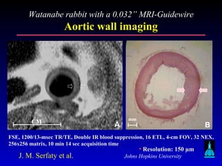

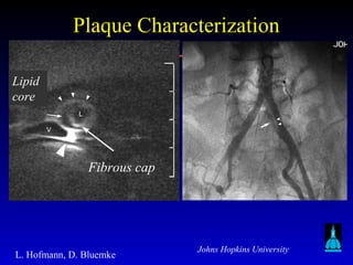
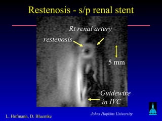
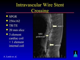
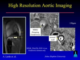
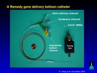
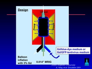
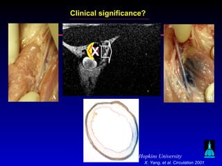
This document discusses vascular interventions guided by MRI, including: 1) Early experiments with intravascular MRI to image arteries and plaques. 2) The technical challenges of MRI-guided balloon angioplasty and stent placement, such as developing MRI-compatible devices and monitoring procedures. 3) Examples of MRI-guided balloon angioplasty, stent placement, and plaque imaging in human studies. 4) The potential for MRI to guide new treatments like gene therapy delivery to diseased arteries.


![PERI-PROSTHETIC FRACTURE NAIL-PLATE CONSTRUCT [NPC].pptx](https://cdn.slidesharecdn.com/ss_thumbnails/drarunkumardrmohamedashrafperiprostheticfrasturenail-plateconstructnpc-260209164459-7e9d15a1-thumbnail.jpg?width=640&height=640&fit=bounds)


