2. Introduction, properties, nomenclature and IUB classification of
enzymes; Enzyme kinetics (Michaelis plot, Line Weaver Burke plot);
Enzyme inhibitors with examples; Regulation of enzymes: enzyme
induction and repression, allosteric enzymes regulation; Therapeutic and
diagnostic applications of enzymes and isoenzymes; Coenzymes –
Structure and biochemical functions.
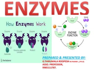
Enzymes may be defined as biocatalysts synthesized by living cells.
They are protein in nature (exception – RNA acting as ribozyme),
colloidal and thermolabile in character, and specific in their action.
3. Berzelius in 1836 coined the term catalysis (Greek : to
dissolve).
In 1878, Kuhne used the word enzyme (Greek : in yeast)
to indicate the catalysis taking place in the biological
systems. Isolation of enzyme system from cell-free extract
of yeast was achieved in 1883 by Buchner.
He named the active principle as zymase (later found to
contain a mixture of enzymes),which could convert sugar
to alcohol.
In 1926,James Sumner first achieved the isolation and
crystallization of the enzyme urease from jack bean and
identified it as a protein.
HISTORICAL BACKGROUND
4. Introduction
Enzymes are biocatalysts – the catalysts of life. A catalyst
is defined as a substance that increases the velocity or
rate of a chemical reaction without itself undergoing any
change in the overall process.
The student-teacher relationship may be a good
example to understand how a catalyst works.
5. NOMENCLATURE AND CLASSIFICATION
Enzymes are sometimes considered under two broad categories :
(a) Intracellular enzymes –They are functional within cells where they
aresynthesized.
(b) Extracellular enzymes – These enzymes are active outside the cell;
all the digestive enzymes belong to this group.
6. 1. Oxidoreductases : Enzymes involved in oxidation-reduction reactions.
2. Transferases : Enzymes that catalyse the transfer of functional
groups.
3. Hydrolases : Enzymes that bring about hydrolysis of various
compounds.
4. Lyases : Enzymes specialised in the addition or removal of water,
ammonia, CO2 etc.
5. Isomerases : Enzymes involved in all the isomerization reactions.
6. Ligases : Enzymes catalysing the synthetic reactions (Greek : ligate—
to bind) where two molecules are joined together and ATP is used.
[The word OTHLIL (first letter in each class) may be
memorised to remember the six classes of enzymes in the
correct order].
Each class in turn is subdivided into many sub-classes which are further divided. A four
digit Enzyme Commission (E.C.) number is assigned to each enzyme representing the
class (first digit), sub-class (second digit), sub-sub class (third digit) and the individual
enzyme (fourth digit). Each enzyme is given a specific name indicating the substrate,
coenzyme (if any) and the type of the reaction catalysed by the enzyme.
ENZYMES CLASSIFICATION
7. 1. Oxidoreductases Oxidation Reduction AH2 + B A + BH2
Alcohol dehydrogenase (alcohol : NAD+ oxidoreductase E.C. 1.1.1.1.), cytochrome
oxidase, L- and D-amino acid oxidases
2. Transferases Group transfer A – X + B A + B – X
Hexokinase (ATP : D-hexose 6-phosphotransferase, E.C. 2.7.1.1.), transaminases,
transmethylases, phosphorylase
3. Hydrolases Hydrolysis A – B + H2O AH + BOH
Lipase (triacylglycerol acyl hydrolase E.C. 3.1.1.3), choline esterase, acid
and alkaline phosphatases, pepsin, urease
4. Lyases Addition Elimination A – B + X – Y AX – BY
Aldolase (ketose 1-phosphate aldehyde lyase, E.C. 4.1.2.7), fumarase, histidase
5. Isomerases Interconversion of isomers A A-
Triose phosphate isomerase (D-glyceraldehyde 3-phosphate ketoisomerase, E.C. 5.3.1.1), retinol
isomerase, phosphohexose isomerase
6. Ligases Condensation (usually dependent on ATP) A + B ATP ADP + Pi A – B
Glutamine synthetase (L-glutamate ammonia ligase, E.C. 6.3.1.2),
acetyl CoA carboxylase, succinate thiokinase *For one enzyme in each class, systematic name
along with E.C. number is given in the brackets.
Classification of enzymes Enzyme class with examples Reaction
catalysed
8. CHEMICAL NATURE AND PROPERTIES OF ENZYMES
The functional unit of the enzyme is known as holoenzyme which is often made up of
apoenzyme (the protein part) and a coenzyme (non-protein organic part).
Holoenzyme Apoenzyme + Coenzyme
(active enzyme) (protein part) (non-protein part)
The term prosthetic group is used when the
non-protein moiety tightly (covalently) binds
with the apoenzyme.
**The coenzyme can be separated by
dialysis from the enzyme while the
prosthetic group cannot be.
The word monomeric
enzyme is used if it is made up of a
single polypeptide e.g. ribonuclease,
trypsin.
Some of the enzymes which possess more than one polypeptide (subunit) chain are known
as oligomeric enzymes e.g. lactate dehydrogenase, aspartate transcarbamoylase etc.
There are certain multienzyme complexes possessing specific sites to catalyse different
reactions in a sequence.
9. FACTORS AFFECTING ENZYME ACTIVITY
The important factors that influence the
velocity of the enzyme reaction are discussed
hereunder
1.Concentration of enzyme
2.Concentration of substrate
3.Effect of temperature
4.Effect of PH
5.Effect of product concentration
6.Effect of activators
7.Effect of time
8.Effect of light and radiation
10. 1.Concentration of enzyme
As the concentration of the enzyme is increased, the velocity of the reaction proportionately
increases (Fig.6.1).
2. Concentration of substrate
Increase in the substrate concentration gradually increases the velocity of enzyme reaction
within the limited range of substrate levels. A rectangular hyperbola is obtained when velocity
is plotted against the substrate concentration (Fig.6.2).
Three distinct phases of the reaction are observed in the graph (A-linear; B-curve; C-almost
unchanged).
11. Enzyme kinetics and Km value
The enzyme (E) and substrate (S) combine with each other to form an unstable enzymesubstrate
complex (ES) for the formation of product (P).
Here k1, k2 and k3 represent the velocity constants for the respective reactions, as indicated by
arrows.
Km, the Michaelis-Menten constant (or Brig’s and Haldane’s constant), is given by the formula
The following equation is obtained after suitable algebraic manipulation.
where v = Measured velocity,
Vmax = Maximum velocity,
S = Substrate concentration,
Km = Michaelis – Menten constant.
Let us assume that the measured velocity (v) is equal to 1/2 Vmax. Then the equation (1) may
be substituted as follows
12. K stands for a constant and m stands for Michaelis (in Km).
Km or the Michaelis-Menten constant is defined as the substrate concentration
(expressed in moles/l) to produce half-maximum velocity in an enzyme catalysed
reaction. It
indicates that half of the enzyme molecules (i.e. 50%) are bound with the substrate
molecules
when the substrate concentration equals the Km value.
Km value is a constant and a characteristic feature of a given enzyme (comparable to
a thumb impression or signature). It is a representative for measuring the strength of
ES complex.
A low Km value indicates a strong affinity between enzyme and substrate, whereas a
high Km value reflects a weak affinity between them. For majority of enzymes, the Km
values are in the range of 10–5 to 10–2 moles. It may however, be noted that Km is not
dependent on the concentration of enzyme.
13. Lineweaver-Burk double reciprocal plot : For the
determination of Km value, the substrate saturation curve (Fig.6.2) is not very accurate since
Vmax is approached asymptotically. By taking the reciprocals of the equation (1), a straight line
graphic representation is obtained.
The above equation is similar to y = ax + b.
Therefore, a plot of the reciprocal of the velocity [1/v] vs. the reciprocal of the substrate
concentration [1/s] gives a straight line.
Here the slope is Km/Vmax and whose y intercept is 1/Vmax.
The Lineweaver-Burk plot is shown in Fig.6.3.
It is much easier to calculate the Km from the intercept on x-axis which is –(1/Km). Further, the
double reciprocal plot is useful in understanding the effect of various inhibitions .
Enzyme reactions with two or moresubstrates : The above discussion is based on the
presumption of a single substrate-enzyme reaction. In fact, a majority of the enzymecatalysed
reactions involve two or more substrates. Even in case of multisubstrate enzymes, despite the
complex mathematical expressions, the fundamental principles conform to Michaelis-Menten
Kinetics.
14. 3. Effect of temperature
Velocity of an enzyme reaction increases with increase in temperature up to a maximum and
then declines. A bell-shaped curve is usually observed (Fig.6.4).
Temperature coefficient or Q10 is defined as increase in enzyme velocity when
the temperature is increased by 10°C. For a majority of enzymes, Q10 is 2 between 0°C and
40°C. Increase in temperature results in higher activation energy of the molecules and more
molecular (enzyme and substrate) collision and interaction for the reaction to proceed faster.
The optimum temperature for most of the enzymes is between 35°C–40°C. However, a few
enzymes (e.g. Taq DNA polymerase, muscle adenylate kinase) are active even at 100°C.
Some plant enzymes like urease have optimum activity around 60°C. This may be
due to very stable structure and conformation of these enzymes.In general, when the enzymes
are exposed to a temperature above 50°C, denaturation leading to derangement in the native
(tertiary) structure of the protein and active site are seen.
Majority of the enzymes become inactive at higher temperature (above 70°C).
Clinical significance : Foods can be preserved in refrigerators (at low temperatures)
due to reduced bacterial enzyme activities. Certain surgeries are carried out by
lowering the patient’s body temperature (induced hyporthermia), and thus the
metabolic rate.
15. 4. Effect of pH
Increase in the hydrogen ion concentration (pH) considerably influences the enzyme
activity and a bell-shaped curve is normally obtained (Fig.6.5).
Each enzyme has an optimum pH at which the velocity is maximum. Below and above
this pH, the enzyme activity is much lower and at extreme pH, the enzyme becomes
totally inactive.
Most of the enzymes of higher organisms show optimum activity around neutral pH
(6-8).
There are, however, many exceptions like pepsin (1-2), acid phosphatase (4-5) and
alkaline phosphatase (10-11). Enzymes from fungi and plants are most active in acidic
pH (4-6).
Hydrogen ions influence the enzyme activity by altering the ionic charges on the amino
acids (particularly at the active site), substrate, ES complex etc.
16. 5. Effect of product concentration
The accumulation of reaction products generally decreases the enzyme
velocity.
6. Effect of activators
Some of the enzymes require certain inorganic metallic cations like Mg2+, Mn2+,
Zn2+, Ca2+, Co2+, Cu2+, Na+, K+ etc. for their optimum activity. Rarely, anions are also
needed for enzyme activity e.g. chloride ion (Cl–) for amylase. Metals function as
activators of enzyme velocity through various mechanisms— combining with the
substrate, formation of ES-metal complex, direct participation in the reaction and
bringing a conformational change in the enzyme.
Two categories of enzymes requiring metals for their activity are distinguished
l. Metal-activated enzymes : The metal is not tightly held by the enzyme and can be
exchanged easily with other ions.
e.g. ATPase (Mg2+ and Ca2+) Enolase (Mg2+)
Il. Metalloenzymes : These enzymes hold the metals rather tightly which are not
readily exchanged. e.g. alcohol dehydrogenase, carbonic anhydrase, alkaline
phosphatase, carboxypeptidase and aldolase contain zinc.
Phenol oxidase (copper);
Pyruvate oxidase (manganese);
Xanthine oxidase (molybdenum);
Cytochrome oxidase (iron and copper).
17. 7. Effect of time
Under ideal and optimal conditions (like Ph,temperature etc.),
the time required for an enzyme reaction is less. Variations in
the time of the reaction are generally related to the
alterations in pH and temperature.
8. Effect of light and radiation
Exposure of enzymes to ultraviolet, beta, gamma and
X-rays inactivates certain enzymes due to the
formation of peroxides. e.g. UV rays inhibit salivary
amylase activity.
18.
19. ACTIVE SITE
Salient features of active site
1. The existence of active site is due to the tertiary structure
of protein resulting in three dimensional native conformation.
2. The active site is made up of amino acids (known as
catalytic residues) which are far from each other in the linear
sequence of amino acids (primary structure of protein).
For instance, the enzyme lysozyme has 129 amino acids. The
active site is formed by the contribution of amino acid
residues numbered 35, 52, 62, 63 and 101.
3. Active sites are regarded as clefts or crevices or pockets
occupying a small region in a big enzyme molecule.
The active site (or active centre) of an enzyme
represents as the small region at which the
substrate(s) binds and participates in the catalysis.
20. 4. The active site is not rigid in structure and shape. It is rather flexible
to promote the specific substrate binding.
5. Generally, the active site possesses a substrate binding site and a
catalytic site. The latter is for the catalysis of the specific reaction.
6. The coenzymes or cofactors on which some enzymes depend are
present as a part of the catalytic site.
7. The substrate(s) binds at the active site by weak non-covalent bonds.
8. Enzymes are specific in their function due to the existence of active
sites.
9. The commonly found amino acids at the active sites are serine,
aspartate, histidine, cysteine, lysine, arginine, glutamate, tyrosine etc.
Among these amino acids, serine is the most frequently found.
10. The substrate[S] binds the enzyme (E) at the active site to form
enzyme-substrate complex
(ES). The product (P) is released after the catalysis and the enzyme is
available for reuse.
E + S ES E + P
21. MECHANISM OF ENZYME ACTION
Enzyme-substrate complex formation
The prime requisite for enzyme catalysis is that the substrate (S) must
combine with the enzyme (E) at the active site to form enzyme
substrate complex (ES) which ultimately results in the product
formation (P).
Lock and key
model
or Fischer’s
template theory
Induced fit
theory
or Koshland’s
model
Substrate
strain
theory
MECHANISM OF ENZYME ACTION
E + S ES E + P
22. Lock and key model or Fischer’s template theory
According to this model, the structure or conformation of the enzyme is rigid. The
substrate fits to the binding site (now active site) just as a key fits into the proper lock
or a hand into the proper glove. Thus the active site of an enzyme is a rigid and pre-
shaped template where only a specific substrate can bind. This model does not give
any scope for the flexible nature of enzymes, hence the model totally fails to explain
many facts of enzymatic reactions, the most important being the effect of allosteric
modulators.
23. Induced fit theory or Koshland’s model
Koshland, in 1958, proposed a more acceptable and realistic model for enzyme
substrate complex formation. As per this model, the active site is not rigid and pre-
shaped. The essential features of the substrate binding site are present at the nascent
active site. The interaction of the substrate with the enzyme induces a fit or a
conformation change in the enzyme, resulting in the formation of a strong substrate
binding site. Further, due to induced fit, the appropriate amino acids of the enzyme
are repositioned to form the active site and bring about the catalysis. Induced fit
model has sufficient experimental evidence from the X-ray diffraction studies.
Koshland’s model also explains the action of allosteric modulators and competitive
inhibition on enzymes.
24. Substrate strain theory
In this model, the substrate is strained due to the induced conformation
change in the enzyme. It is also possible that when a substrate binds to
the preformed active site, the enzyme induces a strain to the substrate.
The strained substrate leads to the formation of product. In fact, a
combination of the induced fit model with the substrate strain is
considered to be operative in the enzymatic action.
25.
26. Enzyme inhibitor is defined as a substance which binds with the enzyme and brings about
a decrease in catalytic activity of that enzyme.
The inhibitor may be organic or inorganic in nature. There are three broad categories of enzyme
inhibition
1. Reversible inhibition.
2. Irreversible inhibition.
3. Allosteric inhibition.
1. Reversible inhibition
The inhibitor binds non-covalently with enzyme and the enzyme inhibition can be reversed if the
inhibitor is removed. The reversible inhibition is further sub-divided into
I. Competitive inhibition (Fig.6.7A) II. Non-competitive inhibition (Fig.6.7B)
27. 1.Competitive inhibition :
The inhibitor (I) which closely resembles the real substrate (S) is regarded as a substrate
analogue. The inhibitor competes with substrate and binds at the active site of the enzyme
but does not undergo any catalysis. As long as the competitive inhibitor holds the active
site, the enzyme is not available for the substrate to bind. During the reaction, ES and EI
complexes are formed as shown below
The relative concentration of the substrate and inhibitor and their respective affinity with the
enzyme determines the degree of competitive inhibition. The inhibition could be overcome by
a high substrate concentration. In competitive inhibition, the Km value increases whereas
Vmax remains unchanged (Fig.6.8).
The enzyme succinate dehydrogenase (SDH) is a classical example of competitive inhibition with
succinic acid as its substrate. The compounds, namely, malonic acid, glutaric acid and oxalic acid,
have structural similarity with succinic acid and compete with the substrate for binding at the
active site of SDH.
Methanol is toxic to the body when it is converted to formaldehyde by the enzyme alcohol
dehydrogenase (ADH). Ethanol can compete with methanol for ADH. Thus, ethanol can be used
in the treatment of methanol poisoning.
Some more examples of the enzymes with substrates and competitive inhibitors (of clinical and
pharmacological significance) are given in Table 6.2.
Competitive inhibition
28.
29. The enzyme succinate dehydrogenase (SDH) is a classical example of competitive
inhibition with succinic acid as its substrate. The compounds, namely, malonic
acid, glutaric acid and oxalic acid, have structural similarity with succinic acid and
compete with the substrate for binding at the active site of SDH.
Competitive inhibition
Competitive inhibition
30. II. Non-competitive inhibition : The inhibitor binds at a site other than the active site on the
enzyme surface. This binding impairs the enzyme function. The inhibitor has no structural
resemblance with the substrate. However, there usually exists a strong affinity for the inhibitor
to bind at the second site. In fact, the inhibitor does not interfere with the enzyme-substrate
binding. But the catalysis is prevented, possibly due to a distortion in the enzyme conformation.
The inhibitor generally binds with the enzyme as well as the ES complex. The overall relation in
non-competitive inhibition is represented below
For non-competitive inhibition, the Km value is unchanged while Vmax is
lowered (Fig.6.9).
Heavy metal ions (Ag+, Pb2+, Hg2+ etc.) can non-competitively inhibit the enzymes by binding
with cysteinyl sulfhydryl groups. The general reaction for Hg2+ is shown below.
Heavy metals also lead to the formation of covalent bonds with carboxyl groups and histidine,
often resulting in irreversible inhibition.
Non-competitive inhibition
31. Non-competitive inhibition
Heavy metals also lead to the formation of covalent bonds with carboxyl groups
and histidine, often resulting in irreversible inhibition.
32. 2. Irreversible inhibition
The inhibitors bind covalently with the enzymes and
inactivate them, which is irreversible. These inhibitors are
usually toxic substances that poison enzymes. Iodoacetate is
an irreversible inhibitor of the enzymes like papain and
glyceraldehyde 3-phosphate dehydrogenase. Iodoacetate
combines with sulfhydryl ( SH) groups at the active site of
these enzymes and makes them inactive.
Diisopropyl fluorophosphate (DFP) is a nerve gas developed by the
Germans during Second World War. DFP irreversibly binds with
enzymes containing serine at the active site, e.g. serine proteases,
acetylcholine esterase.
Many organophosphorus insecticides like melathion are toxic to animals
(including man) as they block the activity of acetylcholine esterase
(essential for nerve conduction), resulting in paralysis of vital body
functions.
33. Disulfiram (Antabuse®) is a drug used in the treatment of alcoholism.
It irreversibly inhibits the enzyme aldehyde dehydrogenase. Alcohol
addicts, when treated with disulfiram become sick due to the
accumulation of acetaldehyde, leading to alcohol avoidance.
(Note : Alcohol is metabolized by two enzymes. It is first acted upon by
alcohol dehydrogenase to yield acetaldehyde. The enzyme aldehyde
dehydrogenase converts acetaldehyde to acetic acid.)
The penicillin antibiotics act as irreversible inhibitors of
serine – containing enzymes, and block the bacterial cell wall
synthesis. Cyanide inhibits cytochrome oxidase (binds to iron
atom) of electron transport chain. Fluoride inhibits enolase
(by removing manganese), and thus glycolysis.
34. Suicide inhibition
Suicide inhibition is a specialized form of irreversible
inhibition. In this case, the original inhibitor (the structural
analogue/competitive inhibitor) is converted to a more potent
form by the same enzyme that ought to be inhibited. The so
formed inhibitor binds irreversibly with the enzyme. This is in
contrast to the original inhibitor which binds reversibly. A
good example of suicide inhibition is allopurinol (used in the
treatment of gout). Allopurinol, an inhibitor of xanthine
oxidase, gets converted to alloxanthine, a more effective
inhibitor of this enzyme.
The use of certain purine and pyrimidine analogues in cancer
therapy is also explained on the basis suicide inhibition. For
instance, 5-fluorouracil gets converted to
fluorodeoxyuridylate which inhibits the enzyme thymidylate
synthase, and thus nucleotide synthesis.
35. 3. Allosteric inhibition
Some of the enzymes possess additional sites, known as allosteric sites (Greek : allo–other),
besides the active site. Such enzymes are known as allosteric enzymes. The allosteric sites are
unique places on the enzyme molecule.
Allosteric effectors : Certain substances referred to as allosteric modulators (effectors or
modifiers) bind at the allosteric site and regulate the enzyme activity. The enzyme activity is
increased when a positive (+) allosteric effector binds at the allosteric site known as activator
site. On the other hand, a negative (–) allosteric effector binds at the allosteric site called
inhibitor site and inhibits the enzyme activity.
Classes of allosteric enzymes : Enzymes that
are regulated by allosteric mechanism are
referred to as allosteric enzymes. They are
divided into two classes based on the
influence
of allosteric effector on Km and Vmax.
K-class of allosteric enzymes, the effector
changes the Km and not the Vmax. Double
reciprocal plots, similar to competitive
inhibition are obtained e.g. phosphofructokinase.
V-class of allosteric enzymes, the effector alters
the Vmax and not the Km. Double reciprocal
plots resemble that of non-competitive inhibition
e.g. acetyl CoA carboxylase.
36. Enzyme inhibition by drugs
Enzymes are the natural targets for development of pharmacologic agents. Many of the drugs
used in the treatment of diseases act as enzyme inhibitors.
For example :
Cholesterol loweing statin drugs (lovastatin) inhibit the enzyme HMG CoA reductase.
Drugs (Tenofovir, emtricitabine) employed to block HIV replication inhibit the enzyme viral
reverse transcriptase.
Hypertension is often treated by the drugs (Captopril, Enalapril)which inhibit angiotensin
converting enzyme.
37. ENZYME SPECIFICITY
Enzymes are highly specific in their action when compared with the
chemical catalysts. The occurrence of thousands of enzymes in the
biological system might be due to the specific nature of enzymes. Three
types of enzyme specificity are well-recognised–stereospecificity,
reaction specificity, and substrate specificity. Specificity is a
characteristic property of the active site.
1. Stereospecificity or optical specificity : Stereoisomers are the
compounds which have the same molecular formula, but differ in their
structural configuration.
The enzymes act only on one isomer and, therefore, exhibit stereospecificity
e.g. L-amino acid oxidase and D-amino acid oxidase act on L- and D-amino acids
respectively; hexokinase acts on D-hexoses; glucokinase on D-glucose; amylase acts on
α-glycosidic linkages; cellulase cleaves β-glycosidic bonds.
Stereospecificity is explained by considering three distinct regions
of substrate molecule specifically binding with three
complementary regions on the surface of the enzyme (Fig.6.10).
The class of enzymes belonging to isomerases do not exhibit
stereospecificity, since they are specialized in the interconversion of
isomers.
38. 2. Reaction specificity : The same substrate can undergo different types of
reactions, each catalysed by a separate enzyme and this is referred to as reaction
specificity. An amino acid can undergo transamination, oxidative deamination,
decarboxylation, racemization etc.
3. Substrate specificity : The substrate specificity varies from enzyme to enzyme.
It may be either absolute, relative or broad.
Absolute substrate specificity : Certain enzymes act only on one substrate e.g.
glucokinase acts on glucose to give glucose 6- phosphate, urease cleaves urea to
ammonia and carbon dioxide.
Relative substrate specificity : Some enzymes act on structurally related substances.
This, in turn, may be dependent on the specific group or a bond present. The action of
trypsin is a good example for group specificity.
39. Trypsin hydrolyses peptide linkage involving arginine or
lysine. Chymotrypsin cleaves peptide bonds attached to
aromatic amino acids (phenylalanine, tyrosine and
tryptophan). Examples of bond specificityglycosidases acting
on glycosidic bonds of carbohydrates, lipases cleaving ester
bonds of lipids etc.
Broad specificity : Some enzymes act on closely related
substrates which is commonly known as broad substrate
specificity, e.g. hexokinase acts on glucose, fructose,
mannose and glucosamine and not on galactose. It is
possible that some structural similarity among the first four
compounds makes them a common substrate for the
enzyme hexokinase.
41. The protein part of the enzyme, on its own, is not always adequate to
bring about the catalytic activity. Many enzymes require certain
nonprotein small additional factors, collectively referred to as cofactors
for catalysis. The cofactors may be organic or inorganic in nature.
DEFINITION: The non-protein, organic, low molecular weight
and dialysable substance associated with enzyme function is
known as coenzyme.
The functional enzyme is referred to as holoenzyme which is made up
of a protein part (apoenzyme) and a non-protein part (coenzyme).
The term prosthetic group is used when a nonprotein moiety is tightly
bound to the enzyme which is not easily separable by dialysis. The term
activator is referred to the inorganic cofactor (like Ca2+, Mg2+, Mn2+
etc.) necessary to enhance enzyme activity. It may, however, be noted
that some authors make no distinction between the terms cofactor,
coenzyme and prosthetic group and use them interchangeably.
42. Coenzymes are second substrates :
Coenzymes are often regarded as the second substrates or co-
substrates, since they have affinity with the enzyme comparable with
that of the substrates. Coenzymes undergo alterations during the
enzymatic reactions, which are later regenerated. This is in contrast to
the substrate which is converted to the product.
Coenzymes participate in various reactions involving transfer of atoms
or groups like hydrogen, aldehyde, keto, amino, acyl, methyl, carbon
dioxide etc. Coenzymes play a decisive role in enzyme function.
Coenzymes from B-complex vitamins :
Most of the coenzymes are the derivatives of water soluble B-complex vitamins. In
fact, the biochemical functions of B-complex vitamins are exerted through their
respective coenzymes.
Non-vitamin coenzymes : Not all coenzymes are vitamin derivatives. There
are some other organic substances, which have no relation with vitamins but function
as coenzymes. They may be considered as non-vitamin coenzymes e.g. ATP, CDP, UDP
etc. The important non-vitamin coenzymes along with their functions are given in
Table 6.4.
43. Protein coenzymes : Thioredoxin is a protein that
serves as a coenzyme for the enzyme ribonucleotide
reductase.
Coenzymes do not decide enzyme specificity :
A particular coenzyme may participate in catalytic reactions
along with different enzymes.
For instance, NAD+ acts as a coenzyme for lactate
dehydrogenase and alcohol dehydrogenase. In both the
enzymatic reactions, NAD+ is involved in hydrogen transfer.
The specificity of the enzyme is mostly dependent on the
apoenzyme and not on the coenzyme.
Nucleotide coenzymes : Some of the coenzymes possess
nitrogenous base, sugar and phosphate. Such coenzymes are, therefore,
regarded as nucleotides e.g. NAD+, NADP+, FMN, FAD, coenzyme A,
UDPG etc.
44. NON-PROTEIN ENZYMES
Ribozymes
Ribozymes are a group of ribonucleic acids that function as biological
catalysts, and they are regarded as non-protein enzymes. Altman and
his coworkers, in 1983, found that ribonuclease P – an enzyme till then
known to cleave precursors of tRNAs to give tRNAs – was functional due
to RNA component present in the enzyme and not the protein part of
the enzyme.
The RNA part isolated from ribonuclease P exhibited a true enzyme
activity and also obeyed Michaelis-Menten kinetics.
Later studies have proved that RNA, in fact, can function as an enzyme
and bring about the catalysis.RNA molecules are known to adapt a
tertiary structure just as in the case of proteins (i.e. enzymes). The
specific conformation of RNA may be responsible for its function as
biocatalyst. It is believed that ribozymes (RNAs) were functioning as
catalysts before the occurrence of protein enzymes during evolution.
46. Detailed information on the diagnostic enzymes including reference values is provided in Table
6.9.
47. APPLICATIONS OF ENZYMES
Enzymes as therapeutic agents
1. Streptokinase prepared from streptococcus is useful for clearing the
blood clots. Streptokinase activates plasma plasminogen to plasmin
which, in turn, attacks fibrin to convert
into soluble products.
2. The enzyme asparaginase is used in the treatment of leukemias.
Tumor cells are dependent on asparagine of the host’s plasma for their
multiplication. By administering asparaginase, the host’s plasma levels
of asparagine are drastically reduced. This leads to depression in the
viability of tumor cells.
Enzymes as analytical reagents
Some enzymes are useful in the clinical laboratory for the measurement of substrates,
drugs, and even the activities of other enzymes. The biochemical compounds (e.g.
glucose, urea, uric acid, cholesterol) can be more accurately and specifically estimated
by enzymatic procedures compared to the conventional chemical methods. A good
example is the estimation of plasma glucose by glucose oxidase and peroxidase
method.
48. Immobilized enzymes
Enzymes can be used as catalytic agents in industrial and medical
applications. Some of these enzymes are immobilized by binding them
to a solid, insoluble matrix which will not affect the enzyme stability or
its catalytic activity. Beaded gels and cyanogen bromide activated
sepharose are commonly used for immobilization of enzymes. The
bound enzymes can be preserved for long periods without loss of
activity.
Glucose oxidase and peroxidase, immobilized and coated on a strip of
paper, are used in the clinical laboratory for the detection of glucose in
urine. Glucose Oxidase Gluconic acid + H2O2
H2O2 Peroxidase H2O
o-Toluidine Oxidized toluidine
(colourless) (blue colour)
O
The intensity of the blue colour depends on the concentration of
glucose. Hence, the strip method is useful for semi-quantitative
estimation of glucose in urine.
49. DIAGNOSTIC IMPORTANCE OF ENZYMES
Estimation of enzyme activities in biological fluids (particularly plasma/serum) is of great clinical
importance. Enzymes in the circulation are divided into two groups – plasma functional and
plasma non-functional.
1. Plasma specific or plasma functional enzymes
Certain enzymes are normally present in the plasma and they have specific functions
to perform. Generally, these enzyme activities are higher in plasma than in the tissues.
They are mostly synthesized in the liver and enter the circulation e.g. lipoprotein
lipase, plasmin, thrombin, choline esterase, ceruloplasmin etc.
2. Non-plasma specific or plasma non-functional enzymes
These enzymes are either totally absent or present at a low
concentration in plasma compared to their levels found in the tissues.
The digestive enzymes of the gastrointestinal tract (e.g. amylase,
pepsin, trypsin, lipase etc.) present in the plasma are known as
secretory enzymes. All the other plasma enzymes associated with
metabolism of the cell are collectively referred to as constitutive
enzymes (e.g. lactate dehydrogenase, transaminases, acid and alkaline
phosphatases, creatine phosphokinase). Estimation of the activities of
non-plasma specific enzymes is very important for the diagnosis and
50. The normal serum level of an enzyme indicates the balance between its synthesis and
release in the routine cell turnover. The raised enzyme levels could be due to cellular
damage, increased rate of cell turnover, proliferation of cells, increased synthesis of
enzymes etc.
Serum enzymes are conveniently used as markers to detect the cellular damage
which ultimately helps in the diagnosis of diseases.
(Note : Ther term biomarker refers to any laboratory analyte (enzyme, protein,
antigen, antibody, metabolite etc.) that is useful for the diagnosis/prognosis of any
disease. Biomarker is a vague term, and less frequently used by biochemists.)
A summary of the important enzymes useful for the diagnosis of specific diseases is
given in Table 6.8.
51. A brief account of selected diagnostic enzymes is
discussed…………….
Amylase : The activity of serum amylase is increased in
acute pancreatitis (reference 80-180 Somogyi units/dl).
(A measure of the activity of the enzyme amylase in blood
serum, defined in 1938 by Michael Somogyi.
Symbol SU Thus, 1 Somogyi unit
is equivalent to 1 milligram per deciliter of amylase)
The peak value is observed within 8-12 hours after the onset of disease
which returns to normal by 3rd or 4th day. Elevated activity of amylase
is also found in urine of the patients of acute pancreatitis. Serum
amylase is also important for the diagnosis of chronic pancreatitis, acute
parotitis (mumps) and obstruction of pancreatic duct.
52. Alanine transaminase (ALT/SGPT) : SGPT is elevated
in acute hepatitis of viral or toxic origin, jaundice and
cirrhosis of liver (reference 3-40 IU/l).
Aspartate transaminase (AST/SGOT) :
SGOT activity in serum is increased
in myocardial infarction and also
in liver diseases (reference 4-45 IU/l).
It may be noted that SGPT is more specific for the diagnosis
of liver diseases while SGOT is for heart diseases. This is
mainly because of their cellular distribution – SGPT is a
cytosomal enzyme while SGOT is found in cytosol and
mitochondria.
53. Alkaline phosphatase (ALP) : It is elevated in certain bone and
liver diseases (reference 3-13 KA units/dl).
ALP is useful for the diagnosis of rickets, hyperparathyroidism,
carcinoma of bone, and obstructive jaundice.
As many as six isoenzymes of alkaline phosphatase (ALP) have been identified. ALP is
a monomer, the isoenzymes are due to the difference in the carbohydrate content
(sialic acid residues).
The most important ALP isoenzymes are α1-ALP, α2-heat labile ALP, α2-
heat stable ALP, pre- β ALP, γ–ALP etc.
Increase in α2-heat labile ALP suggests hepatitis whereas pre β-ALP
indicates bone diseases.
Acid phosphatase (ACP) : It is increased in the cancer of prostate
gland (reference 0.5-4 KA units/dl). The tartarate labile ACP (reference
<1 KA units/dl) is useful for the diagnosis and prognosis of prostate
cancers i.e. ACP is a good tumor marker.
54. Lactate dehydrogenase (LDH) : LDH is useful for the diagnosis of
myocardial infarction, infective hepatitis, leukemia and muscular
dystrophy (serum LDH reference 50-200 IU/l).
LDH has five isoenzymes--------- LDH1,2,3,4, & 5
In healthy individuals, the activity of LDH2 is higher than that of LDH1
in serum. In the case of myocardial infarction, LDH1 is much greater
than LDH2 and this happens within 12 to 24 hours after infarction.
Increased activity of LDH5 in serum is an indicator of liver diseases.
LDH activity in the RBC is 80–100 times more than that in the serum.
55. Creatine kinase (CK) OR creatine phosphokinase(CPK):
It is elevated in myocardial infarction (early detection) and muscular
dystrophy (reference 10-50 IU/l).
CK has three isoenzymes------ CPK1,2 &3
Therefore, estimation of the enzyme CPK2 (MB) is the earliest reliable
indication of myocardial infarction.
56. Glutamyl transpeptidase (GGT) : It is a sensitive
diagnostic marker for the detection of
alcoholism. GGT is also increased in infective
hepatitis and obstructive jaundice.
Decreased plasma enzyme activities
Sometimes, the plasma activities of the enzymes may be lower than
normal which could be due to decreased enzyme synthesis or congenital
deficiency. In Table 6.10, the decreased plasma enzymes in certain
disorders are given.
57. Isoenzymes of Alcohol dehydrogenase
Alcohol dehydrogenase (ADH) has two heterodimer
isoenzymes. Among the white Americans and
Europeans, αβ1 isoenzyme is predominant whereas in
Japanese and Chinese (Orientals) αβ2 is mostly
present. The isomer αβ2 more rapidly converts
alcohol to acetaldehyde.
Accumulation of
acetaldehyde is associated with tachycardia (increase
in heart rate) and facial flushing among Orientals
which is not commonly seen in whites. It is believed
that Japanese and Chinese have increased sensitivity
to alcohol due to the presence of αβ2–isoenzyme of
ADH.
58. ENZYME PATTERN IN DISEASES
For the right diagnosis of a particular disease, it is always better to estimate a few (three or
more) serum enzymes, instead of a single enzyme. Examples of enzyme patterns in important
diseases are given here.
Enzymes in myocardial infarction
The enzymes – Namely 1.creatine phosphokinase (CPK), 2.aspartate transaminase
(AST) and 3.lactate dehydrogenase (LDH)—are important for the diagnosis of
myocardial infarction (MI).
The elevation of these enzymes in serum in relation to hours/days of MI is given in the Fig.6.16.
1.Creatine phosphokinase (precisely isoenzyme MB) is the first enzyme to be
released into circulation within 6-18 hours after the infarction. Therefore, CPK estimation is
highly useful for the early diagnosis of MI. This enzyme reaches a peak value within 24-30 hours,
and returns to normal level by the 2nd or 3rd day.
2.Aspartate transaminase (AST or SGOT) rises sharply after CPK, and reaches a
peak within 48 hours of the myocardial infarction. AST takes 4-5 days to return to normal level.
3.Lactate dehydrogenase (LDH1) generally rises from the second day after
infarction, attains a peak by the 3rd or 4th day and takes about 10-15 days to reach normal level.
Thus, LDH is the last enzyme to rise and also the last enzyme to return to normal level in MI.
59. Cardiac troponins (CT) :
Although not enzymes, the proteins cardiac troponins are highly useful
for the early diagnosis of MI. Among these, troponin I (inhibitory
element of actomysinATPase) and troponin T (tropomysin binding
element) are important.
Cardiac troponin I (CTI) is released into circulation within four hours
after the onset of MI, reaches a peak value by 12–24 hours, and remains
elevated for about a week.
The protein myoglobin is also an early marker for the diagnosis of
MI.
However, it is not specific to cardiac diseases. High serum
concentration of brain natriuretic peptide is a marker for congestive
cardiac failure.
In the Table 6.12, a summary of the diagnostic markers used in MI is
given.
Table 6.13 gives enzyme patterns in various diseases.
60.
61. Enzymes in liver diseases
The following enzymes—when elevated in serum – are useful
for the diagnosis of liver dysfunction due to viral hepatitis
(jaundice), toxic hepatitis, cirrhosis and hepatic necrosis
1. Alanine transaminase
2. Aspartate transaminase
3. Lactate dehydrogenase.
The enzymes that markedly increase in intrahepatic and
extrahepatic cholestasis are :
(1) Alkaline phosphatase,
(2) 5’-Nucleotidase Serum-γ-glutamyl transpeptidase is
useful in the diagnosis of alcoholic liver diseases.
62. Enzymes in muscle diseases
In the muscular dystrophies, serum levels of certain muscle
enzymes are increased. These include creatine
phosphokinase, aldolase and aspartate transaminase.
Enzymes in cancers
Increase in the serum acid phosphatase (tartarate labile) is specific for
the detection of prostatic carcinoma.
[Note : Prostate specific antigen (PSA; mol wt. 32 KD), though not an
enzyme, is a more reliable marker for the detection of prostate cancer.
Normal serum concentration of PSA is 1-4 ng/ml].
Neuron–specific enolase serves as a marker
for lung cancer, neuroblastoma, pheochromocytoma etc.
63. DIAGNOSTIC IMPORTANCE OF ENZYMES IN OTHER BODY FLUIDS AND TISSUES
Besides serum/plasma enzymes, enzyme estimations in other body
fluids, and tissues also have some diagnostic importance. A few
examples are listed.
Urine : Urinary amylase is increased in acute pencreatitis. β-N-
Acetylgalactosidase in urine is elevated in renal graft dysfunction.
β-Glucuronidase is increased in the cancers of urinary bladder,
pancreas etc.
Cerebrospinal fluid : Lactate dehydrogenase is increased in CSF in meningitis.
Gastric juice : β-Glucuronidase activity is increased in gastric carcinoma.
Feces : Fetal trypsin levels are decreased in cystic fibrosis.
Liver : Glucose 6-phosphatase in liver is significantly lower in type I glycogen storage
disease.
Muscle : Phosphorylase activity in muscle is decreased in McArdle’s disease.
Erythrocytes : Glucose 6-phosphate delydrogenase deficiency in RBC causes
hemolytic anemia. Decreased transketolase activity in erythrocytes is used for
diagnosis of thiamine deficiency.
In addition to the above, cultured fibroblasts, and amniotic cells are
frequently used for the diagnosis of inborn errors of metabolism e.g. phenylalanine
hydroxylase deficiency in phenylkonuria (in cultured amniotic cells).
64. Estimation of serum enzymes is of great help in the
diagnosis of several diseases. Serum amylase and lipase are
increased in acute pancreatitis; Alanine transaminase in
hepatitis; Aspartate transaminase, Lactate dehydrogenase
(LDH) and Creatine phosphokinase (CPK) in myocardial
infarction; Alkaline phosphatase in rickets and
hyperparathyroidism; Acid phosphatase in prostatic
carcinoma; γ-glutamyl transpeptidase in alcoholism.
Isoenzymes are the multiple forms of an enzyme catalysing
the same reaction which however, differ in their physical and
chemical properties.
LDH has five isoenzymes
CPK has three.
LDH1 and CPK2 are very important in the diagnosis of MI.
65. Isoenzymes
Multiple forms of the same enzyme will also help in the regulation of enzyme activity, Many of
the isoenzymes are tissue-specific. Although isoenzymes of a given enzyme catalyse the same
reaction, they differ in Km, Vmax or both. e.g. isoenzymes of LDH and CPK.
66. The multiple forms of an enzyme catalysing the same reaction are
isoenzymes or isozymes.
They, however, differ in their physical and chemical properties which
include the structure, electrophoretic and immunological properties,
Km and Vmax values, pH optimum, relative susceptibility to inhibitors
and degree of denaturation.
Explanation for the existence of isoenzymes
Many possible reasons are offered to explain the presence of
isoenzymes in the living systems.
1. Isoenzymes synthesized from different genes e.g. malate
dehydrogenase of cytosol is different from that found in mitochondria.
2. Oligomeric enzymes consisting of more than one type of subunits e.g.
lactate dehydrogenase and creatine phosphokinase.
3. An enzyme may be active as monomer or oligomer e.g. glutamate
dehydrogenase.
4. In glycoprotein enzymes, differences in carbohydrate content may be
responsible for isoenzymes e.g. alkaline phosphatase.
Isoenzymes or Isozymes
67. Isoenzymes of lactate dehydrogenase (LDH)
Among the isoenzymes, LDH has been the most thoroughly investigated.
LDH whose systematic name is L-lactate- NAD+ oxidoreductase (E.C.
1.1.1.27) catalyses the interconversion of lactate and pyruvate as shown
below
LDH has five distinct isoenzymes LDH1, LDH2, LDH3, LDH4 and LDH5.
They can be separated by electrophoresis (cellulose or starch gel or
agarose gel). LDH1 has more positive charge and fastest in
electrophoretic mobility while LDH5 is the slowest.
68. Structure of LDH isoenzymes :
LDH is an Oligomeric (tetrameric) enzyme made up of
four polypeptide subunits. Two types of subunits namely M
(for muscle) and H (for heart) are produced by different
genes. M–subunit is basic while H subunit is acidic. The
isoenzymes contain either one or both the subunits giving
LDH1 to LDH5. The characteristic features of LDH isoenzymes
are given in Table 6.11.
69. Significance of differential catalytic activity :
LDH1 (H4) is predominantly found in heart muscle and is
inhibited by pyruvate – the substrate. Hence, pyruvate is not
converted to lactate in cardiac muscle but is converted to
acetyl CoA which enters citric acid cycle. LDH5 (M4) is mostly
present in skeletal muscle and the inhibition of this enzyme
by pyruvate is minimal, hence pyruvate is converted to
lactate.
Further, LDH5 has low Km (high affinity) while LDH1
has high Km (low affinity) for pyruvate. The differential
catalytic activities of LDH1 and LDH5 in heart and skeletal
muscle, respectively, are well suited for the aerobic (presence
of oxygen) and anaerobic (absence of oxygen) conditions,
prevailing in these tissues.
70. Diagnostic importance of LDH :
Isoenzymes of LDH have immense value in the diagnosis of
heart and liver related disorders (Fig.6.15). In healthy
individuals, the activity of LDH2 is higher than that of LDH1
in serum. In the case of myocardial infarction, LDH1 is much
greater than LDH2 and this happens within 12 to 24 hours
after infarction. Increased activity of LDH5 in serum is an
indicator of liver diseases. LDH activity in the RBC is 80–100
times more than that in the serum. Hence for estimation of
LDH or its isoenzymes, serum should be totally free from
hemolysis or else false positive results will be obtained.
71. Isoenzymes of creatine phosphokinase- Creatine kinase (CK)
or creatine phosphokinase (CPK) catalyses the inter-conversion of
phosphocreatine (or creatine phosphate) to creatine.
CPK exists as three isoenzymes. Each isoenzyme is a dimer composed of
two subunits—M (muscle) or B (brain) or both.
In healthy individuals, the isoenzyme CPK2 (MB) is almost undetectable in serum with less than
2% of total CPK. After the myocardial infarction (MI), within the first 6-18 hours, CPK2 increases
in the serum to as high as 20% (against 2% normal). CPK2 isoenzyme is not elevated in skeletal
muscle disorders. Therefore, estimation of the enzyme CPK2 (MB) is the earliest reliable
indication of myocardial infarction.
72. Isoenzymes of alkaline phosphatase
As many as six isoenzymes of alkaline phosphatase
(ALP) have been identified. ALP is a monomer, the
isoenzymes are due to the difference in the
carbohydrate content (sialic acid residues).
The most important ALP isoenzymes are α1-ALP, α2-heat
labile ALP, α2-heat stable ALP, pre- β ALP, γ–ALP etc.
Increase in α2-heat labile ALP suggests hepatitis whereas pre
β-ALP indicates bone diseases.
73. Isoenzymes of alcohol dehydrogenase
Alcohol dehydrogenase (ADH) has two heterodimer
isoenzymes. Among the white Americans and Europeans, αβ1
isoenzyme is predominant whereas in Japanese and Chinese
(Orientals) αβ2 is mostly present. The isomer αβ2 more
rapidly converts alcohol to acetaldehyde.
Accumulation of acetaldehyde is
associated with tachycardia (increase in heart rate) and facial
flushing among Orientals which is not commonly seen in
whites. It is believed that Japanese and Chinese have
increased sensitivity to alcohol due to the presence of αβ2–
isoenzyme of ADH.