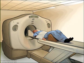
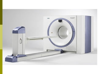
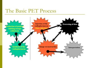
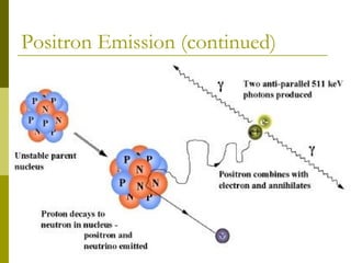
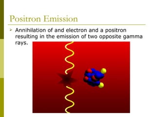
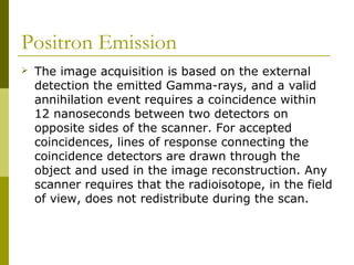
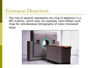
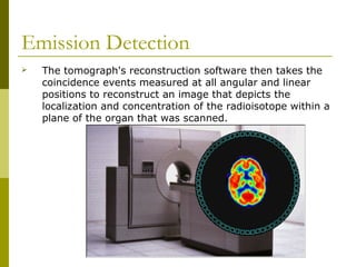
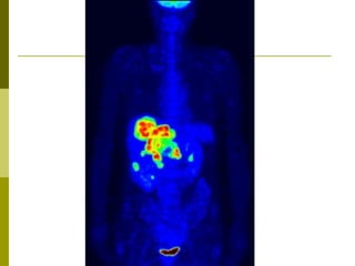
Positron emission tomography (PET) is an imaging technique that uses radiolabeled tracers to produce images showing their distribution in the body. During a PET scan, a tracer containing a radioactive isotope is injected and decays, emitting positrons. The positrons interact with electrons, producing pairs of gamma rays detected by the PET scanner to reconstruct images. PET scans are used to study brain function, detect and characterize cancers, and examine heart disease. Advantages include showing tissue function, but disadvantages include expense and limited availability.


![Pet appilcation[1]](https://cdn.slidesharecdn.com/ss_thumbnails/petappilcation1-191002015502-thumbnail.jpg?width=640&height=640&fit=bounds)


