1. A Novel Concept for a New Positron Emission Tomography Scanner
Jeena Khatri (PHYS), Rupak Mahapatra (Professor, PHYS)
PHYS 489
PET Scanner helps to take the metabolic image by tracing radioactive drug that is
ingested or inhaled into patients body. This is useful to reveal conditions like
cancer, heart disease and brain disorders. Present PET Scanners are further out in
MRI (Magnetic Resonance Imaging) which takes longer time to get data and loses
most of its data before reaching the scanner. The PET Scanner that we are
developing will fit inside the MRI and positioned to interact with patients more
efficiently. This will decrease the scan time in contrast increasing the amount of
data, which will decrease the radioactive drug ingested into the patient. The higher
amount of data gives us the better profile of the image than what we get in the
present days.
Abstract How does it work?
Importance
Existing Vs New
New
•Long cylindrical
•Fits in MRI
•Closer to patients – longer exposer
– less radioactive drug
•Organic (plastic) scintillator
• Much cheaper
• Faster response time
•Silicon Photomultipliers (SiPM)
• Cheaper
• Fast rise time
Present
•Huge size - further out in MRI - more
drug intake – small resolution
•Combined modalities difficult
•Inorganic Scintillators
• High stopping power
• Fast response time
• Very expensive!
•Photomultiplier Tubes (PMT)
• Fast, sensitive response
• Also very expensive!
A patient is administered
pharmaceuticals marked with
radioactive isotope emitting
photons.
Isotopes experience β+ (positron)
emission decay.
e+/e- annihilation results in two
back-to-back 511 keV gamma
particles.
Back to back photons emitted from
the isotope are detected with
scintillating material which re-emits
several lower energy photons that
activate a photomultiplier tube
(PMT).
Back-to-back photons detected
within ring create a line of response
(LOR).
Full body PET/CT scan of a mouse
Diagnosis of tumor
malignancy
Analysis of muscle
activation
Assessing neuroactivity
Observation of drug uptake
and concentration in
pharmaceutics
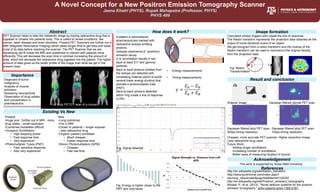
• Energy measurements
• Timing measurements
Image formation
θ r
Coincident photon triggers and create the line of response.
The Radon transform represents the projection data obtained as the
output of cross-sectional scans of an object.
We get sinogram from a radon transform and the inverse of the
Radon transform can be used to reconstruct the original density
from the projection data.
θ
r
Result and conclusion
Original image Gaussian filtered normal PET scan.
Gaussian filtered strip PET scan
500ps timing resolution.
Gaussian filtered strip PET scan
100ps timing resolution.
Cheaper, more accurate PET scanner; Higher resolution image.
Less radioactive drug used.
Future Work:
Adding longer scintillators
Increasing number of scintillators
Better ways of measuring location of source
References
Acknowledgement
This work is supported by Texas A&M University
http://en.wikipedia.org/wiki/Radon_transform
http://www.auntminnie.com/index.aspx?
sec=sup_n&sub=adv&pag=dis&itemId=104252
http://en.wikipedia.org/wiki/Positron_emission_tomography
Moskal, P., et al. (2013). "Novel detector systems for the positron
emission tomography." arXiv preprint arXiv:1305.5187.
Fig: Signal obtained
Fig: Energy is higher closer to the
PMT and vice-versa
Fig: Radon
Transformation