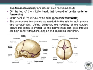
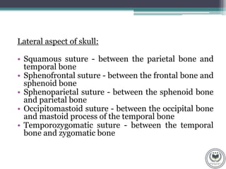
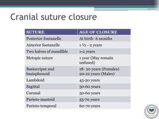
SKULL SUTURES
FORENSIC SIGNIFICANCE
Made by: Dr. Pankti Patel
M.Sc. Forensic Odontology
Volunteer at AFRS
CONTENTS
• What are cranial sutures
• Types of cranial sutures
• Cranial suture closure
• Age estimation from cranial suture
CRANIAL SUTURES
• Cranial sutures are fibrous type of connective tissue
that connect the bones of the skull.
• The dense fibrous tissue that connects the sutures
is made mostly out of collagen. These joints are
fixed, immovable, and they have no cavity. They are
also referred to as the synarthroses.
• In fetal skull the sutures are wide and allow slight
movement during birth, but later they become rigid
and fixed just like in the adults.
• An infant's skull is made up of 6 separate cranial (skull)
bones:
• Frontal bone
• Occipital bone
• Two parietal bones
• Two temporal bones
• The spaces between the bones that remain open in babies
and young children are called fontanelles. Sometimes, they
are called soft spots. These spaces are a part of normal
development.
• They then grow together as part of normal growth. They stay
connected throughout adulthood.
• Two fontanelles usually are present on a newborn's skull:
• On the top of the middle head, just forward of center (anterior
fontanelle)
• In the back of the middle of the head (posterior fontanelle)
• The sutures and fontanelles are needed for the infant's brain growth
and development. During childbirth, the flexibility of the sutures
allows the bones to overlap so the baby's head can pass through
the birth canal without pressing on and damaging their brain.
Types of cranial sutures
Anterior aspect of skull:
• Frontonasal suture - between the frontal bone and
nasal bones
• Frontozygomatic suture - between the frontal bone
and zygomatic bone
• Zygomaticomaxillary suture - between zygoma and
the maxilla
• Intermaxillary suture - between two maxilla
• Metopic suture - found in children; on the midline
of the frontal bone
Posterior aspect of skull:
• Sagittal suture - between two parietal bones
• Lambdoid suture - between the parietal bone
and occipital bone
Superior aspect of skull:
• Coronal suture - between the frontal bone and
the parietal bone
Lateral aspect of skull:
• Squamous suture - between the parietal bone and
temporal bone
• Sphenofrontal suture - between the frontal bone and
sphenoid bone
• Sphenoparietal suture - between the sphenoid bone
and parietal bone
• Occipitomastoid suture - between the occipital bone
and mastoid process of the temporal bone
• Temporozygomatic suture - between the temporal
bone and zygomatic bone
Inferior aspect of skull:
• Median palatine suture - between the hotizontal plates of the
palatines
• Transverse palatine suture - between the palatine process of
the maxillary bone and the palatine bone
• Petro-occipital suture - between the occipital bone and
petrous part of the temporal bone
• Spheno-occipital suture - between the sphenoid bone and
occipital bone
• Petrosquamous suture - between the petrous and squamous
parts of the temporal bone
• Petrotympanic suture - between the temporomandibular joint
and the tympanic cavity
Image source- https://commons.wikimedia.org/
Cranial suture closure
SUTURE AGE OF CLOSURE
Posterior fontanelle At birth- 6 months
Anterior fontanelle 1 ½ - 2 years
Two halves of mandible 1-2 years
Metopic suture 1 year (May remain
unfused)
Basiocciput and
basisphenoid
18- 20 years (Females)
20-22 years (Males)
Lambdoid 45-50 years
Sagittal 50-60 years
Coronal 50-60 years
Parieto-mastoid 55-70 years
Parieto-temporal 60-70 years
Age estimation from cranial suture
• Acsadi and Nemeskeri scoring system
SCORE DESCRIPTION
0 Open suture. Little space left between edges and
adjoining bones
1 Incipient closure. Suture is closed but clearly visible
as a continuous zigzag line
2 Closure in process. Suture line thinner, less zigzag
may be interrupted by complete closure
3 Advanced closure. Only pits indicate where the
suture is located
4 Closed suture. Suture completely obliterated
• Masset scoring criteria:
(1)Open, with no evidence of bony bridging across the
suture.
(2)Minimal closure, indicated by any evidence of bony
bridging up to 50% closure.
(3)Significant closure, indicated by evidence of bony
bridging of greater than 50%.
(4)Obliteration, with no trace remaining of the suture
margins
Image source- sciencedirect.com
• Meindl and Lovejoy scoring system
SCORE DESCIPTION
0 Open.There is no evidence of any ectocranial closure at
the site
1 Minimal closure. Given for minimal to moderate
closure-from single bony bridge across the suture to
about 50% synostosis at the site
2 Significant closure. Marked degree of closure but some
portion of the site still not completely fused
3 Advanced closure. Only pits indicate where the suture is
located
SKULL SUTURES.pptx

SKULL SUTURES.pptx

  • 2.
    SKULL SUTURES FORENSIC SIGNIFICANCE Madeby: Dr. Pankti Patel M.Sc. Forensic Odontology Volunteer at AFRS
  • 3.
    CONTENTS • What arecranial sutures • Types of cranial sutures • Cranial suture closure • Age estimation from cranial suture
  • 4.
    CRANIAL SUTURES • Cranialsutures are fibrous type of connective tissue that connect the bones of the skull. • The dense fibrous tissue that connects the sutures is made mostly out of collagen. These joints are fixed, immovable, and they have no cavity. They are also referred to as the synarthroses. • In fetal skull the sutures are wide and allow slight movement during birth, but later they become rigid and fixed just like in the adults.
  • 5.
    • An infant'sskull is made up of 6 separate cranial (skull) bones: • Frontal bone • Occipital bone • Two parietal bones • Two temporal bones • The spaces between the bones that remain open in babies and young children are called fontanelles. Sometimes, they are called soft spots. These spaces are a part of normal development. • They then grow together as part of normal growth. They stay connected throughout adulthood.
  • 6.
    • Two fontanellesusually are present on a newborn's skull: • On the top of the middle head, just forward of center (anterior fontanelle) • In the back of the middle of the head (posterior fontanelle) • The sutures and fontanelles are needed for the infant's brain growth and development. During childbirth, the flexibility of the sutures allows the bones to overlap so the baby's head can pass through the birth canal without pressing on and damaging their brain.
  • 7.
    Types of cranialsutures Anterior aspect of skull: • Frontonasal suture - between the frontal bone and nasal bones • Frontozygomatic suture - between the frontal bone and zygomatic bone • Zygomaticomaxillary suture - between zygoma and the maxilla • Intermaxillary suture - between two maxilla • Metopic suture - found in children; on the midline of the frontal bone
  • 8.
    Posterior aspect ofskull: • Sagittal suture - between two parietal bones • Lambdoid suture - between the parietal bone and occipital bone Superior aspect of skull: • Coronal suture - between the frontal bone and the parietal bone
  • 9.
    Lateral aspect ofskull: • Squamous suture - between the parietal bone and temporal bone • Sphenofrontal suture - between the frontal bone and sphenoid bone • Sphenoparietal suture - between the sphenoid bone and parietal bone • Occipitomastoid suture - between the occipital bone and mastoid process of the temporal bone • Temporozygomatic suture - between the temporal bone and zygomatic bone
  • 10.
    Inferior aspect ofskull: • Median palatine suture - between the hotizontal plates of the palatines • Transverse palatine suture - between the palatine process of the maxillary bone and the palatine bone • Petro-occipital suture - between the occipital bone and petrous part of the temporal bone • Spheno-occipital suture - between the sphenoid bone and occipital bone • Petrosquamous suture - between the petrous and squamous parts of the temporal bone • Petrotympanic suture - between the temporomandibular joint and the tympanic cavity
  • 11.
  • 12.
    Cranial suture closure SUTUREAGE OF CLOSURE Posterior fontanelle At birth- 6 months Anterior fontanelle 1 ½ - 2 years Two halves of mandible 1-2 years Metopic suture 1 year (May remain unfused) Basiocciput and basisphenoid 18- 20 years (Females) 20-22 years (Males) Lambdoid 45-50 years Sagittal 50-60 years Coronal 50-60 years Parieto-mastoid 55-70 years Parieto-temporal 60-70 years
  • 13.
    Age estimation fromcranial suture • Acsadi and Nemeskeri scoring system SCORE DESCRIPTION 0 Open suture. Little space left between edges and adjoining bones 1 Incipient closure. Suture is closed but clearly visible as a continuous zigzag line 2 Closure in process. Suture line thinner, less zigzag may be interrupted by complete closure 3 Advanced closure. Only pits indicate where the suture is located 4 Closed suture. Suture completely obliterated
  • 14.
    • Masset scoringcriteria: (1)Open, with no evidence of bony bridging across the suture. (2)Minimal closure, indicated by any evidence of bony bridging up to 50% closure. (3)Significant closure, indicated by evidence of bony bridging of greater than 50%. (4)Obliteration, with no trace remaining of the suture margins
  • 15.
  • 16.
    • Meindl andLovejoy scoring system SCORE DESCIPTION 0 Open.There is no evidence of any ectocranial closure at the site 1 Minimal closure. Given for minimal to moderate closure-from single bony bridge across the suture to about 50% synostosis at the site 2 Significant closure. Marked degree of closure but some portion of the site still not completely fused 3 Advanced closure. Only pits indicate where the suture is located