The four major sutures are the coronal
•Download as DOCX, PDF•
19 likes•101,182 views
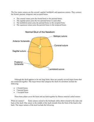
The four major sutures of the skull are the coronal, sagittal, lambdoid, and squamous sutures. Sutures connect the frontal, parietal, temporal, and occipital bones of the skull and allow movement during birth by acting as expansion joints that enable even growth of the bones and brain. There are also two fontanelles between the skull bones, the anterior and posterior fontanelles, which are soft spots in an infant's skull that close during the first two years of life.
Report
Share
Report
Share

Recommended
Axial skeleton parts 1 2
The document summarizes the axial skeleton and its components. The axial skeleton includes the skull, vertebral column, and thoracic cage. The skull is made up of 29 bones divided into 5 areas: the cranium (8 bones), facial bones (14 bones), hyoid bone (1 bone), ossicles of the ear (6 bones), and Wormian bones (? bones). The cranium contains the frontal, parietal, occipital, temporal, sphenoid, and ethmoid bones. The facial bones include the nasal, maxilla, zygomatic, mandible, lacrimal, palatine, inferior nasal conchae, and vomer bones.
Axial skeleton parts 3 5
The document summarizes the organization and components of the human skeleton. It describes the axial skeleton, which includes the skull and vertebral column, and the appendicular skeleton. It provides details on the bones and structures of the skull, vertebral column, thoracic cage, and their functions in supporting the body and vital organs. Key bones mentioned include the cranium, ribs, sternum, and vertebrae.
Skull
The skull is composed of 22 bones that form the cranium (housing the brain) and face. The 8 bones of the cranium are the frontal, occipital, sphenoid, ethmoid, and two each of the parietal and temporal bones. These bones protect the brain and support structures like the spinal cord. The 14 bones of the face are the mandible, maxillae, zygomatic, lacrimal, nasal, inferior conchae, palatine, and vomer bones, which form structures like the nose and jaws.
Topic 5 bone of skull neck
The document summarizes the bones that make up the human skull and their functions. It discusses the 22 bones that comprise the skull, which can be divided into cranial bones that form the cranial cavity to protect the brain, and facial bones that form the framework of the face. Key bones described include the frontal, parietal, temporal, occipital, sphenoid, ethmoid, nasal, maxilla, zygomatic, lacrimal and palatine bones. Each bone has a specific shape and features that contribute to its role in housing and protecting important structures of the head.
Muscular system
The document summarizes the structure and function of the muscular system. It describes the three types of muscles, with a focus on skeletal muscles. Skeletal muscles produce movement by pulling on tendons attached to bones. The document then details the anatomy of skeletal muscles including their gross anatomy, blood supply, and microanatomy down to the level of the sarcomere. It explains how skeletal muscle contraction occurs through the sliding filament theory involving the interaction of actin and myosin filaments.
thoracic cage, rib cage, thoracic cavity by dr shahid alam
The thoracic cage is composed of the thoracic vertebrae, ribs, and sternum. It forms the skeleton of the chest and protects the organs of the thoracic cavity. There are 12 pairs of ribs, which are divided into true ribs attached to the sternum and false ribs not directly attached. The sternum is a flat bone in the midline of the chest with three parts - the manubrium, body, and xiphoid process. The ribs and costal cartilages connect to the sternum and vertebrae, allowing for respiration.
Skeletal system. anatomy and physiology of skeletal system. appendicular skel...
SKELETAL SYSTEM
bones, cartilage and ligaments are tightly joined to form a strong, flexible framework called skeletal system
anatomy and physiology of axial and appendicular skeletal system
Axial Skeleton: The axial skeleton includes the skull, spine, ribs and sternum.
Appendicular Skeleton:
The appendicular skeleton includes the appendages of the body, which are the shoulders, arms, hips, and legs.
General physiology
This document provides an overview of general physiology concepts including:
- Physiology is the study of how cells, tissues, and organisms function
- Shivering occurs when we feel cold to help warm the body through involuntary muscle contractions
- The hypothalamus detects a fall in temperature and causes shivering to increase body temperature
- Homeostasis refers to maintaining a relatively constant internal environment through feedback mechanisms like negative feedback which acts to reverse changes and positive feedback which accelerates changes.
Central Nervous system of human body
central nervous system of human body, it includes brain and its part, spinal cord, autonomic and parasympathetic system
Skeletal System 1
The skeletal system is composed of bones and connective tissues that provide structure, support, protection, movement, and mineral storage. There are four main bone types - long bones in the limbs, short bones in the wrists and ankles, flat bones in the skull and trunk, and irregular bones like the vertebrae. Bones have external features like ridges and internal structures like compact and spongy bone. The spinal column consists of 33 vertebrae that are specialized in different regions and enable movement while protecting the spinal cord.
Know about Muscular tissue
Muscle is a contractile tissue found throughout the body that produces movement when stimulated. There are three main types of muscle tissue: smooth, cardiac, and skeletal muscle. Skeletal muscle is striated, voluntary muscle attached to bones that allows for conscious control of movement. It comprises over 600 muscles in the musculoskeletal system, including axial muscles that control facial expression and posture, and appendicular muscles of the limbs. Muscle fibers contract when stimulated by motor nerves, producing movement through their interaction with bones and tendons.
Ossification
Ossification is the term use to describe a process of bone fermentation by deposition of calcium in the fetal hyaline cartilage.
The Muscular System
The document summarizes the key components and functions of the muscular system. It describes the three main types of muscle tissue - skeletal, smooth, and cardiac muscle - and their distinct characteristics. Skeletal muscle is voluntary and attached to bones, controlling movement, posture, and respiration. Smooth muscle is involuntary and within organs and blood vessels, roles include peristalsis and vasoconstriction. Cardiac muscle is only found in the heart, its automatic contractions pump blood throughout the body. The document also outlines the cellular structure of muscles and the proteins involved in muscle contraction.
Bones
The document discusses the musculoskeletal system and provides an overview of bone structure and classification. It identifies the main functions of bones as support, protection, movement, storage of minerals/fats, and blood cell formation. Bones are classified based on location in the body, shape, and internal structure as either compact or spongy bone.
Axial Skeleton - Skull
The skull has two main parts - the cranium and facial bones. The cranium is made up of eight flat bones that protect the brain, while the facial bones form the eyes, nose, and lower jaw. There are also small bones in the skull like the sphenoid, ethmoid, and hyoid bone. The skull features various markings like foramina for blood vessels, sinuses filled with air, and processes for muscle attachments.
Endocrine system
The endocrine system is a system that transfers information around the body via chemical messengers called hormones. It helps maintain homeostasis. The endocrine system contains several ductless glands that synthesize, store, and release hormones like insulin, glucagon, estrogen, testosterone, thyroid hormones, cortisol, and others. These hormones then travel through the bloodstream to target tissues and organs to regulate processes like metabolism, stress response, reproduction, and development.
Cell physiology
The document discusses the structure and function of cells, tissues, organs and systems in the human body. It begins by defining cells as the basic functional units that make up tissues like blood, muscle and bone. Tissues combine to form organs like the heart, stomach and brain. Organs work together in organ systems to carry out important body functions. The document then provides detailed descriptions of cell structures such as the cell membrane, cytoplasm, organelles, and the functions they perform. It also discusses how cells, tissues and organ systems are organized in the body.
Cartilage
Cartilage is a connective tissue that provides support and flexibility to various regions of the body. There are three main types of cartilage - hyaline, elastic, and fibrocartilage. Hyaline cartilage is the most abundant and is characterized by chondrocytes embedded in a matrix with collagen fibers and proteoglycans. It is found in locations like the fetal skeleton, nose, and joints. Elastic cartilage contains elastic fibers that allow for flexibility and is present in the ear and epiglottis. Fibrocartilage consists of thick collagen fibers and is found in joints like the pubic symphysis where it can withstand compressive forces.
Bones
This document provides an overview of bones, including their introduction, types, contents, and advantages. It notes that bones are rigid organs that make up the vertebrate endoskeleton. There are two main types of bones: soft bones like the nose and ear cartilage, and hard bones that make up the rest of the skeleton. Bone mainly consists of three parts - an outer compact bone, inner soft bone, and yellow marrow at the center. The key advantages of bones are that they protect internal organs, are the main source of blood production, give the body its structure, and bear the total body weight.
Osteology
This document provides an introduction to osteology, the study of bones. It defines osteology and discusses the classification, structure, and microscopic features of bones. The key points covered are:
1. Bones are classified based on their position in the body as either axial or appendicular, and based on their shape as long, short, flat, irregular, or sesamoid.
2. Bones are composed of compact cortical bone and cancellous spongy bone. Long bones specifically have a diaphysis and two epiphyses.
3. Bones are living tissues with osteoblasts, osteoclasts, and osteocytes that remodel the matrix of collagen fibers, hydroxyapatite crystals,
Physiology of respiration
Respiration is the process of gas exchange between an organism and the environment. It consists of external respiration, which is breathing and the exchange of gases between the lungs and environment. And internal respiration, which is the exchange of oxygen and carbon dioxide between the blood and tissues via cellular respiration. The document defines the key terms and processes involved in respiration, explores factors affecting breathing rate, and how to measure lung volumes and vital capacity.
Anatomy of nervous system
The nervous system is divided into the central nervous system (CNS) and peripheral nervous system (PNS). The CNS contains the brain and spinal cord, while the PNS connects them to sensory receptors and effector organs. Neurons are the basic functional units and transmit electrical signals through axons and dendrites. Communication occurs at synapses between neurons. The brain is divided into regions that control functions like breathing, movement coordination, homeostasis, and cognition. The brain and spinal cord are protected by meninges and cerebrospinal fluid.
Sensory organs eyes
The document summarizes the anatomy and function of the human eye. It describes how light enters through the cornea and is focused on the retina by the lens. The retina contains photoreceptor cells called rods and cones that convert light into nerve impulses which are sent to the brain for processing and interpretation as vision. The iris controls the size of the pupil to regulate the amount of light entering the eye.
Surface Anatomy
This document provides a chapter outline on surface anatomy, describing prominent anatomical landmarks that can be observed or felt on the external surfaces of the body. It covers surface features of the head and neck, thorax, back, abdomen, upper limbs, gluteal region, and thighs. Key points include descriptions of bones, muscles, blood vessels, and other structures that can be identified by visual inspection, palpation, percussion, or auscultation on the surface of various body regions. The document is intended to familiarize readers with techniques for examining surface anatomy and identifying important anatomical structures.
Synovial joints
synovial joint, definition of synovial joint, diarthrodial joints, components of synovial joint, types of synovial joints, hinge joint with examples, pivot joint with examples, condyloid joint with examples, saddle joint with examples, ball and socket joint with examples, gliding joint with examples, features of synovial joint, synovial membrane, synovial fluid, components of synovial membrane, meniscus, true and accessory ligament of synovial joint, bursae, blood supply of synovial joint, innervation of synovial joint
The Muscular System
This document provides information on muscles, joints, and movement. It defines the three main types of muscle - skeletal, smooth, and cardiac - and their functions. It describes muscle attachments via tendons and ligaments connecting bones. Various joints are defined, including hinge and ball-and-socket joints, and the basic movements they allow including flexion, extension, and rotation. Key muscles of the upper and lower body are identified along with their actions.
Hindbrain
This document provides information about the hindbrain, which consists of the medulla oblongata, pons, and cerebellum. It describes the anatomy and functions of each part. The medulla oblongata extends from the foramen magnum to the pons and controls vital functions like breathing, blood pressure, and heart rate. The pons relays signals between the cerebrum and cerebellum and is involved in processes like sleep, respiration, and hearing. The cerebellum, located above the pons, aids in balance, muscle coordination, and motor skills.
Axial Skeleton Skull
Slides to accompany an anatomy lecture to introduce axial skeleton and go through each bone of the skull
Sutures of the Skull
The document discusses sutures of the human skull. It identifies the primary cranial sutures, including the coronal, sagittal, lambdoid, and metopic sutures. It also discusses the midpalatal suture and presents a classification system with 5 stages of maturation based on CBCT imaging. The classification aims to assess midpalatal suture morphology for determining candidacy for rapid maxillary expansion. The sutures most commonly affected by craniosynostosis are also identified.
Skull bones, features and markings II
Inferior (mandible removed) and superior view (top of cranium removed - cranial floor / sphenoid view) skull anatomy warm-up for Anatomy and Physiology students. Bones, features and markings are shown
More Related Content
What's hot
Central Nervous system of human body
central nervous system of human body, it includes brain and its part, spinal cord, autonomic and parasympathetic system
Skeletal System 1
The skeletal system is composed of bones and connective tissues that provide structure, support, protection, movement, and mineral storage. There are four main bone types - long bones in the limbs, short bones in the wrists and ankles, flat bones in the skull and trunk, and irregular bones like the vertebrae. Bones have external features like ridges and internal structures like compact and spongy bone. The spinal column consists of 33 vertebrae that are specialized in different regions and enable movement while protecting the spinal cord.
Know about Muscular tissue
Muscle is a contractile tissue found throughout the body that produces movement when stimulated. There are three main types of muscle tissue: smooth, cardiac, and skeletal muscle. Skeletal muscle is striated, voluntary muscle attached to bones that allows for conscious control of movement. It comprises over 600 muscles in the musculoskeletal system, including axial muscles that control facial expression and posture, and appendicular muscles of the limbs. Muscle fibers contract when stimulated by motor nerves, producing movement through their interaction with bones and tendons.
Ossification
Ossification is the term use to describe a process of bone fermentation by deposition of calcium in the fetal hyaline cartilage.
The Muscular System
The document summarizes the key components and functions of the muscular system. It describes the three main types of muscle tissue - skeletal, smooth, and cardiac muscle - and their distinct characteristics. Skeletal muscle is voluntary and attached to bones, controlling movement, posture, and respiration. Smooth muscle is involuntary and within organs and blood vessels, roles include peristalsis and vasoconstriction. Cardiac muscle is only found in the heart, its automatic contractions pump blood throughout the body. The document also outlines the cellular structure of muscles and the proteins involved in muscle contraction.
Bones
The document discusses the musculoskeletal system and provides an overview of bone structure and classification. It identifies the main functions of bones as support, protection, movement, storage of minerals/fats, and blood cell formation. Bones are classified based on location in the body, shape, and internal structure as either compact or spongy bone.
Axial Skeleton - Skull
The skull has two main parts - the cranium and facial bones. The cranium is made up of eight flat bones that protect the brain, while the facial bones form the eyes, nose, and lower jaw. There are also small bones in the skull like the sphenoid, ethmoid, and hyoid bone. The skull features various markings like foramina for blood vessels, sinuses filled with air, and processes for muscle attachments.
Endocrine system
The endocrine system is a system that transfers information around the body via chemical messengers called hormones. It helps maintain homeostasis. The endocrine system contains several ductless glands that synthesize, store, and release hormones like insulin, glucagon, estrogen, testosterone, thyroid hormones, cortisol, and others. These hormones then travel through the bloodstream to target tissues and organs to regulate processes like metabolism, stress response, reproduction, and development.
Cell physiology
The document discusses the structure and function of cells, tissues, organs and systems in the human body. It begins by defining cells as the basic functional units that make up tissues like blood, muscle and bone. Tissues combine to form organs like the heart, stomach and brain. Organs work together in organ systems to carry out important body functions. The document then provides detailed descriptions of cell structures such as the cell membrane, cytoplasm, organelles, and the functions they perform. It also discusses how cells, tissues and organ systems are organized in the body.
Cartilage
Cartilage is a connective tissue that provides support and flexibility to various regions of the body. There are three main types of cartilage - hyaline, elastic, and fibrocartilage. Hyaline cartilage is the most abundant and is characterized by chondrocytes embedded in a matrix with collagen fibers and proteoglycans. It is found in locations like the fetal skeleton, nose, and joints. Elastic cartilage contains elastic fibers that allow for flexibility and is present in the ear and epiglottis. Fibrocartilage consists of thick collagen fibers and is found in joints like the pubic symphysis where it can withstand compressive forces.
Bones
This document provides an overview of bones, including their introduction, types, contents, and advantages. It notes that bones are rigid organs that make up the vertebrate endoskeleton. There are two main types of bones: soft bones like the nose and ear cartilage, and hard bones that make up the rest of the skeleton. Bone mainly consists of three parts - an outer compact bone, inner soft bone, and yellow marrow at the center. The key advantages of bones are that they protect internal organs, are the main source of blood production, give the body its structure, and bear the total body weight.
Osteology
This document provides an introduction to osteology, the study of bones. It defines osteology and discusses the classification, structure, and microscopic features of bones. The key points covered are:
1. Bones are classified based on their position in the body as either axial or appendicular, and based on their shape as long, short, flat, irregular, or sesamoid.
2. Bones are composed of compact cortical bone and cancellous spongy bone. Long bones specifically have a diaphysis and two epiphyses.
3. Bones are living tissues with osteoblasts, osteoclasts, and osteocytes that remodel the matrix of collagen fibers, hydroxyapatite crystals,
Physiology of respiration
Respiration is the process of gas exchange between an organism and the environment. It consists of external respiration, which is breathing and the exchange of gases between the lungs and environment. And internal respiration, which is the exchange of oxygen and carbon dioxide between the blood and tissues via cellular respiration. The document defines the key terms and processes involved in respiration, explores factors affecting breathing rate, and how to measure lung volumes and vital capacity.
Anatomy of nervous system
The nervous system is divided into the central nervous system (CNS) and peripheral nervous system (PNS). The CNS contains the brain and spinal cord, while the PNS connects them to sensory receptors and effector organs. Neurons are the basic functional units and transmit electrical signals through axons and dendrites. Communication occurs at synapses between neurons. The brain is divided into regions that control functions like breathing, movement coordination, homeostasis, and cognition. The brain and spinal cord are protected by meninges and cerebrospinal fluid.
Sensory organs eyes
The document summarizes the anatomy and function of the human eye. It describes how light enters through the cornea and is focused on the retina by the lens. The retina contains photoreceptor cells called rods and cones that convert light into nerve impulses which are sent to the brain for processing and interpretation as vision. The iris controls the size of the pupil to regulate the amount of light entering the eye.
Surface Anatomy
This document provides a chapter outline on surface anatomy, describing prominent anatomical landmarks that can be observed or felt on the external surfaces of the body. It covers surface features of the head and neck, thorax, back, abdomen, upper limbs, gluteal region, and thighs. Key points include descriptions of bones, muscles, blood vessels, and other structures that can be identified by visual inspection, palpation, percussion, or auscultation on the surface of various body regions. The document is intended to familiarize readers with techniques for examining surface anatomy and identifying important anatomical structures.
Synovial joints
synovial joint, definition of synovial joint, diarthrodial joints, components of synovial joint, types of synovial joints, hinge joint with examples, pivot joint with examples, condyloid joint with examples, saddle joint with examples, ball and socket joint with examples, gliding joint with examples, features of synovial joint, synovial membrane, synovial fluid, components of synovial membrane, meniscus, true and accessory ligament of synovial joint, bursae, blood supply of synovial joint, innervation of synovial joint
The Muscular System
This document provides information on muscles, joints, and movement. It defines the three main types of muscle - skeletal, smooth, and cardiac - and their functions. It describes muscle attachments via tendons and ligaments connecting bones. Various joints are defined, including hinge and ball-and-socket joints, and the basic movements they allow including flexion, extension, and rotation. Key muscles of the upper and lower body are identified along with their actions.
Hindbrain
This document provides information about the hindbrain, which consists of the medulla oblongata, pons, and cerebellum. It describes the anatomy and functions of each part. The medulla oblongata extends from the foramen magnum to the pons and controls vital functions like breathing, blood pressure, and heart rate. The pons relays signals between the cerebrum and cerebellum and is involved in processes like sleep, respiration, and hearing. The cerebellum, located above the pons, aids in balance, muscle coordination, and motor skills.
Axial Skeleton Skull
Slides to accompany an anatomy lecture to introduce axial skeleton and go through each bone of the skull
What's hot (20)
Viewers also liked
Sutures of the Skull
The document discusses sutures of the human skull. It identifies the primary cranial sutures, including the coronal, sagittal, lambdoid, and metopic sutures. It also discusses the midpalatal suture and presents a classification system with 5 stages of maturation based on CBCT imaging. The classification aims to assess midpalatal suture morphology for determining candidacy for rapid maxillary expansion. The sutures most commonly affected by craniosynostosis are also identified.
Skull bones, features and markings II
Inferior (mandible removed) and superior view (top of cranium removed - cranial floor / sphenoid view) skull anatomy warm-up for Anatomy and Physiology students. Bones, features and markings are shown
Different Types of Sutures
Sutures can be divided into absorbable sutures, non absorbable sutures, mono filament, multi filament, natural and synthetic sutures according to their type of material, raw material used, absoprtion nature.
01 skull
This document provides an overview of the bones in the human skull and face that will be covered in the Biology 71 Human Anatomy lab on skull anatomy. It lists the specific bones that will be examined, including the cranial bones (occipital, frontal, sphenoid, ethmoid, temporal), facial bones (mandible, zygomatic, maxilla, palatine, vomer, nasal), and associated bones (hyoid, malleus, incus, stapes). It also notes that the first quiz for the class will be next week.
Anatomy of Skull
The document lists and describes various features and norms of the human skull, including the norma verticalis, temporal lines, norma occipitalis, occipital point, frontal region, orbital openings, zygoma, and norma laterialis. It also details the boundaries, walls, and contents of the temporal fossa, noting structures like the temporalis muscle, arteries, and bones that form its borders.
Age estimation by bones
This document discusses age estimation through analysis of bone development visible on x-rays. It begins by describing the different types of bones and how they develop. Next, it reviews early studies on using bone age to determine chronological age. It then discusses factors like race, nutrition, and climate that can cause variation between populations. The document proceeds to examine bone development timelines from several studies and populations. It concludes by outlining the typical chronological order of appearance and fusion of epiphyses in males and females.
Positioning and radiographic anatomy of the skull
This document provides information on positioning and radiographic anatomy of the skull. It discusses the anatomy of the skull and lists the 8 cranial bones. It then describes various positioning considerations for skull radiography including erect vs recumbent positioning, patient comfort, hygiene, exposure factors, SID and radiation protection. Several common skull radiographic projections are outlined including the AP, lateral, PA, submentovertex and oblique projections. For each projection, the demonstrated pathology, positioning, central ray angle and structures shown are described.
Carpal Tunnel Massage
This document provides information about carpal tunnel massage techniques. It discusses what carpal tunnel is, the benefits of massage for carpal tunnel and achy muscles. It lists muscles benefited by the massage. It describes the items and techniques used in the massage, including kneading, compression and skin rolling. Steps for performing the massage are outlined, such as sanitizing hands, placing towels, and using lotion while rubbing fingers and palms. The arm is also massaged from elbow to wrist.
Skull
The document outlines the bones that make up the human skull, including the frontal, parietal, occipital, temporal, sphenoid, ethmoid, nasal, maxilla, palatine, zygomatic, and mandible bones. It describes how the skull can be viewed from different angles including superior, posterior, anterior, and lateral views. It also notes the major sutures of the skull and provides brief descriptions of the shape and bones that make up the different parts of the skull.
Cta 201 module 5 cranium bones and facial bones (1)
This document provides an overview of the cranium and facial bones, structures of the ear, sinuses, and orbit. It describes the 8 cranial bones, 14 facial bones, and key features of the maxilla, mandible, nasal bones, sphenoid bone, temporal bone, and paranasal sinuses. The ethmoid, frontal, maxillary, and sphenoid sinuses are discussed in more detail. Structures of the ear including the external auditory meatus, tympanic membrane, middle ear ossicles, and internal ear are also summarized. Finally, the document reviews the bones that make up the orbit, as well as its imaging and openings.
Skull bones, features and markings 3
Introduction to bones and features of the infant human skull and the the features and markings of the mandible for Anatomy and Physiology students.
Ch07 a.skeletal
The document summarizes the bones that make up the axial skeleton, including the skull, vertebral column, and thoracic cage. It describes in detail the individual bones of the skull, including the cranial and facial bones. It identifies the frontal, parietal, occipital, temporal, sphenoid, ethmoid, and facial bones. It highlights key features of each bone such as their articulation points and major markings. Diagrams are included to illustrate the anatomical features.
14 bones of the face
The document lists 14 bones that make up the human skull: the zygomatic, ethmoid, temporal, maxilla, frontal, sphenoid, occipital, mandible, parietal, palatine, lacrimal, vomer, nasal bones, and the inferior nasal conchae. These bones protect the brain and form the structure of the face.
14 bones of the face
This document lists 14 bones that make up the human skull: the zygomatic, ethmoid, temporal, maxilla, frontal, sphenoid, occipital, mandible, parietal, palatine, lacrimal, vomer, nasal bones, and inferior nasal conchae.
The nervous system 2014
This document provides information about the nervous system and reflex actions. It defines key terms like stimulus, receptors, central nervous system, effector, and the three types of neurons. It explains that the nervous system coordinates and regulates bodily functions through sensory neurons that detect stimuli, relay neurons that transmit signals within the central nervous system, and motor neurons that activate effectors. It describes reflex arcs and gives examples of spinal and cranial reflexes like the knee jerk reaction and pupil reflex. Finally, it outlines the sequence of events that occur in a reflex action from stimulus detection to response.
Upper limb skeleton 5th october 2013
This is a presentation for 1st year medical students, Anatomy course, Mansoura Faculty of Medicine,Mansoura University,Mansoura,Egypt.
We are talking about the upper limb skeleton, starting with general features of three bones which are:
Clavicle, Scapula & Humerus.
I hope you can get benefit of it.
Enjoy my friends.....
Emergency Radiology: Shoulder & Upper Extremities
This document provides an overview of orthopedic radiology of the shoulder and upper extremity. It begins with basic concepts in radiographic diagnosis of fractures. It then reviews standard and supplementary views of the shoulder, elbow, wrist, hand and fingers. Specific injuries are discussed for each area, along with relevant anatomy, imaging findings and radiologic principles. Case examples are presented to demonstrate various fractures and dislocations. Key imaging signs are emphasized for accurate diagnosis.
Sutures material
This document discusses different types of suture materials. It describes both absorbable and non-absorbable natural and synthetic suture materials. Absorbable sutures such as surgical gut, Monocryl, Vicryl and PDS are made from collagen or synthetic polymers and are absorbed by the body over time. Non-absorbable sutures like silk, nylon and Prolene are made from materials that resist absorption and provide long-term wound support. The document outlines the properties, characteristics and appropriate uses of various commonly used suture materials.
Joints articulations
This document provides an overview of human joint anatomy and classification. It discusses the different types of joints including fibrous joints, cartilaginous joints, and synovial joints. For synovial joints, it describes the general anatomy including the articular capsule, joint cavity, synovial fluid, articular cartilage, and ligaments. It also classifies and describes the movements permitted at different joints, such as gliding, angular, rotational, and circumduction. Finally, it discusses the specific structures that comprise a synovial joint, including articular bony surfaces, articular cartilage, the joint cavity, and articular capsule.
Skull bones and features 1
The skull is composed of 22 bones, including 8 cranial bones and 14 facial bones. The cranial bones are the frontal, occipital, ethmoid, sphenoid, and two parietal and temporal bones each. The facial bones include the vomer, mandible, two maxillae, two zygomatic, two nasal, two nasal concha, two lacrimal, and two palatine bones. Key skull features include the sagittal, coronal, lambdoidal, and squamosal sutures, as well as the orbit.
Viewers also liked (20)
Cta 201 module 5 cranium bones and facial bones (1)
Cta 201 module 5 cranium bones and facial bones (1)
Similar to The four major sutures are the coronal
fetal skull
The fetal skull consists of 3 main parts - the vault, face, and base/brow. The vault contains 2 frontal bones, 2 parietal bones, 1 occipital bone, and 2 temporal bones, separated by sutures including the frontal, coronal, sagittal, and lambdoid sutures. There are 6 fontanelles but the 2 important ones in obstetrics are the anterior and posterior fontanelles. Understanding the diameters, landmarks, and molding process of the fetal skull is important for midwives to evaluate labor progression and fetal well-being.
The skeletal system (slide show)
The document discusses the anatomy and features of the human skeletal system. It describes the main components and functions of the skeletal system, including support, protection, movement, mineral storage, blood cell production, and energy storage. It provides details on the types of bones in the human body, including long bones, short bones, flat bones, and irregular bones. It also summarizes the main parts of the axial and appendicular skeleton, focusing on the skull, vertebral column, ribs, and bones of the upper and lower limbs.
SWERNY SKELETAL 2.pdf
The document provides an overview of the axial and appendicular skeleton. The axial skeleton consists of the skull, vertebral column, and rib cage. It describes the bones that make up the skull in detail. The functions of the skull are to protect the brain and sense organs. The vertebral column consists of vertebrae and provides structure, protection of the spinal cord, and allows for movement. The thoracic cage surrounds the heart and lungs and its bones provide attachment points for muscles. The appendicular skeleton includes the shoulder girdle and pelvic girdle bones that attach the arms and legs.
Axial skeletal
The document summarizes key aspects of the axial skeleton. It is divided into bones of the skull - cranium (occipital, parietal, temporal, frontal) and facial bones. It describes each bone's functions, articulations, regions/landmarks and foramina. It also covers the vertebral column, ribs, sternum and functions of the axial skeleton in respiration and stabilization of structures.
Head and neck anat fin
The document provides an overview of the anatomy of the head and neck, focusing on the bones that make up the skull. It describes each of the cranial bones - the frontal, parietal, occipital, temporal, and sphenoid bones - listing their distinguishing features and articulations with other bones. It also briefly discusses the cervical vertebrae of the neck. The document is intended to teach medical students the osteology and key structures of the skull.
Fetal skull
The fetal skull contains the delicate brain and must adapt to pass through the birth canal. It is large relative to the true pelvis. The skull is divided into the vault, base, and face. Key regions include the occiput, vertex, brow, and face. The skull contains bones like the occipital, parietal, and frontal bones joined by sutures. Fontanelles form where sutures meet. Key diameters that allow the skull to mold during birth include the biparietal, suboccipitobregmatic, and mentovertical diameters.
Axial skeleton ANATOMY AND PHYSIOLOGY
The bones of the skeleton are divided into two groups:
the axial skeleton and the appendicular skeleton
Fetal head diameters
This document summarizes the anatomy of the fetal head, including its bones, sutures, landmarks, and diameters. It describes how the cranial bones are thin and overlapping at birth, allowing molding to the maternal pelvis. The sutures between bones are identified. Landmarks from front to back are listed. Four anteroposterior diameters and two transverse diameters are defined. The average fetal head circumference at term is provided.
a_4_skull.ppt
The skull is composed of 28 bones that protect the brain and provide attachment points for muscles. The cranium consists of 8 bones that form the cranial vault surrounding the brain. The facial skeleton consists of 15 irregular bones that form the face and hold the teeth. Several sutures connect the cranial bones, including sagittal, coronal, squamosal, and lambdoidal sutures. In infants, fontanelles between bones allow the skull to flex during birth and accommodate brain growth.
Lec 5 skull
The skull is composed of 22 bones that make up the cranium and face. The 8 bones of the cranium are the frontal, two parietal, occipital, two temporal, sphenoid, and ethmoid bones. Each of these bones has characteristic structures and parts that were formed through either intramembranous or endochondral ossification. The 14 bones of the face are the two zygomatic, two maxillae, two nasal, two lacrimal, vomer, two palatine, two inferior conchae, and single mandible. Together, these bones protect the brain and form cavities for sensory structures.
Osseous System Part I.pptx
It is skeletal system of human body in detail description. In this ppt gives axial skeleton of body cranium thoracic cage and Vertibral coloumn . i gave structure and function of the bone , parts of axial skeleton with diagram
SKELETAL system- axial.ppt
This document provides information about the skeletal system, specifically focusing on the axial skeleton. It defines the main types of bones and classifies them as long, short, flat, irregular, or sesamoid. It then describes the functions of bones and divides the skeletal system into the axial and appendicular skeleton. The majority of the document describes the bones that make up the axial skeleton, including the skull, vertebral column, ribs, and sternum. It provides details on the individual bones of the skull and vertebral column, including their features and locations.
anatomy of human skull
This document discusses the anatomy of the skull and cranium. It describes the 22 bones that make up the skull, including the 8 bones of the cranium that protect the brain, and the 14 facial bones. It details the neurocranium and viscerocranium, and provides pictures and names of the individual cranial bones. It also discusses the sutures between bones, sinuses in the skull, and fontanelles in infants. Finally, it briefly mentions diseases that can affect the fontanelles such as bulging, sunken, or enlarged fontanelles.
Osteology of facial skeleton
The document provides an overview of osteology and classification of bones in the facial skeleton. It begins with an introduction to bone structure and function. It then classifies bones based on shape and development. The document outlines the bones that make up the skull, including the calvarium (paired and unpaired bones), and provides details on the norma views (verticalis, occipitalis, lateralis, frontalis, basalis). It describes each individual bone, its landmarks and clinical relevance. In summary, the document categorizes and describes in detail the bones that make up the skull and facial skeleton.
Skull, neck and muscle
The document discusses the anatomy of the skull, its bones, and their functions. It describes the 22 bones that make up the skull, divided into cranial bones that form the cranial cavity protecting the brain, and facial bones that form the framework of the face. Each of the 8 cranial bones (frontal, parietal, temporal, occipital, sphenoid, ethmoid) and their features are then discussed in detail. The skull bones provide protection, sites for muscle attachment, and support for sensory organs.
Skull of rabbit
this powerpoint presentation explains the structure and function of various parts of a rabbit skull.......
The Skeletal Sytem
The document summarizes the skeletal system, specifically the axial skeleton. It describes the main bones that make up the skull, vertebral column, and ribs. It discusses the classification of bones based on their microscopic appearance, developmental origin, shape, and location in the body. For each bone, it provides a brief definition and identifies its key features.
skeletal system
The musculoskeletal system consists of the skeletal and muscular systems. The skeletal system includes 206 bones that make up the axial skeleton (skull, vertebral column, rib cage) and appendicular skeleton (shoulder and pelvic girdles, upper and lower limbs). Bones develop through the processes of ossification and remodeling. The skeletal system works with muscles to allow movement and protect organs.
Axial skeleton 2.pptx
The axial skeleton comprises the skull, vertebral column, ribs, and sternum. The skull includes the cranial bones that enclose the brain (occipital, parietal, interparietal, temporal, frontal, sphenoid, ethmoid) and facial bones. The occipital bone forms the caudal part of the cranium and articulates with the atlas vertebra. The parietal bones form the roof of the cranium. The temporal bone forms part of the lateral skull and includes the squamous, tympanic, and petrous portions.
Similar to The four major sutures are the coronal (20)
Osteology of the Skull by DR. KENNEDY MUNISI, MD, MMed, MSc. PhD..pptx
Osteology of the Skull by DR. KENNEDY MUNISI, MD, MMed, MSc. PhD..pptx
The four major sutures are the coronal
- 1. The four major sutures are the coronal, sagittal, lambdoid, and squamous sutures. They connect the frontal, parietal, temporal, and occipital bones. The coronal suture joins the frontal bone to the parietal bones. The sagittal suture joins the two parietal bones to each other. The lambdoid suture joins the parietal bones to the occipital bone. The squamous suture joins the parietal bones to the temporal bones. Although the skull appears to be one large bone, there are actually several major bones that are connected together. The major bones that compose the skull of a newborn include the following: 2 frontal bones 2 parietal bones 1 occipital bone These bony plates cover the brain and are held together by fibrous material called sutures. What are sutures? Some sutures extend to the forehead, while others extend to the sides and back of the skull. One suture in the middle of the skull extends from the front of the head to the back. The major sutures of the skull include the following:
- 2. metopic suture - extends from the top of the head down the middle of the forehead, toward the nose. The two frontal bone plates meet at the metopic suture. coronal suture - extends from ear to ear. Each frontal bone plate meets with a parietal bone plate at the coronal suture. sagittal suture - extends from the front of the head to the back, down the middle of the top of the head. The two parietal bone plates meet at the sagittal suture. lambdoid suture - extends across the back of the head. Each parietal bone plate meets the occipital bone plate at the lambdoid suture. Sutures allow the bones to move during the birth process. They act like an expansion joint, allowing the bone to enlarge evenly as the brain grows and the skull expands, resulting in a symmetrically shaped head. However, if any of the sutures close too early (fuse prematurely), there may be no growth in that area. This may force growth to occur in another area or direction, resulting in an abnormal head shape. What are fontanelles? There are two fontanelles (the space between the bones of an infant's skull where the sutures intersect) that are covered by tough membranes. The fontanelles include: anterior fontanelle (Also called soft spot.) - the junction where the two frontal and two parietal bones meet. The anterior fontanelle remains soft until about 2 years of age. posterior fontanelle - the junction of the two parietal bones and the occipital bone. The posterior fontanelle usually closes first, before the anterior fontanelle, during the first several months of an infant's life.