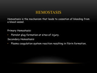
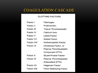
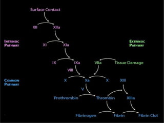
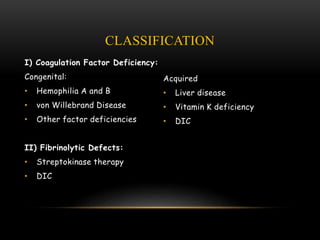
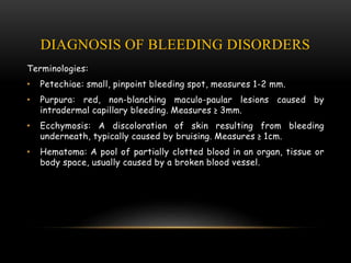
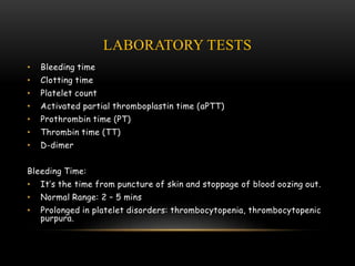
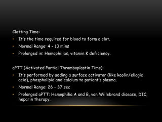
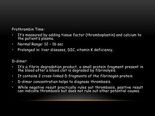
Presented by:
Rajat Hegde
BLEEDING DISORDERS:
CLASSIFICATION AND DIAGNOSIS
CONTENTS
 Introduction
 Hemostasis
 Coagulation cascade
 Classification
 Diagnosis
 Laboratory tests
 Conclusion
 References
INTRODUCTION
• Bleeding disorders are rare disorders affecting the way the body
controls bleeding.
• Excessive bleeding can result from:
1. Increased fragility of vessels
2. Platelet deficiency or dysfunction
3. Derangement of coagulation
• The normal hemostatic response involves the blood vessel wall, the
platelets and the clotting cascade.
• Various laboratory tests are used in evaluation of a bleeding
diathesis.
Blood vessel injury triggers the following sequence:
1. Vessel constriction.
2. Circulating platelets adhere to vessel.
3. A series of enzymatic reactions involving coagulation proteins,
produces fibrin to form a stable hemostatic plug.
HEMOSTASIS
Hemostasis is the mechanism that leads to cessation of bleeding from
a blood vessel.
Primary Hemostasis:
• Platelet plug formation at sites of injury.
Secondary Hemostasis:
• Plasma coagulation system reaction resulting in fibrin formation.
COAGULATION CASCADE
CLASSIFICATION
I) Coagulation Factor Deficiency:
Congenital:
• Hemophilia A and B
• von Willebrand Disease
• Other factor deficiencies
II) Fibrinolytic Defects:
• Streptokinase therapy
• DIC
Acquired
• Liver disease
• Vitamin K deficiency
• DIC
III) Vascular Disorders:
• Scurvy
• Purpura
• Hereditary hemorrhagic telangiectasia
• Cushing syndrome
• Ehlers Danlos syndrome
IV) Platelet Disorders:
1] Quantitative Disorders (Thrombocytopenia):
2] Qualitative Disorders:
Congenital:
• Glanzmann thrombasthenia
• von Willebrand disease
Acquired:
• Liver disease
• Drug-induced
• Alcoholism
Immune Mediated:
• Idiopathic
• Collagen vascular disease
• Sarcoidosis
Non-Immune Mediated:
• Microangiopathic hemolytic anemia
• Leukemia
• Myelofibrosis
DIAGNOSIS OF BLEEDING DISORDERS
Terminologies:
• Petechiae: small, pinpoint bleeding spot, measures 1-2 mm.
• Purpura: red, non-blanching maculo-paular lesions caused by
intradermal capillary bleeding. Measures ≥ 3mm.
• Ecchymosis: A discoloration of skin resulting from bleeding
underneath, typically caused by bruising. Measures ≥ 1cm.
• Hematoma: A pool of partially clotted blood in an organ, tissue or
body space, usually caused by a broken blood vessel.
• It’s important to have relevant clinical information before
interpreting results.
• From the history, it’s important to differentiate hereditary
disorders from acquired.
• Hereditary disorders generally start from an early age or
recurrent in nature and may have family history.
• Acquired disorders on the other hand occur at any age and have an
underlying predisposing cause.
• Depending on type of bleeding, it’s important to differentiate
between platelet and factor bleeds.
• History of medication should include replacement therapy, use of
anticoagulants like heparin besides intake of analgesics and
antibiotics etc.
• In general, symptoms of:
1. Platelet bleeds: petechial hemorrhage, mucosal bleed.
2. Coagulation factor disorders: deep abdominal bleeds, muscle
bleeds, hemostasis, ecchymotic patches.
LABORATORY TESTS
• Bleeding time
• Clotting time
• Platelet count
• Activated partial thromboplastin time (aPTT)
• Prothrombin time (PT)
• Thrombin time (TT)
• D-dimer
Bleeding Time:
• It’s the time from puncture of skin and stoppage of blood oozing out.
• Normal Range: 2 – 5 mins
• Prolonged in platelet disorders: thrombocytopenia, thrombocytopenic
purpura.
Clotting Time:
• It’s the time required for blood to form a clot.
• Normal Range: 4 - 10 mins
• Prolonged in: Hemophilias, vitamin K deficiency.
aPTT (Activated Partial Thromboplastin Time):
• It’s performed by adding a surface activator (like kaolin/ellagic
acid), phospholipid and calcium to patient’s plasma.
• Normal Range: 26 – 37 sec
• Prolonged aPTT: Hemophilia A and B, von Willebrand disease, DIC,
heparin therapy.
Prothrombin Time:
• It’s measured by adding tissue factor (thromboplastin) and calcium to
the patient’s plasma.
• Normal Range: 12 – 16 sec
• Prolonged in: liver diseases, DIC, vitamin K deficiency,
D-dimer:
• It’s a fibrin degradation product, a small protein fragment present in
the blood after a blood clot is degraded by fibrinolysis.
• It contains 2 cross-linked D fragments of the fibrinogen protein.
• D-dimer concentration helps to diagnose thrombosis.
• While negative result practically rules out thrombosis, positive result
can indicate thrombosis but does not rule out other potential causes.
CONCLUSION
• The medical and family history is very important in evaluating
patients with bleeding disorders.
• Dental extractions are very common major stresses of hemostatic
mechanism, and a prior history is very important.
• So, a thorough understanding and knowledge about bleeding
disorders is very much needed for dental professionals to minimise
the complications of many treatment procedures.
REFERENCES
 Robbins and Cottran Pathologic Basis of Disease.
 Davidson’s Essentials of Medicine.
THANK YOU

Bleeding Disorders: Classification and Diagnosis

  • 1.
    Presented by: Rajat Hegde BLEEDINGDISORDERS: CLASSIFICATION AND DIAGNOSIS
  • 2.
    CONTENTS  Introduction  Hemostasis Coagulation cascade  Classification  Diagnosis  Laboratory tests  Conclusion  References
  • 3.
    INTRODUCTION • Bleeding disordersare rare disorders affecting the way the body controls bleeding. • Excessive bleeding can result from: 1. Increased fragility of vessels 2. Platelet deficiency or dysfunction 3. Derangement of coagulation • The normal hemostatic response involves the blood vessel wall, the platelets and the clotting cascade. • Various laboratory tests are used in evaluation of a bleeding diathesis.
  • 4.
    Blood vessel injurytriggers the following sequence: 1. Vessel constriction. 2. Circulating platelets adhere to vessel. 3. A series of enzymatic reactions involving coagulation proteins, produces fibrin to form a stable hemostatic plug.
  • 5.
    HEMOSTASIS Hemostasis is themechanism that leads to cessation of bleeding from a blood vessel. Primary Hemostasis: • Platelet plug formation at sites of injury. Secondary Hemostasis: • Plasma coagulation system reaction resulting in fibrin formation.
  • 6.
  • 8.
    CLASSIFICATION I) Coagulation FactorDeficiency: Congenital: • Hemophilia A and B • von Willebrand Disease • Other factor deficiencies II) Fibrinolytic Defects: • Streptokinase therapy • DIC Acquired • Liver disease • Vitamin K deficiency • DIC
  • 9.
    III) Vascular Disorders: •Scurvy • Purpura • Hereditary hemorrhagic telangiectasia • Cushing syndrome • Ehlers Danlos syndrome
  • 10.
    IV) Platelet Disorders: 1]Quantitative Disorders (Thrombocytopenia): 2] Qualitative Disorders: Congenital: • Glanzmann thrombasthenia • von Willebrand disease Acquired: • Liver disease • Drug-induced • Alcoholism Immune Mediated: • Idiopathic • Collagen vascular disease • Sarcoidosis Non-Immune Mediated: • Microangiopathic hemolytic anemia • Leukemia • Myelofibrosis
  • 11.
    DIAGNOSIS OF BLEEDINGDISORDERS Terminologies: • Petechiae: small, pinpoint bleeding spot, measures 1-2 mm. • Purpura: red, non-blanching maculo-paular lesions caused by intradermal capillary bleeding. Measures ≥ 3mm. • Ecchymosis: A discoloration of skin resulting from bleeding underneath, typically caused by bruising. Measures ≥ 1cm. • Hematoma: A pool of partially clotted blood in an organ, tissue or body space, usually caused by a broken blood vessel.
  • 12.
    • It’s importantto have relevant clinical information before interpreting results. • From the history, it’s important to differentiate hereditary disorders from acquired. • Hereditary disorders generally start from an early age or recurrent in nature and may have family history. • Acquired disorders on the other hand occur at any age and have an underlying predisposing cause. • Depending on type of bleeding, it’s important to differentiate between platelet and factor bleeds.
  • 13.
    • History ofmedication should include replacement therapy, use of anticoagulants like heparin besides intake of analgesics and antibiotics etc. • In general, symptoms of: 1. Platelet bleeds: petechial hemorrhage, mucosal bleed. 2. Coagulation factor disorders: deep abdominal bleeds, muscle bleeds, hemostasis, ecchymotic patches.
  • 14.
    LABORATORY TESTS • Bleedingtime • Clotting time • Platelet count • Activated partial thromboplastin time (aPTT) • Prothrombin time (PT) • Thrombin time (TT) • D-dimer Bleeding Time: • It’s the time from puncture of skin and stoppage of blood oozing out. • Normal Range: 2 – 5 mins • Prolonged in platelet disorders: thrombocytopenia, thrombocytopenic purpura.
  • 15.
    Clotting Time: • It’sthe time required for blood to form a clot. • Normal Range: 4 - 10 mins • Prolonged in: Hemophilias, vitamin K deficiency. aPTT (Activated Partial Thromboplastin Time): • It’s performed by adding a surface activator (like kaolin/ellagic acid), phospholipid and calcium to patient’s plasma. • Normal Range: 26 – 37 sec • Prolonged aPTT: Hemophilia A and B, von Willebrand disease, DIC, heparin therapy.
  • 16.
    Prothrombin Time: • It’smeasured by adding tissue factor (thromboplastin) and calcium to the patient’s plasma. • Normal Range: 12 – 16 sec • Prolonged in: liver diseases, DIC, vitamin K deficiency, D-dimer: • It’s a fibrin degradation product, a small protein fragment present in the blood after a blood clot is degraded by fibrinolysis. • It contains 2 cross-linked D fragments of the fibrinogen protein. • D-dimer concentration helps to diagnose thrombosis. • While negative result practically rules out thrombosis, positive result can indicate thrombosis but does not rule out other potential causes.
  • 17.
    CONCLUSION • The medicaland family history is very important in evaluating patients with bleeding disorders. • Dental extractions are very common major stresses of hemostatic mechanism, and a prior history is very important. • So, a thorough understanding and knowledge about bleeding disorders is very much needed for dental professionals to minimise the complications of many treatment procedures.
  • 18.
    REFERENCES  Robbins andCottran Pathologic Basis of Disease.  Davidson’s Essentials of Medicine.
  • 19.