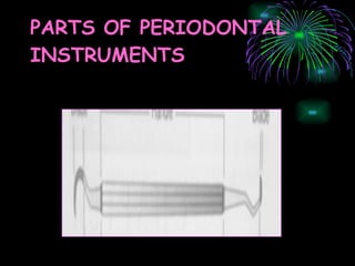
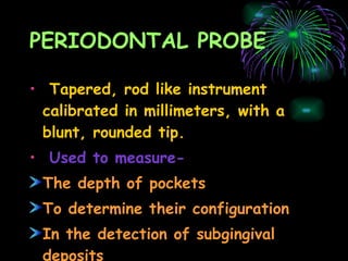
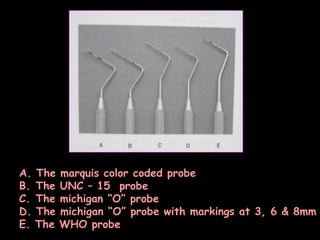
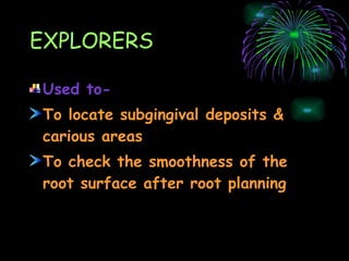
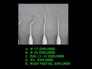
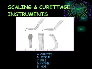
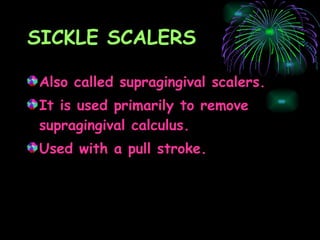
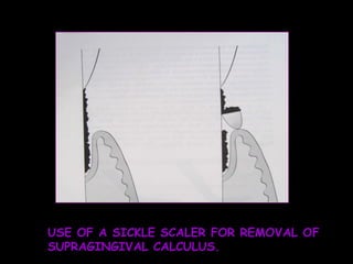
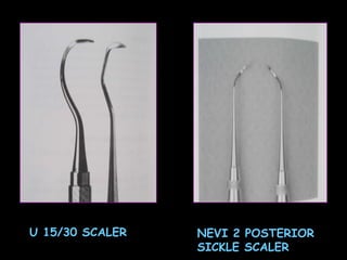
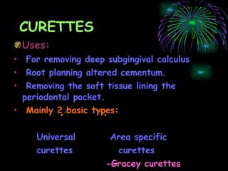
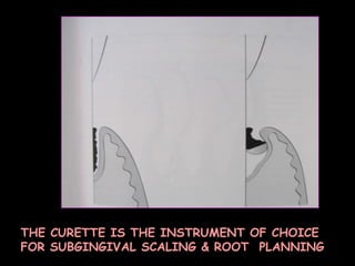
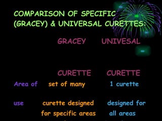
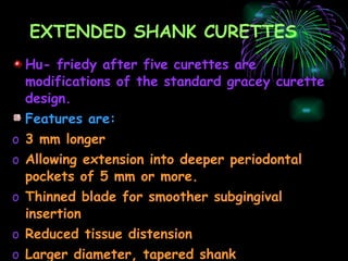
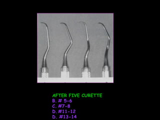
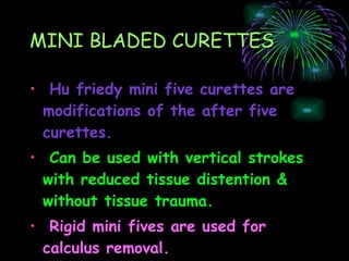
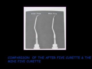
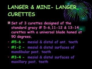
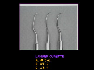
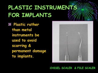
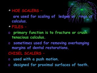
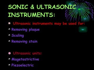
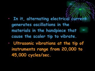
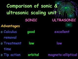
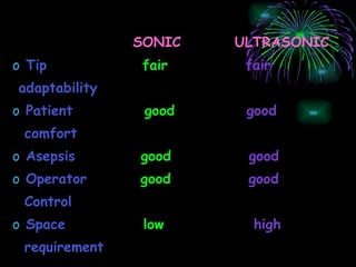
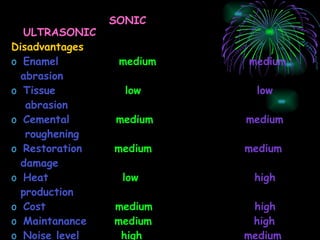
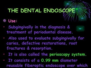
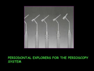
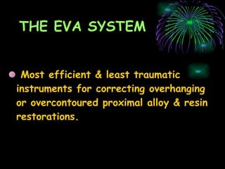
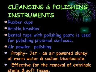
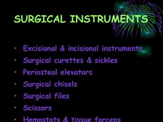
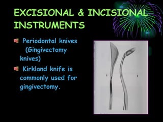
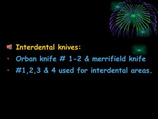
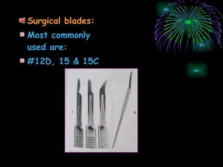
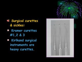
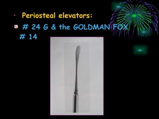
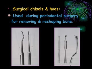
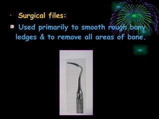
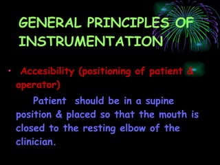
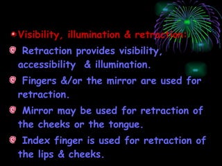
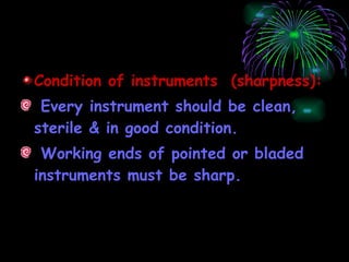
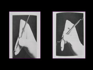
PERIODONTAL  INSTRUMENTATION
Peridontal instruments are designed for specific purposes- Removing calculus Planning root surfaces Curetting the gingiva Removing diseased tissue
CLASSIFICATION 1.PERIODONTAL PROBE – is used to locate, measure & mark pockets. 2.EXPLORER – is used to locate calculus deposits & caries.
3.SCALING,ROOT PLANNING & CURETTAGE INSTRUMENTS – To removal of plaque  & calcified deposits from the crown & root of a tooth. Removal of altered cementum from the subgingival root surface. Debridement of the soft tissue lining the pocket.
Scaling & curetting instruments used to remove supragingival calculus. i.Sickel scalers –   used to remove supragingival calculus. ii.Curettes –   used for subgingival scaling, root lining & removal of soft tissue lining the pocket.
iii.Hoe,Chisel & file scalers –   Used to remove subgingival calculus & altered cementum. iv.Ultrasonic & sonic instruments- Used for scaling & cleaning tooth surfaces & curetting the soft tissue wall of the periodontal pocket.
4.THE PERIODONTAL ENDOSCOPE - is used to visualize deep subgingivally in pockets & furcations. 5.CLEANSING & POLISHING INSTRUMENTS- such as rubber cups,brushes & dental cap are used to clean & polishing tooth surface.
PARTS OF PERIODONTAL INSTRUMENTS
Stainless steel are most commonly used metal for manufacturing instruments. High carbon steel are also available.
PERIODONTAL PROBE Tapered, rod like instrument calibrated in millimeters, with a blunt, rounded tip. Used to measure- The depth of pockets To determine their configuration In the detection of subgingival deposits
A. The marquis color coded probe The UNC – 15  probe The michigan “O” probe The michigan “O” probe with markings at 3, 6 & 8mm E. The WHO probe
EXPLORERS Used to- To locate subgingival deposits & carious areas To check the smoothness of the root surface after root planning
# 17 EXPLORER # 23 EXPLORER C. EXD 11-12 EXPLORER #3  EXPLORER E. #3CH PIGTAIL EXPLORER
SCALING & CURETTAGE INSTRUMENTS CURETTE SICKLE FILE D.CHISEL E. HOE
SICKLE SCALERS Also called supragingival scalers. It is used primarily to remove supragingival calculus. Used with a pull stroke.
Sickles with straight shanks are designed for use on anterior teeth & premolars. Sickles with contra-angled shanks are used for post.teeth. Different types of scalers are: 204SD - small, curved sickle scaler U15/30 - large scalers Jaquette sickles #1,2 & 3 - medium sized Curved 204 sickles - large,medium or small blades Nevi 2 post. sickle  scaler
USE OF A SICKLE SCALER FOR REMOVAL OF SUPRAGINGIVAL CALCULUS.
U 15/30 SCALER NEVI 2 POSTERIOR SICKLE SCALER
CURETTES Uses: For removing deep subgingival calculus Root planning altered cementum.  Removing the soft tissue lining the periodontal pocket. Mainly 2 basic types: Universal  Area specific  curettes  curettes -Gracey curettes
THE CURETTE IS THE INSTRUMENT OF CHOICE FOR SUBGINGIVAL SCALING & ROOT  PLANNING
COMPARISON OF SPECIFIC (GRACEY) & UNIVERSAL CURETTES: GRACEY  UNIVESAL  CURETTE  CURETTE Area of   set of many   1 curette   use  curette designed   designed for for specific areas   all areas & surfaces.   & surfaces.
GRACEY  UNIVERSAL  CURETTE  CURETTE   Cutting edge Use    1 cutting edge     both cutting     used     edges used   Curvature   curved in  2     curved in 1 planes    Plane Blade angle   Offset blade:   Not offset:   face of blade     face of blade   beveled at 60     beveled at 90    degrees to shank    degrees to shank.
EXTENDED SHANK CURETTES Hu- friedy after five curettes are modifications of the standard gracey curette design. Features are: 3 mm longer Allowing extension into deeper periodontal pockets of 5 mm or more. Thinned blade for smoother subgingival insertion Reduced tissue distension Larger diameter, tapered shank Use:   for heavy or tenacious calculus
AFTER FIVE CURETTE # 5-6  #7-8 #11-12 D. #13-14
MINI BLADED CURETTES Hu friedy mini five curettes are modifications of the after five curettes. Can be used with vertical strokes with reduced tissue distention & without tissue trauma. Rigid mini fives are used for calculus removal.
COMPARISON  OF THE AFTER FIVE CURETTE & THE MINI FIVE CURETTE
LANGER & MINI- LANGER CURETTES Set of 3 curettes designed of the standard gracy # 5-6,11-12 & 13-14 curettes with a universal blade honed at 90 degrees. #5-6 -   mesial & distal of ant. teeth #1-2 -   mesial & distal surfaces of mandibular post. teeth #3-4 -   mesial & distal surfaces of maxillary post. teeth
LANGER CURETTE A. # 5-6 B. #1-2 C. #3-4
PLASTIC INSTRUMENTS FOR IMPLANTS Plastic rather than metal instruments be used to avoid scarring & permanent damage to implants. CHISEL SCALER  & FILE SCALER
HOE SCALERS –  are used for scaling of  ledges or  rings of calculus. FILES –  primary function is to fracture or crush tenacious calculus. sometimes used for removing overhanging margins of dental restorations. CHISEL SCALERS – used with a push motion. designed for proximal surfaces of teeth.
SONIC & ULTRASONIC INSTRUMENTS: Ultrasonic instruments may be used for:  Removing plaque Scaling  Removing stain  Ultrasonic units: Magetostrictive  Piezoelectric
ULTRASONIC & SONIC TIPS CAVITRON FSI SLIM LINE ULTRASONIC TIPS
In it, alternating electrical current generates oscillations in the materials in the handpiece that cause the scaler tip to vibrate. Ultrasonic vibrations at the tip of instruments range from 20,000 to 45,000 cycles/sec.
Comparison of sonic & ultrasonic scaling unit : SONIC  ULTRASONIC   Advantages  Calculus   good  excellent removal Treatment   low  low time Tip action   orbital  magneto:elliptical piezo: linear
SONIC  ULTRASONIC Tip  fair  fair  adaptability Patient   good   good comfort Asepsis   good   good Operator  good   good Control Space  low   high requirement
SONIC  ULTRASONIC Disadvantages Enamel  medium   medium  abrasion Tissue  low  low abrasion Cemental  medium  medium roughening Restoration  medium  medium  damage Heat  low  high  production Cost  medium  high Maintanance  medium   high Noise level   high  medium
THE DENTAL ENDOSCOPE Use: Subgingivally in the diagnosis & treatment of periodontal disease. Also used to evaluate subgingivally for caries, defective restorations, root fractures & resorption. It is also called the  perioscopy system. It consists of a  0.99 mm  diameter  reusable fiberoptic endoscope over which is fitted a disposable, sterile sheath.
PERIODONTAL EXPLORERS FOR THE PERIOSCOPY SYSTEM
THE EVA SYSTEM Most efficient & least traumatic  instruments for correcting overhanging or overcontoured proximal alloy & resin restorations.
CLEANSING & POLISHING INSTRUMENTS Rubber cups Bristle brushes Dental tape with polishing paste is used for polishing proximal surfaces. Air powder  polishing Prophy- Jet – an air powered slurey of warm water & sodium bicarbonate. Effective for the removal of extrinsic stains & soft tissue.
SURGICAL INSTRUMENTS Excisional & incisional instruments Surgical curettes & sickles Periosteal elevators Surgical chisels  Surgical files Scissors Hemostats & tissue forceps
EXCISIONAL & INCISIONAL INSTRUMENTS Periodontal knives  (Gingivectomy  knives) Kirkland knife is commonly used for gingivectomy.
Interdental knives: Orban knife # 1-2 & merrifield knife #1,2,3 & 4 used for interdental areas.
Surgical blades:  Most commonly used are: #12D, 15 & 15C
Surgical curettes & sickles: Kramer curettes  #1,2 & 3 Kirlkand surgical instruments are heavy curettes.
Periosteal elevators: # 24 G & the GOLDMAN FOX # 14
Surgical chisels & hoes: Used  during periodontal surgery for removing & reshaping bone.
Surgical files: Used primarily to smooth rough bony ledges & to remove all areas of bone.
Scissors & Nippers: Used for: Removing tabs of tissue during gingivectomy Trimming the margins of flaps Enlarging incisions in periodontal abscesses Removing muscle attachments in mucogingival surgery. Needle holders : are used to suture the flap at the desired position after the surgical procedure.
CONVENTIONAL NEEDLE HOLDER CASTROVIEJO NEEDLE HOLDER
GENERAL PRINCIPLES OF  INSTRUMENTATION Accesibility (positioning of patient & operator) Patient  should be in a supine position & placed so that the mouth is closed to the resting elbow of the clinician.
Visibility, illumination & retraction: Retraction provides visibility, accessibility  & illumination. Fingers &/or the mirror are used for retraction. Mirror may be used for retraction of the cheeks or the tongue. Index finger is used for retraction of the lips & cheeks.
Condition of instruments  (sharpness): Every instrument should be clean, sterile & in good condition. Working ends of pointed or bladed instruments must be sharp.
Maintaining a clean field: Adequate suction is essential for ejecting a saliva. Blood & debris can be removed from the operative field with suction & by wiping or blotting with gauze squares.
Instrument stabilization: Stability  & control are essential for effective instrumentation & avoidance of injury to the patient or clinician. 2 major factors are: 1. instrument grasp 2. finger rest
1. Instrument grasp:   More effective  & stable grasp is modified pen grasp. Also standard pen grasp ensures the greatest control. Palm & thumb grasp :   useful for stabilizing instruments during sharpening for manipulating air & water syringe.
MODIFIED PEN GRASP STANDARD PEN GRASP
PALM & THUMB GRASP
2. Finger rest : Intraoral Extraoral fulcrum Intraoral finger rests are: Conventional:   finger rest is established on tooth surfaces immediately adjacent to the working area. Cross – arch:   finger rest is established on tooth surfaces on the other side of the same arch. Opposite arch:   finger rest is established on tooth surfaces on the opposite arch. Finger on finger:   finger rest is established on the index finger or thumb of the non operating hand.
Extraoral fulcrum are: Palm up Palm down
Instrument activation: Adaptation: refers to the manner in which working end of a periodontal instrument is placed against the surface of a tooth. objective-  to make the working end of the instrument conform to the contour of the tooth surface.
Angulation : refers to angle between the face of a bladed instrument & the tooth surface. may also be called the “tooth blade relationship”. Lateral pressure: refers to the pressure created when force is applied against the surface of a tooth with the cutting edge of a bladed instrument.
Strokes: Exploratory stroke Scaling stroke Root planning stroke Vertical & oblique stroke are used most frequently. Exploratory stroke: to evaluate the dimensions of the pocket & to detect calculus & irregularities of the tooth surface. Scaling stroke: used for removal of both supragingival & subgingival calculus.
Root planning stroke: used for final smoothing & planning of the root surfaces.
CHAIR POSITIONS It may be describe by the location of the operator or by the location of the operator’s arm. For a right – handed operator, there are essentially 3 positions -   right front, right & right rear. These are sometimes referred to as the   7, 9 & 11 o’clock positions. For the left handed operator, the 3 positions are   left front, left, & left rear or the 5,3 & 1 o’clock positions. A 4 th  position, direct rear or 12 o’clock position.
Right front position: It facilitates examination & work on   mandibular anterior teeth, mandibular posterior teeth (especially on the right side) & maxillary anterior teeth.
Right position: In this, the operator is directly to the right of the patient. This position is convenient for operating   on the facial surfaces of the maxillary & mandibular right posterior teeth & the occlusal surfaces of the mandibular right posterior teeth.
Right rear position: It is the position of the choice for most of the procedure. Most of the areas of the mouth are accessible & can be viewed directly or indirectly using mouth mirror. The lingual & incisal (occlusal) surfaces of the maxillary teethare viewed in the mouth mirror. Direct vision may be used on mandibular teeth, particularly on the left side.
Direct rear position: It has limited application & primarily used for operating on the lingual surfaces of mandibular anterior teeth.

Periodontal instrumentation

  • 1.
  • 2.
    Peridontal instruments aredesigned for specific purposes- Removing calculus Planning root surfaces Curetting the gingiva Removing diseased tissue
  • 3.
    CLASSIFICATION 1.PERIODONTAL PROBE– is used to locate, measure & mark pockets. 2.EXPLORER – is used to locate calculus deposits & caries.
  • 4.
    3.SCALING,ROOT PLANNING &CURETTAGE INSTRUMENTS – To removal of plaque & calcified deposits from the crown & root of a tooth. Removal of altered cementum from the subgingival root surface. Debridement of the soft tissue lining the pocket.
  • 5.
    Scaling & curettinginstruments used to remove supragingival calculus. i.Sickel scalers – used to remove supragingival calculus. ii.Curettes – used for subgingival scaling, root lining & removal of soft tissue lining the pocket.
  • 6.
    iii.Hoe,Chisel & filescalers – Used to remove subgingival calculus & altered cementum. iv.Ultrasonic & sonic instruments- Used for scaling & cleaning tooth surfaces & curetting the soft tissue wall of the periodontal pocket.
  • 7.
    4.THE PERIODONTAL ENDOSCOPE- is used to visualize deep subgingivally in pockets & furcations. 5.CLEANSING & POLISHING INSTRUMENTS- such as rubber cups,brushes & dental cap are used to clean & polishing tooth surface.
  • 8.
  • 9.
    Stainless steel aremost commonly used metal for manufacturing instruments. High carbon steel are also available.
  • 10.
    PERIODONTAL PROBE Tapered,rod like instrument calibrated in millimeters, with a blunt, rounded tip. Used to measure- The depth of pockets To determine their configuration In the detection of subgingival deposits
  • 11.
    A. The marquiscolor coded probe The UNC – 15 probe The michigan “O” probe The michigan “O” probe with markings at 3, 6 & 8mm E. The WHO probe
  • 12.
    EXPLORERS Used to-To locate subgingival deposits & carious areas To check the smoothness of the root surface after root planning
  • 13.
    # 17 EXPLORER# 23 EXPLORER C. EXD 11-12 EXPLORER #3 EXPLORER E. #3CH PIGTAIL EXPLORER
  • 14.
    SCALING & CURETTAGEINSTRUMENTS CURETTE SICKLE FILE D.CHISEL E. HOE
  • 15.
    SICKLE SCALERS Alsocalled supragingival scalers. It is used primarily to remove supragingival calculus. Used with a pull stroke.
  • 16.
    Sickles with straightshanks are designed for use on anterior teeth & premolars. Sickles with contra-angled shanks are used for post.teeth. Different types of scalers are: 204SD - small, curved sickle scaler U15/30 - large scalers Jaquette sickles #1,2 & 3 - medium sized Curved 204 sickles - large,medium or small blades Nevi 2 post. sickle scaler
  • 17.
    USE OF ASICKLE SCALER FOR REMOVAL OF SUPRAGINGIVAL CALCULUS.
  • 18.
    U 15/30 SCALERNEVI 2 POSTERIOR SICKLE SCALER
  • 19.
    CURETTES Uses: Forremoving deep subgingival calculus Root planning altered cementum. Removing the soft tissue lining the periodontal pocket. Mainly 2 basic types: Universal Area specific curettes curettes -Gracey curettes
  • 20.
    THE CURETTE ISTHE INSTRUMENT OF CHOICE FOR SUBGINGIVAL SCALING & ROOT PLANNING
  • 21.
    COMPARISON OF SPECIFIC(GRACEY) & UNIVERSAL CURETTES: GRACEY UNIVESAL CURETTE CURETTE Area of set of many 1 curette use curette designed designed for for specific areas all areas & surfaces. & surfaces.
  • 22.
    GRACEY UNIVERSAL CURETTE CURETTE Cutting edge Use 1 cutting edge both cutting used edges used Curvature curved in 2 curved in 1 planes Plane Blade angle Offset blade: Not offset: face of blade face of blade beveled at 60 beveled at 90 degrees to shank degrees to shank.
  • 23.
    EXTENDED SHANK CURETTESHu- friedy after five curettes are modifications of the standard gracey curette design. Features are: 3 mm longer Allowing extension into deeper periodontal pockets of 5 mm or more. Thinned blade for smoother subgingival insertion Reduced tissue distension Larger diameter, tapered shank Use: for heavy or tenacious calculus
  • 24.
    AFTER FIVE CURETTE# 5-6 #7-8 #11-12 D. #13-14
  • 25.
    MINI BLADED CURETTESHu friedy mini five curettes are modifications of the after five curettes. Can be used with vertical strokes with reduced tissue distention & without tissue trauma. Rigid mini fives are used for calculus removal.
  • 26.
    COMPARISON OFTHE AFTER FIVE CURETTE & THE MINI FIVE CURETTE
  • 27.
    LANGER & MINI-LANGER CURETTES Set of 3 curettes designed of the standard gracy # 5-6,11-12 & 13-14 curettes with a universal blade honed at 90 degrees. #5-6 - mesial & distal of ant. teeth #1-2 - mesial & distal surfaces of mandibular post. teeth #3-4 - mesial & distal surfaces of maxillary post. teeth
  • 28.
    LANGER CURETTE A.# 5-6 B. #1-2 C. #3-4
  • 29.
    PLASTIC INSTRUMENTS FORIMPLANTS Plastic rather than metal instruments be used to avoid scarring & permanent damage to implants. CHISEL SCALER & FILE SCALER
  • 30.
    HOE SCALERS – are used for scaling of ledges or rings of calculus. FILES – primary function is to fracture or crush tenacious calculus. sometimes used for removing overhanging margins of dental restorations. CHISEL SCALERS – used with a push motion. designed for proximal surfaces of teeth.
  • 31.
    SONIC & ULTRASONICINSTRUMENTS: Ultrasonic instruments may be used for: Removing plaque Scaling Removing stain Ultrasonic units: Magetostrictive Piezoelectric
  • 32.
    ULTRASONIC & SONICTIPS CAVITRON FSI SLIM LINE ULTRASONIC TIPS
  • 33.
    In it, alternatingelectrical current generates oscillations in the materials in the handpiece that cause the scaler tip to vibrate. Ultrasonic vibrations at the tip of instruments range from 20,000 to 45,000 cycles/sec.
  • 34.
    Comparison of sonic& ultrasonic scaling unit : SONIC ULTRASONIC Advantages Calculus good excellent removal Treatment low low time Tip action orbital magneto:elliptical piezo: linear
  • 35.
    SONIC ULTRASONICTip fair fair adaptability Patient good good comfort Asepsis good good Operator good good Control Space low high requirement
  • 36.
    SONIC ULTRASONICDisadvantages Enamel medium medium abrasion Tissue low low abrasion Cemental medium medium roughening Restoration medium medium damage Heat low high production Cost medium high Maintanance medium high Noise level high medium
  • 37.
    THE DENTAL ENDOSCOPEUse: Subgingivally in the diagnosis & treatment of periodontal disease. Also used to evaluate subgingivally for caries, defective restorations, root fractures & resorption. It is also called the perioscopy system. It consists of a 0.99 mm diameter reusable fiberoptic endoscope over which is fitted a disposable, sterile sheath.
  • 38.
    PERIODONTAL EXPLORERS FORTHE PERIOSCOPY SYSTEM
  • 39.
    THE EVA SYSTEMMost efficient & least traumatic instruments for correcting overhanging or overcontoured proximal alloy & resin restorations.
  • 40.
    CLEANSING & POLISHINGINSTRUMENTS Rubber cups Bristle brushes Dental tape with polishing paste is used for polishing proximal surfaces. Air powder polishing Prophy- Jet – an air powered slurey of warm water & sodium bicarbonate. Effective for the removal of extrinsic stains & soft tissue.
  • 41.
    SURGICAL INSTRUMENTS Excisional& incisional instruments Surgical curettes & sickles Periosteal elevators Surgical chisels Surgical files Scissors Hemostats & tissue forceps
  • 42.
    EXCISIONAL & INCISIONALINSTRUMENTS Periodontal knives (Gingivectomy knives) Kirkland knife is commonly used for gingivectomy.
  • 43.
    Interdental knives: Orbanknife # 1-2 & merrifield knife #1,2,3 & 4 used for interdental areas.
  • 44.
    Surgical blades: Most commonly used are: #12D, 15 & 15C
  • 45.
    Surgical curettes &sickles: Kramer curettes #1,2 & 3 Kirlkand surgical instruments are heavy curettes.
  • 46.
    Periosteal elevators: #24 G & the GOLDMAN FOX # 14
  • 47.
    Surgical chisels &hoes: Used during periodontal surgery for removing & reshaping bone.
  • 48.
    Surgical files: Usedprimarily to smooth rough bony ledges & to remove all areas of bone.
  • 49.
    Scissors & Nippers:Used for: Removing tabs of tissue during gingivectomy Trimming the margins of flaps Enlarging incisions in periodontal abscesses Removing muscle attachments in mucogingival surgery. Needle holders : are used to suture the flap at the desired position after the surgical procedure.
  • 50.
    CONVENTIONAL NEEDLE HOLDERCASTROVIEJO NEEDLE HOLDER
  • 51.
    GENERAL PRINCIPLES OF INSTRUMENTATION Accesibility (positioning of patient & operator) Patient should be in a supine position & placed so that the mouth is closed to the resting elbow of the clinician.
  • 52.
    Visibility, illumination &retraction: Retraction provides visibility, accessibility & illumination. Fingers &/or the mirror are used for retraction. Mirror may be used for retraction of the cheeks or the tongue. Index finger is used for retraction of the lips & cheeks.
  • 53.
    Condition of instruments (sharpness): Every instrument should be clean, sterile & in good condition. Working ends of pointed or bladed instruments must be sharp.
  • 54.
    Maintaining a cleanfield: Adequate suction is essential for ejecting a saliva. Blood & debris can be removed from the operative field with suction & by wiping or blotting with gauze squares.
  • 55.
    Instrument stabilization: Stability & control are essential for effective instrumentation & avoidance of injury to the patient or clinician. 2 major factors are: 1. instrument grasp 2. finger rest
  • 56.
    1. Instrument grasp: More effective & stable grasp is modified pen grasp. Also standard pen grasp ensures the greatest control. Palm & thumb grasp : useful for stabilizing instruments during sharpening for manipulating air & water syringe.
  • 57.
    MODIFIED PEN GRASPSTANDARD PEN GRASP
  • 58.
  • 59.
    2. Finger rest: Intraoral Extraoral fulcrum Intraoral finger rests are: Conventional: finger rest is established on tooth surfaces immediately adjacent to the working area. Cross – arch: finger rest is established on tooth surfaces on the other side of the same arch. Opposite arch: finger rest is established on tooth surfaces on the opposite arch. Finger on finger: finger rest is established on the index finger or thumb of the non operating hand.
  • 60.
    Extraoral fulcrum are:Palm up Palm down
  • 61.
    Instrument activation: Adaptation:refers to the manner in which working end of a periodontal instrument is placed against the surface of a tooth. objective- to make the working end of the instrument conform to the contour of the tooth surface.
  • 62.
    Angulation : refersto angle between the face of a bladed instrument & the tooth surface. may also be called the “tooth blade relationship”. Lateral pressure: refers to the pressure created when force is applied against the surface of a tooth with the cutting edge of a bladed instrument.
  • 63.
    Strokes: Exploratory strokeScaling stroke Root planning stroke Vertical & oblique stroke are used most frequently. Exploratory stroke: to evaluate the dimensions of the pocket & to detect calculus & irregularities of the tooth surface. Scaling stroke: used for removal of both supragingival & subgingival calculus.
  • 64.
    Root planning stroke:used for final smoothing & planning of the root surfaces.
  • 65.
    CHAIR POSITIONS Itmay be describe by the location of the operator or by the location of the operator’s arm. For a right – handed operator, there are essentially 3 positions - right front, right & right rear. These are sometimes referred to as the 7, 9 & 11 o’clock positions. For the left handed operator, the 3 positions are left front, left, & left rear or the 5,3 & 1 o’clock positions. A 4 th position, direct rear or 12 o’clock position.
  • 66.
    Right front position:It facilitates examination & work on mandibular anterior teeth, mandibular posterior teeth (especially on the right side) & maxillary anterior teeth.
  • 67.
    Right position: Inthis, the operator is directly to the right of the patient. This position is convenient for operating on the facial surfaces of the maxillary & mandibular right posterior teeth & the occlusal surfaces of the mandibular right posterior teeth.
  • 68.
    Right rear position:It is the position of the choice for most of the procedure. Most of the areas of the mouth are accessible & can be viewed directly or indirectly using mouth mirror. The lingual & incisal (occlusal) surfaces of the maxillary teethare viewed in the mouth mirror. Direct vision may be used on mandibular teeth, particularly on the left side.
  • 69.
    Direct rear position:It has limited application & primarily used for operating on the lingual surfaces of mandibular anterior teeth.