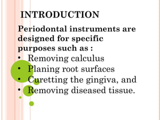
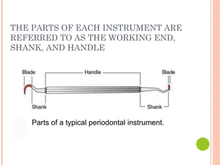
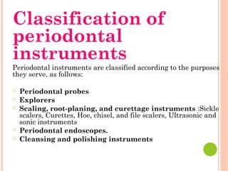
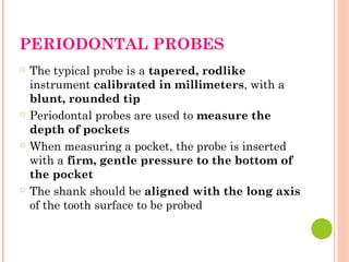
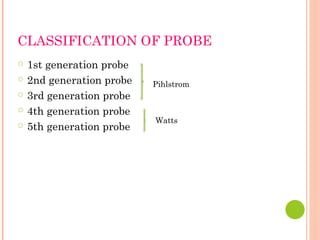
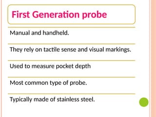
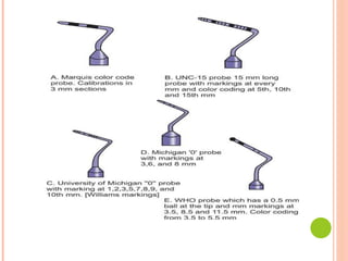
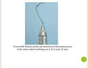
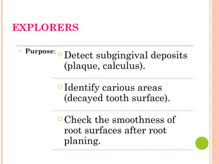
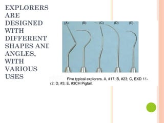
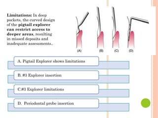
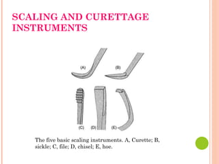
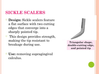
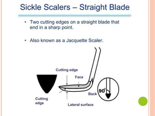
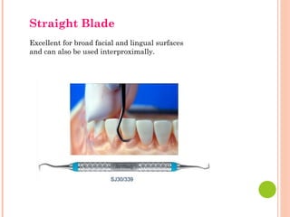
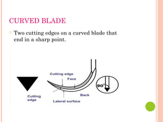
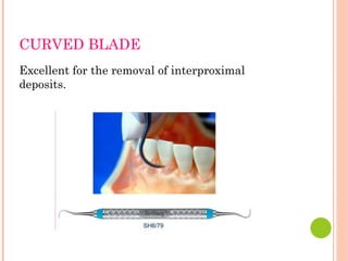
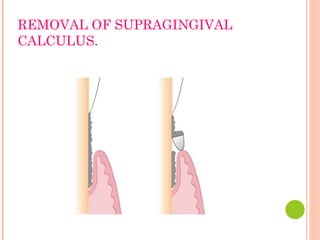
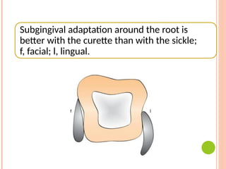
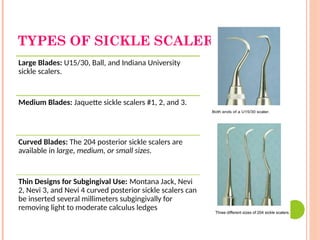
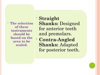
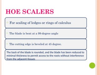
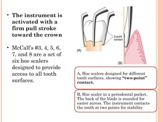
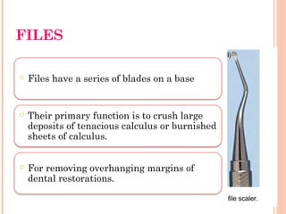
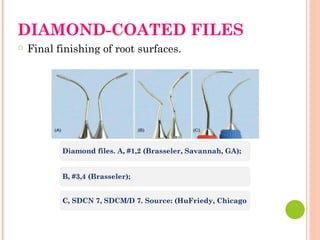
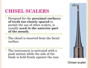
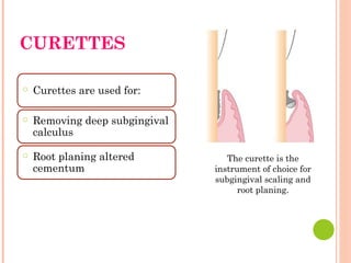
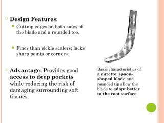
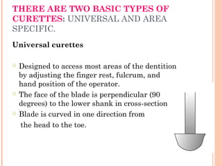
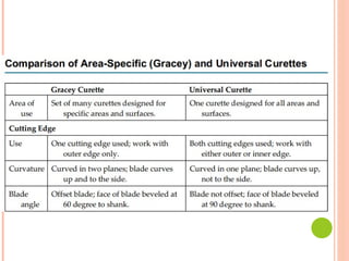
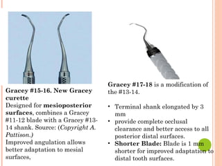
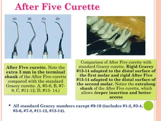
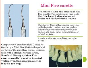
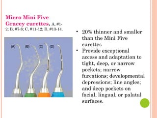
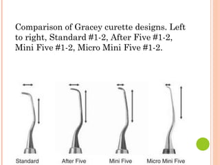
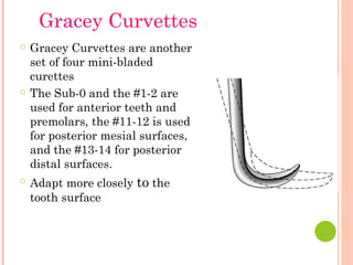
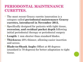
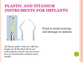
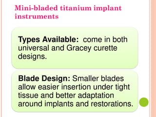
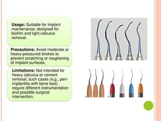
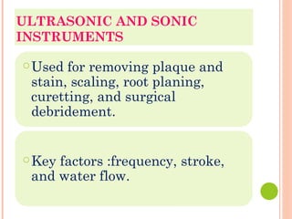
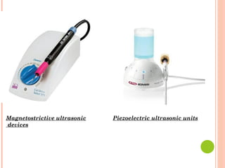
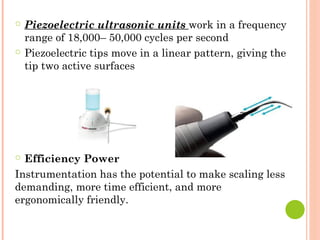
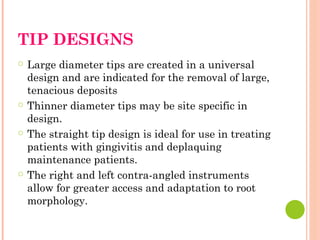
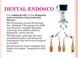
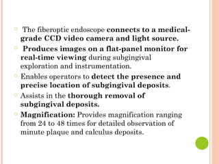
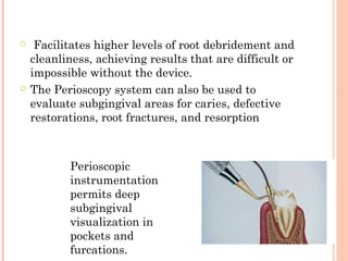
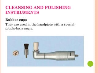
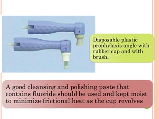
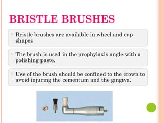
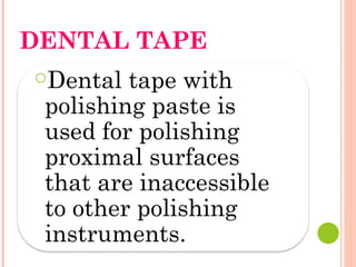
CLASSIFICATION OF
PERIODONTAL INSTRUMENTS
DEPARTMENT OF
PERIODONTOLOGY
SUBMITTED BY:
ANUSHKA DUBEY
BDS 4TH
YEAR
CONTENTS
○ Introduction
○ Parts of Instruments
○ Classification
○ Periodontal probes
○ Explorers
○ Scaling and Curettage Instruments
○ Ultrasonic and Sonic Instruments
○ Dental Endoscopes
○ Cleaning and polishing Instruments
Periodontal instruments are
designed for specific
purposes such as :
• Removing calculus
• Planing root surfaces
• Curetting the gingiva, and
• Removing diseased tissue.
INTRODUCTION
THE PARTS OF EACH INSTRUMENT ARE
REFERRED TO AS THE WORKING END,
SHANK, AND HANDLE
Classification of
periodontal
instruments
Periodontal instruments are classified according to the purposes
they serve, as follows:
○ Periodontal probes
○ Explorers
○ Scaling, root-planing, and curettage instruments :Sickle
scalers, Curettes, Hoe, chisel, and file scalers, Ultrasonic and
sonic instruments
○ Periodontal endoscopes.
○ Cleansing and polishing instruments
PERIODONTAL PROBES
○ The typical probe is a tapered, rodlike
instrument calibrated in millimeters, with a
blunt, rounded tip
○ Periodontal probes are used to measure the
depth of pockets
○ When measuring a pocket, the probe is inserted
with a firm, gentle pressure to the bottom of
the pocket
○ The shank should be aligned with the long axis
of the tooth surface to be probed
CLASSIFICATION OF PROBE
○ 1st generation probe
○ 2nd generation probe
○ 3rd generation probe
○ 4th generation probe
○ 5th generation probe
Pihlstrom
Watts
First Generation probe
Manual and handheld.
They rely on tactile sense and visual markings.
Used to measure pocket depth
Most common type of probe.
Typically made of stainless steel.
Second generation PROBE
Second generation probes are
manual.
Equipped with a built-in
mechanism.
Regulates probing force to 20
grams.
Third Generation PROBE
Electronic probes.
Measure pocket depth.
Display results on a digital screen.
Built-in mechanism to regulate probing
force.
Fourth generation probe
Fourth generation probes are three-
dimensional (3D).
They create detailed maps of periodontal
tissues.
Currently under development.
Potential to revolutionize diagnosis and
treatment of periodontal disease.
Fifth-generation probes
Fifth-generation probes are in
development.
Expected to be more advanced than
fourth-generation.
May utilize artificial intelligence for data
analysis.
Aimed at providing more accurate and
personalized diagnoses.
Curved #2 Nabers probe for detection of furcation areas,
with color-coded markings at 3, 6, 9, and 12 mm.
EXPLORERS
○ Purpose: ○ Detect subgingival deposits
(plaque, calculus).
○ Identify carious areas
(decayed tooth surface).
○ Check the smoothness of
root surfaces after root
planing.
EXPLORERS
ARE
DESIGNED
WITH
DIFFERENT
SHAPES AND
ANGLES,
WITH
VARIOUS
USES
Limitations: In deep
pockets, the curved design
of the pigtail explorer
can restrict access to
deeper areas, resulting
in missed deposits and
inadequate assessments..
A. Pigtail Explorer shows limitations
B. #3 Explorer insertion
C.#3 Explorer limitations
D. Periodontal probe insertion
SCALING AND CURETTAGE
INSTRUMENTS
The five basic scaling instruments. A, Curette; B,
sickle; C, file; D, chisel; E, hoe.
SICKLE SCALERS
○ Design: Sickle scalers feature
a flat surface with two cutting
edges that converge into a
sharply pointed tip.
○ This design provides strength,
making the tip resistant to
breakage during use.
○ Use: removing supragingival
calculus.
Triangular shape,
double-cutting edge,
and pointed tip
BLADE
DESIGN
Straight Curved
Straight Blade
Excellent for broad facial and lingual surfaces
and can also be used interproximally.
CURVED BLADE
○ Two cutting edges on a curved blade that
end in a sharp point.
CURVED BLADE
Excellent for the removal of interproximal
deposits.
REMOVAL OF SUPRAGINGIVAL
CALCULUS.
Subgingival adaptation around the root is
better with the curette than with the sickle;
f, facial; l, lingual.
TYPES OF SICKLE SCALERS:
Large Blades: U15/30, Ball, and Indiana University
sickle scalers.
Medium Blades: Jaquette sickle scalers #1, 2, and 3.
Curved Blades: The 204 posterior sickle scalers are
available in large, medium, or small sizes.
Thin Designs for Subgingival Use: Montana Jack, Nevi
2, Nevi 3, and Nevi 4 curved posterior sickle scalers can
be inserted several millimeters subgingivally for
removing light to moderate calculus ledges
• Straight
Shanks: Designed
for anterior teeth
and premolars.
• Contra-Angled
Shanks: Adapted
for posterior teeth.
○ The selection
of these
instruments
should be
based on the
area to be
scaled.
HOE SCALERS
○ For scaling of ledges or rings of calculus
○ The blade is bent at a 99-degree angle
○ The cutting edge is beveled at 45 degree.
The back of the blade is rounded, and the blade has been reduced to
minimal thickness to permit access to the roots without interference
from the adjacent tissues
A, Hoe scalers designed for different
tooth surfaces, showing “two-point”
contact.
B, Hoe scaler in a periodontal pocket.
The back of the blade is rounded for
easier access. The instrument contacts
the tooth at two points for stability
• The instrument is
activated with a
firm pull stroke
toward the crown
• McCall’s #3, 4, 5, 6,
7, and 8 are a set of
six hoe scalers
designed to provide
access to all tooth
surfaces.
FILES
○ Files have a series of blades on a base
○ Their primary function is to crush large
deposits of tenacious calculus or burnished
sheets of calculus.
○ For removing overhanging margins of
dental restorations.
DIAMOND-COATED FILES
○ Final finishing of root surfaces.
Diamond files. A, #1,2 (Brasseler, Savannah, GA);
B, #3,4 (Brasseler);
C, SDCN 7, SDCM/D 7. Source: (HuFriedy, Chicago
CHISEL SCALERS
○ Designed for the proximal surfaces
of teeth too closely spaced to
permit the use of other scalers, is
usually used in the anterior part
of the mouth.
○ The chisel is inserted from the facial
surface.
○ The instrument is activated with a
push motion while the side of the
blade is held firmly against the root.
CURETTES
○ Curettes are used for:
○ Removing deep subgingival
calculus
○ Root planing altered
cementum
The curette is the
instrument of choice for
subgingival scaling and
root planing.
○ Design Features:
● Cutting edges on both sides of
the blade and a rounded toe.
● Finer than sickle scalers; lacks
sharp points or corners.
○ Advantage: Provides good
access to deep pockets
while reducing the risk of
damaging surrounding soft
tissues.
Basic characteristics of
a curette: spoon-
shaped blade and
rounded tip allow the
blade to adapt better
to the root surface
THERE ARE TWO BASIC TYPES OF
CURETTES: UNIVERSAL AND AREA
SPECIFIC.
Universal curettes
○ Designed to access most areas of the dentition
by adjusting the finger rest, fulcrum, and
hand position of the operator.
○ The face of the blade is perpendicular (90
degrees) to the lower shank in cross-section
○ Blade is curved in one direction from
the head to the toe.
A, Double-ended curette for the
removal of subgingival calculus.
B, Cross-section of the curette
blade (arrow) against the cemental
wall of a deep periodontal pocket.
C, Curette in position at the base of a
periodontal pocket on the facial
surface of a mandibular molar.
D, Curette inserted in a pocket with
the tip directed apically.
E, Curette in position at the base of a
pocket on the distal surface of the
mandibular molar.
○ Examples:
• Barnhart curettes
#1-2 and #5-6
• Columbia
curettes #13-14,
#2R-2L, and #4R-
4L
• Younger-Good
#7-8
• McCall’s #17-18
• Indiana
University #17-18
AREA-SPECIFIC CURETTES
• Instruments designed and angled to
adapt to specific anatomic areas
• Best instruments for subgingival
scaling and root planing
○ Gracey
curettes
#1-2 and #3-4: Anterior teeth
A, #5-6 :Anterior teeth and premolars
B #7-8 and 9-10: Posterior teeth: facial and lingual
C #11-12:Posterior teeth: mesial
D #13-14:Posterior teeth: distal
Gracey #13-14 curette. For distal
surfaces.
Gracey #11-12 curette. For mesial
surfaces
○ Single-Ended Gracey Curette
○ Typically consists of a set of 14 instruments.
○ Area-Specific Use
○ An experienced operator can adapt each instrument
for use in several different areas by altering the
position of his or her hand and the position of the
patient
○ Blade Angulation: Blade is angled 60–70 degrees
from the lower shank
○ Subgingival Scaling: Unique angulation allows for
precise placement
○ Curved Blade from head to toe and along the
cutting edge, allowing only a pull stroke.
Gracey #17-18 is a modification of
the #13-14.
• Terminal shank elongated by 3
mm
• provide complete occlusal
clearance and better access to all
posterior distal surfaces.
• Shorter Blade: Blade is 1 mm
shorter for improved adaptation to
distal tooth surfaces.
Gracey #15-16. New Gracey
curette
Designed for mesioposterior
surfaces, combines a Gracey
#11-12 blade with a Gracey #13-
14 shank. Source: (Copyright A.
Pattison.)
Improved angulation allows
better adaptation to mesial
surfaces,
After Five curette. Note the
extra 3 mm in the terminal
shank of the After Five curette
compared with the standard
Gracey curette. A, #5-6; B, #7-
8; C, #11-12; D, #13- 14.)
Comparison of After Five curette with
standard Gracey curette. Rigid Gracey
#13-14 adapted to the distal surface of
the first molar and rigid After Five
#13-14 adapted to the distal surface of
the second molar. Notice the extralong
shank of the After Five curette, which
allows deeper insertion and better
access
▪ All standard Gracey numbers except #9-10 (includes #1-2, #3-4,
#5-6, #7-8, #11-12, #13-14).
After Five Curette
Comparison of After Five curette and Mini
Five curette. The shorter Mini Five blade
(half the length) allows increased
access and reduced tissue trauma.
The shorter blade allows easier insertion
and adaptation in deep, narrow pockets;
furcations; developmental grooves; line
angles; and deep, tight, facial, lingual, or
palatal pockets.’
Area in which root morphology or tight
tissue
Comparison of standard rigid Gracey #5-
6 with rigid Mini Five #5-6 on the palatal
surfaces of the maxillary central incisors.
used with a straight vertical stroke.
Standard Gracey or After Five
curette usually cannot be inserted
vertically in this area because the
blade is too long.
Mini Five curette
Micro Mini Five
Gracey curettes. A, #1-
2; B, #7-8; C, #11-12; D, #13-14.
• 20% thinner and smaller
than the Mini Five
curettes
• Provide exceptional
access and adaptation to
tight, deep, or narrow
pockets; narrow
furcations; developmental
depressions; line angles;
and deep pockets on
facial, lingual, or palatal
surfaces.
Comparison of Gracey curette designs. Left
to right, Standard #1-2, After Five #1-2,
Mini Five #1-2, Micro Mini Five #1-2.
○ Gracey Curvettes are another
set of four mini-bladed
curettes
○ The Sub-0 and the #1-2 are
used for anterior teeth and
premolars, the #11-12 is used
for posterior mesial surfaces,
and the #13-14 for posterior
distal surfaces.
○ Adapt more closely to the
tooth surface
Gracey Curvettes
PERIODONTAL MAINTENANCE
CURETTES.
○ The most recent Gracey curette innovation is a
category called periodontal maintenance Gracey
curettes, introduced in November 2015.
○ Specifically designed for patients with tight tissue,
recession, and residual pocket depth following
initial periodontal therapy or periodontal surgery
○ Length: 1 mm shorter than standard blades.
○ Thickness: 20% thinner, allowing easier insertion
and access.
○ Blade-to-Shank Angle: Offset at 60 degrees
(standard is 70 degrees) for better adaptation to tight
tissue.
• Reduced tissue
distention increases
patient comfort.
• Improved access to root
surfaces and furcation
areas.
• Helps prevent spanning
across root depressions.
Blade Size: Three-
quarter length, between
standard and mini-bladed
Gracey curettes for better
adaptation.
LANGER AND MINI-LANGER
CURETTES
○ A set of three combining shank
designs of standard Gracey #5-6, #11-
12, and #13-14 with universal blades
honed at 90 degrees.
○ Can adapt to mesial and distal
surfaces without changing
instruments.
○
Langer #5-6: For anterior teeth
○ Langer #1-2: Adapted for
mandibular posterior teeth with
Gracey #11-12 shank.
○ Langer #3-4: Adapted for maxillary
posterior teeth with Gracey #13-14
shank.
QUÉTIN FURCATION
CURETTES
○ The Quétin furcation curettes are actually hoes
with a shallow, halfmoon radius that fits into the
roof or floor of the furcation.
Quétin furcation curettes: BL2 (larger)
and BL1 (smaller).
• These instruments
remove burnished
calculus from recessed
areas of the furcation
• Lessen the likelihood of
root damage.
SCHWARTZ
PERIOTRIEVERS
• Design:Set of two double-
ended instruments.
• Highly magnetized for
effective retrieval
• Function: Retrieving
broken instrument tips from
periodontal pockets.
• They are indispensable
when the clinician has
broken a curette tip in a
furcation or deep pocket.
PLASTIC AND TITANIUM
INSTRUMENTS FOR IMPLANTS
(A) Plastic probe: Colorvue. (B) New
Implacare II Barnhart #5-6 cone
socket plastic curette tips that screw
into an autoclavable stainless steel
handle.
Used to avoid scarring
and damage to imlants
Types Available: come in both
universal and Gracey curette
designs.
Blade Design: Smaller blades
allow easier insertion under tight
tissue and better adaptation
around implants and restorations.
Mini-bladed titanium implant
instruments
Usage: Suitable for implant
maintenance; designed for
biofilm and light calculus
removal.
Precautions: Avoid moderate or
heavy-pressured strokes to
prevent scratching or roughening
of implant surfaces.
Limitations: Not intended for
heavy calculus or cement
removal; such cases (e.g., peri-
implantitis with bone loss)
require different instrumentation
and possible surgical
intervention.
ULTRASONIC AND SONIC
INSTRUMENTS
○Used for removing plaque and
stain, scaling, root planing,
curetting, and surgical
debridement.
○Key factors :frequency, stroke,
and water flow.
○Frequency: Frequency is defined as
the number of times per second an
insert tip moves back and forth
during one cycle in an orbital,
elliptic, or linear stroke path.
○Stroke: Stroke is the maximum
distance the insert tip travels during
one cycle or stoke path
Water Flow in Ultrasonic Scalers:
○ Types: Ultrasonic scalers can be manually or
automatically tuned.
○ Manual-Tuned Units: Feature three control
knobs (water, tuning, power) allowing clinicians
to adjust frequency.
○ Auto-Tuned Units: Have two knobs (water,
power) and maintain a stable frequency through
feedback that adjusts the insert's vibration
automatically.
○ Water contributes to three physiologic effects that enhance
the efficacy of power scalers: acoustic streaming, acoustic
turbulence, and cavitation.
○ Acoustic streaming is the unidirectional fluid flow caused
by ultrasound waves.
○ Acoustic turbulence is created when the movement of the
tip causes the coolant to accelerate, producing an intensified
swirling effect.
○ This turbulence continues until cavitation occurs. Cavitation
is the formation of bubbles in water caused by the high
turbulence.
○ In vitro, the combination of acoustic streaming, acoustic
turbulence, and cavitation has been shown to disrupt
microflora.
TYPE AND BENEFIT OF POWER
INSTRUMENTS
○ Sonic units work at a frequency of 2000–6500
cycles per second and use a high- or low-speed air
source from the dental unit.
○ Water is delivered via same tubing used to deliver
water to a dental handpiece.
○ A sonic scaler tip travels in an elliptical or orbital
stroke pattern.
Magnetostrictive ultrasonic devices work in a
frequency range of 18,000–50,000 cycles per second.
Tips move in an elliptical or orbital stroke pattern.
This allows the tip four active working surfaces.
Magnetostrictive ultrasonic
devices
Piezoelectric ultrasonic units
○ Piezoelectric ultrasonic units work in a frequency
range of 18,000– 50,000 cycles per second
○ Piezoelectric tips move in a linear pattern, giving the
tip two active surfaces
○ Efficiency Power
Instrumentation has the potential to make scaling less
demanding, more time efficient, and more
ergonomically friendly.
TIP DESIGNS
○ Large diameter tips are created in a universal
design and are indicated for the removal of large,
tenacious deposits
○ Thinner diameter tips may be site specific in
design.
○ The straight tip design is ideal for use in treating
patients with gingivitis and deplaquing
maintenance patients.
○ The right and left contra-angled instruments
allow for greater access and adaptation to root
morphology.
DENTAL ENDOSCOPE
○ Use subgingivally in the diagnosis
and treatment of periodontal
disease
○ The Perioscopy system (Perioscopy,
Inc., Oakland, CA) consists of a 0.99-
mm diameter, reusable fiberoptic
endoscope over which is fitted a
disposable, sterile sheath.
○ The fiberoptic endoscope fits onto
periodontal probes and ultrasonic
instruments that have been designed
to accept it
○ Sheath delivers water irrigation that
flushes the pocket , keeping the field
clear.
○ The fiberoptic endoscope connects to a medical-
grade CCD video camera and light source.
○ Produces images on a flat-panel monitor for
real-time viewing during subgingival
exploration and instrumentation.
○ Enables operators to detect the presence and
precise location of subgingival deposits.
○ Assists in the thorough removal of
subgingival deposits.
○ Magnification: Provides magnification ranging
from 24 to 48 times for detailed observation of
minute plaque and calculus deposits.
○ Facilitates higher levels of root debridement and
cleanliness, achieving results that are difficult or
impossible without the device.
○ The Perioscopy system can also be used to
evaluate subgingival areas for caries, defective
restorations, root fractures, and resorption
Perioscopic
instrumentation
permits deep
subgingival
visualization in
pockets and
furcations.
CLEANSING AND POLISHING
INSTRUMENTS
Rubber cups
They are used in the handpiece with a special
prophylaxis angle.
A good cleansing and polishing paste that
contains fluoride should be used and kept moist
to minimize frictional heat as the cup revolves
Disposable plastic
prophylaxis angle with
rubber cup and with
brush.
BRISTLE BRUSHES
○ Bristle brushes are available in wheel and cup
shapes
○ The brush is used in the prophylaxis angle with a
polishing paste.
○ Use of the brush should be confined to the crown to
avoid injuring the cementum and the gingiva.
DENTAL TAPE
○Dental tape with
polishing paste is
used for polishing
proximal surfaces
that are inaccessible
to other polishing
instruments.
AIR-POWDER POLISHING
○ This device, called the Prophy-Jet (Dentsply
International, York, PA), is very effective for the
removal of extrinsic stains and soft deposits
○ It is used with a specially designed handpiece
CONCLUSION
○ Advancements in instrument making and dental
practice have led to a wide variety of instrument
designs.
○ Modern instruments can effectively access nearly
all areas of the teeth and gums.
○ Many efficient instruments from historical sets
have proven durable and continue to be included
in new sets.
REFERENCES
○ Newman and Carranza's Clinical Periodontology
THIRD SOUTH ASIA EDITION
THANKYOU

Classification of Periodontal Instruments

  • 1.
    CLASSIFICATION OF PERIODONTAL INSTRUMENTS DEPARTMENTOF PERIODONTOLOGY SUBMITTED BY: ANUSHKA DUBEY BDS 4TH YEAR
  • 2.
    CONTENTS ○ Introduction ○ Partsof Instruments ○ Classification ○ Periodontal probes ○ Explorers ○ Scaling and Curettage Instruments ○ Ultrasonic and Sonic Instruments ○ Dental Endoscopes ○ Cleaning and polishing Instruments
  • 3.
    Periodontal instruments are designedfor specific purposes such as : • Removing calculus • Planing root surfaces • Curetting the gingiva, and • Removing diseased tissue. INTRODUCTION
  • 4.
    THE PARTS OFEACH INSTRUMENT ARE REFERRED TO AS THE WORKING END, SHANK, AND HANDLE
  • 5.
    Classification of periodontal instruments Periodontal instrumentsare classified according to the purposes they serve, as follows: ○ Periodontal probes ○ Explorers ○ Scaling, root-planing, and curettage instruments :Sickle scalers, Curettes, Hoe, chisel, and file scalers, Ultrasonic and sonic instruments ○ Periodontal endoscopes. ○ Cleansing and polishing instruments
  • 6.
    PERIODONTAL PROBES ○ Thetypical probe is a tapered, rodlike instrument calibrated in millimeters, with a blunt, rounded tip ○ Periodontal probes are used to measure the depth of pockets ○ When measuring a pocket, the probe is inserted with a firm, gentle pressure to the bottom of the pocket ○ The shank should be aligned with the long axis of the tooth surface to be probed
  • 7.
    CLASSIFICATION OF PROBE ○1st generation probe ○ 2nd generation probe ○ 3rd generation probe ○ 4th generation probe ○ 5th generation probe Pihlstrom Watts
  • 8.
    First Generation probe Manualand handheld. They rely on tactile sense and visual markings. Used to measure pocket depth Most common type of probe. Typically made of stainless steel.
  • 9.
    Second generation PROBE Secondgeneration probes are manual. Equipped with a built-in mechanism. Regulates probing force to 20 grams.
  • 10.
    Third Generation PROBE Electronicprobes. Measure pocket depth. Display results on a digital screen. Built-in mechanism to regulate probing force.
  • 11.
    Fourth generation probe Fourthgeneration probes are three- dimensional (3D). They create detailed maps of periodontal tissues. Currently under development. Potential to revolutionize diagnosis and treatment of periodontal disease.
  • 12.
    Fifth-generation probes Fifth-generation probesare in development. Expected to be more advanced than fourth-generation. May utilize artificial intelligence for data analysis. Aimed at providing more accurate and personalized diagnoses.
  • 14.
    Curved #2 Nabersprobe for detection of furcation areas, with color-coded markings at 3, 6, 9, and 12 mm.
  • 15.
    EXPLORERS ○ Purpose: ○Detect subgingival deposits (plaque, calculus). ○ Identify carious areas (decayed tooth surface). ○ Check the smoothness of root surfaces after root planing.
  • 16.
  • 17.
    Limitations: In deep pockets,the curved design of the pigtail explorer can restrict access to deeper areas, resulting in missed deposits and inadequate assessments.. A. Pigtail Explorer shows limitations B. #3 Explorer insertion C.#3 Explorer limitations D. Periodontal probe insertion
  • 18.
    SCALING AND CURETTAGE INSTRUMENTS Thefive basic scaling instruments. A, Curette; B, sickle; C, file; D, chisel; E, hoe.
  • 19.
    SICKLE SCALERS ○ Design:Sickle scalers feature a flat surface with two cutting edges that converge into a sharply pointed tip. ○ This design provides strength, making the tip resistant to breakage during use. ○ Use: removing supragingival calculus. Triangular shape, double-cutting edge, and pointed tip
  • 20.
  • 22.
    Straight Blade Excellent forbroad facial and lingual surfaces and can also be used interproximally.
  • 23.
    CURVED BLADE ○ Twocutting edges on a curved blade that end in a sharp point.
  • 24.
    CURVED BLADE Excellent forthe removal of interproximal deposits.
  • 25.
  • 26.
    Subgingival adaptation aroundthe root is better with the curette than with the sickle; f, facial; l, lingual.
  • 27.
    TYPES OF SICKLESCALERS: Large Blades: U15/30, Ball, and Indiana University sickle scalers. Medium Blades: Jaquette sickle scalers #1, 2, and 3. Curved Blades: The 204 posterior sickle scalers are available in large, medium, or small sizes. Thin Designs for Subgingival Use: Montana Jack, Nevi 2, Nevi 3, and Nevi 4 curved posterior sickle scalers can be inserted several millimeters subgingivally for removing light to moderate calculus ledges
  • 28.
    • Straight Shanks: Designed foranterior teeth and premolars. • Contra-Angled Shanks: Adapted for posterior teeth. ○ The selection of these instruments should be based on the area to be scaled.
  • 29.
    HOE SCALERS ○ Forscaling of ledges or rings of calculus ○ The blade is bent at a 99-degree angle ○ The cutting edge is beveled at 45 degree. The back of the blade is rounded, and the blade has been reduced to minimal thickness to permit access to the roots without interference from the adjacent tissues
  • 30.
    A, Hoe scalersdesigned for different tooth surfaces, showing “two-point” contact. B, Hoe scaler in a periodontal pocket. The back of the blade is rounded for easier access. The instrument contacts the tooth at two points for stability • The instrument is activated with a firm pull stroke toward the crown • McCall’s #3, 4, 5, 6, 7, and 8 are a set of six hoe scalers designed to provide access to all tooth surfaces.
  • 31.
    FILES ○ Files havea series of blades on a base ○ Their primary function is to crush large deposits of tenacious calculus or burnished sheets of calculus. ○ For removing overhanging margins of dental restorations.
  • 32.
    DIAMOND-COATED FILES ○ Finalfinishing of root surfaces. Diamond files. A, #1,2 (Brasseler, Savannah, GA); B, #3,4 (Brasseler); C, SDCN 7, SDCM/D 7. Source: (HuFriedy, Chicago
  • 33.
    CHISEL SCALERS ○ Designedfor the proximal surfaces of teeth too closely spaced to permit the use of other scalers, is usually used in the anterior part of the mouth. ○ The chisel is inserted from the facial surface. ○ The instrument is activated with a push motion while the side of the blade is held firmly against the root.
  • 34.
    CURETTES ○ Curettes areused for: ○ Removing deep subgingival calculus ○ Root planing altered cementum The curette is the instrument of choice for subgingival scaling and root planing.
  • 35.
    ○ Design Features: ●Cutting edges on both sides of the blade and a rounded toe. ● Finer than sickle scalers; lacks sharp points or corners. ○ Advantage: Provides good access to deep pockets while reducing the risk of damaging surrounding soft tissues. Basic characteristics of a curette: spoon- shaped blade and rounded tip allow the blade to adapt better to the root surface
  • 36.
    THERE ARE TWOBASIC TYPES OF CURETTES: UNIVERSAL AND AREA SPECIFIC. Universal curettes ○ Designed to access most areas of the dentition by adjusting the finger rest, fulcrum, and hand position of the operator. ○ The face of the blade is perpendicular (90 degrees) to the lower shank in cross-section ○ Blade is curved in one direction from the head to the toe.
  • 37.
    A, Double-ended curettefor the removal of subgingival calculus. B, Cross-section of the curette blade (arrow) against the cemental wall of a deep periodontal pocket. C, Curette in position at the base of a periodontal pocket on the facial surface of a mandibular molar. D, Curette inserted in a pocket with the tip directed apically. E, Curette in position at the base of a pocket on the distal surface of the mandibular molar.
  • 38.
    ○ Examples: • Barnhartcurettes #1-2 and #5-6 • Columbia curettes #13-14, #2R-2L, and #4R- 4L • Younger-Good #7-8 • McCall’s #17-18 • Indiana University #17-18
  • 39.
    AREA-SPECIFIC CURETTES • Instrumentsdesigned and angled to adapt to specific anatomic areas • Best instruments for subgingival scaling and root planing ○ Gracey curettes #1-2 and #3-4: Anterior teeth A, #5-6 :Anterior teeth and premolars B #7-8 and 9-10: Posterior teeth: facial and lingual C #11-12:Posterior teeth: mesial D #13-14:Posterior teeth: distal
  • 40.
    Gracey #13-14 curette.For distal surfaces. Gracey #11-12 curette. For mesial surfaces
  • 41.
    ○ Single-Ended GraceyCurette ○ Typically consists of a set of 14 instruments. ○ Area-Specific Use ○ An experienced operator can adapt each instrument for use in several different areas by altering the position of his or her hand and the position of the patient ○ Blade Angulation: Blade is angled 60–70 degrees from the lower shank ○ Subgingival Scaling: Unique angulation allows for precise placement ○ Curved Blade from head to toe and along the cutting edge, allowing only a pull stroke.
  • 43.
    Gracey #17-18 isa modification of the #13-14. • Terminal shank elongated by 3 mm • provide complete occlusal clearance and better access to all posterior distal surfaces. • Shorter Blade: Blade is 1 mm shorter for improved adaptation to distal tooth surfaces. Gracey #15-16. New Gracey curette Designed for mesioposterior surfaces, combines a Gracey #11-12 blade with a Gracey #13- 14 shank. Source: (Copyright A. Pattison.) Improved angulation allows better adaptation to mesial surfaces,
  • 44.
    After Five curette.Note the extra 3 mm in the terminal shank of the After Five curette compared with the standard Gracey curette. A, #5-6; B, #7- 8; C, #11-12; D, #13- 14.) Comparison of After Five curette with standard Gracey curette. Rigid Gracey #13-14 adapted to the distal surface of the first molar and rigid After Five #13-14 adapted to the distal surface of the second molar. Notice the extralong shank of the After Five curette, which allows deeper insertion and better access ▪ All standard Gracey numbers except #9-10 (includes #1-2, #3-4, #5-6, #7-8, #11-12, #13-14). After Five Curette
  • 45.
    Comparison of AfterFive curette and Mini Five curette. The shorter Mini Five blade (half the length) allows increased access and reduced tissue trauma. The shorter blade allows easier insertion and adaptation in deep, narrow pockets; furcations; developmental grooves; line angles; and deep, tight, facial, lingual, or palatal pockets.’ Area in which root morphology or tight tissue Comparison of standard rigid Gracey #5- 6 with rigid Mini Five #5-6 on the palatal surfaces of the maxillary central incisors. used with a straight vertical stroke. Standard Gracey or After Five curette usually cannot be inserted vertically in this area because the blade is too long. Mini Five curette
  • 46.
    Micro Mini Five Graceycurettes. A, #1- 2; B, #7-8; C, #11-12; D, #13-14. • 20% thinner and smaller than the Mini Five curettes • Provide exceptional access and adaptation to tight, deep, or narrow pockets; narrow furcations; developmental depressions; line angles; and deep pockets on facial, lingual, or palatal surfaces.
  • 47.
    Comparison of Graceycurette designs. Left to right, Standard #1-2, After Five #1-2, Mini Five #1-2, Micro Mini Five #1-2.
  • 48.
    ○ Gracey Curvettesare another set of four mini-bladed curettes ○ The Sub-0 and the #1-2 are used for anterior teeth and premolars, the #11-12 is used for posterior mesial surfaces, and the #13-14 for posterior distal surfaces. ○ Adapt more closely to the tooth surface Gracey Curvettes
  • 49.
    PERIODONTAL MAINTENANCE CURETTES. ○ Themost recent Gracey curette innovation is a category called periodontal maintenance Gracey curettes, introduced in November 2015. ○ Specifically designed for patients with tight tissue, recession, and residual pocket depth following initial periodontal therapy or periodontal surgery ○ Length: 1 mm shorter than standard blades. ○ Thickness: 20% thinner, allowing easier insertion and access. ○ Blade-to-Shank Angle: Offset at 60 degrees (standard is 70 degrees) for better adaptation to tight tissue.
  • 50.
    • Reduced tissue distentionincreases patient comfort. • Improved access to root surfaces and furcation areas. • Helps prevent spanning across root depressions. Blade Size: Three- quarter length, between standard and mini-bladed Gracey curettes for better adaptation.
  • 51.
    LANGER AND MINI-LANGER CURETTES ○A set of three combining shank designs of standard Gracey #5-6, #11- 12, and #13-14 with universal blades honed at 90 degrees. ○ Can adapt to mesial and distal surfaces without changing instruments. ○ Langer #5-6: For anterior teeth ○ Langer #1-2: Adapted for mandibular posterior teeth with Gracey #11-12 shank. ○ Langer #3-4: Adapted for maxillary posterior teeth with Gracey #13-14 shank.
  • 52.
    QUÉTIN FURCATION CURETTES ○ TheQuétin furcation curettes are actually hoes with a shallow, halfmoon radius that fits into the roof or floor of the furcation. Quétin furcation curettes: BL2 (larger) and BL1 (smaller). • These instruments remove burnished calculus from recessed areas of the furcation • Lessen the likelihood of root damage.
  • 53.
    SCHWARTZ PERIOTRIEVERS • Design:Set oftwo double- ended instruments. • Highly magnetized for effective retrieval • Function: Retrieving broken instrument tips from periodontal pockets. • They are indispensable when the clinician has broken a curette tip in a furcation or deep pocket.
  • 54.
    PLASTIC AND TITANIUM INSTRUMENTSFOR IMPLANTS (A) Plastic probe: Colorvue. (B) New Implacare II Barnhart #5-6 cone socket plastic curette tips that screw into an autoclavable stainless steel handle. Used to avoid scarring and damage to imlants
  • 55.
    Types Available: comein both universal and Gracey curette designs. Blade Design: Smaller blades allow easier insertion under tight tissue and better adaptation around implants and restorations. Mini-bladed titanium implant instruments
  • 56.
    Usage: Suitable forimplant maintenance; designed for biofilm and light calculus removal. Precautions: Avoid moderate or heavy-pressured strokes to prevent scratching or roughening of implant surfaces. Limitations: Not intended for heavy calculus or cement removal; such cases (e.g., peri- implantitis with bone loss) require different instrumentation and possible surgical intervention.
  • 57.
    ULTRASONIC AND SONIC INSTRUMENTS ○Usedfor removing plaque and stain, scaling, root planing, curetting, and surgical debridement. ○Key factors :frequency, stroke, and water flow.
  • 58.
    ○Frequency: Frequency isdefined as the number of times per second an insert tip moves back and forth during one cycle in an orbital, elliptic, or linear stroke path. ○Stroke: Stroke is the maximum distance the insert tip travels during one cycle or stoke path
  • 59.
    Water Flow inUltrasonic Scalers: ○ Types: Ultrasonic scalers can be manually or automatically tuned. ○ Manual-Tuned Units: Feature three control knobs (water, tuning, power) allowing clinicians to adjust frequency. ○ Auto-Tuned Units: Have two knobs (water, power) and maintain a stable frequency through feedback that adjusts the insert's vibration automatically.
  • 60.
    ○ Water contributesto three physiologic effects that enhance the efficacy of power scalers: acoustic streaming, acoustic turbulence, and cavitation. ○ Acoustic streaming is the unidirectional fluid flow caused by ultrasound waves. ○ Acoustic turbulence is created when the movement of the tip causes the coolant to accelerate, producing an intensified swirling effect. ○ This turbulence continues until cavitation occurs. Cavitation is the formation of bubbles in water caused by the high turbulence. ○ In vitro, the combination of acoustic streaming, acoustic turbulence, and cavitation has been shown to disrupt microflora.
  • 61.
    TYPE AND BENEFITOF POWER INSTRUMENTS ○ Sonic units work at a frequency of 2000–6500 cycles per second and use a high- or low-speed air source from the dental unit. ○ Water is delivered via same tubing used to deliver water to a dental handpiece. ○ A sonic scaler tip travels in an elliptical or orbital stroke pattern. Magnetostrictive ultrasonic devices work in a frequency range of 18,000–50,000 cycles per second. Tips move in an elliptical or orbital stroke pattern. This allows the tip four active working surfaces.
  • 62.
  • 63.
    ○ Piezoelectric ultrasonicunits work in a frequency range of 18,000– 50,000 cycles per second ○ Piezoelectric tips move in a linear pattern, giving the tip two active surfaces ○ Efficiency Power Instrumentation has the potential to make scaling less demanding, more time efficient, and more ergonomically friendly.
  • 64.
    TIP DESIGNS ○ Largediameter tips are created in a universal design and are indicated for the removal of large, tenacious deposits ○ Thinner diameter tips may be site specific in design. ○ The straight tip design is ideal for use in treating patients with gingivitis and deplaquing maintenance patients. ○ The right and left contra-angled instruments allow for greater access and adaptation to root morphology.
  • 65.
    DENTAL ENDOSCOPE ○ Usesubgingivally in the diagnosis and treatment of periodontal disease ○ The Perioscopy system (Perioscopy, Inc., Oakland, CA) consists of a 0.99- mm diameter, reusable fiberoptic endoscope over which is fitted a disposable, sterile sheath. ○ The fiberoptic endoscope fits onto periodontal probes and ultrasonic instruments that have been designed to accept it ○ Sheath delivers water irrigation that flushes the pocket , keeping the field clear.
  • 66.
    ○ The fiberopticendoscope connects to a medical- grade CCD video camera and light source. ○ Produces images on a flat-panel monitor for real-time viewing during subgingival exploration and instrumentation. ○ Enables operators to detect the presence and precise location of subgingival deposits. ○ Assists in the thorough removal of subgingival deposits. ○ Magnification: Provides magnification ranging from 24 to 48 times for detailed observation of minute plaque and calculus deposits.
  • 67.
    ○ Facilitates higherlevels of root debridement and cleanliness, achieving results that are difficult or impossible without the device. ○ The Perioscopy system can also be used to evaluate subgingival areas for caries, defective restorations, root fractures, and resorption Perioscopic instrumentation permits deep subgingival visualization in pockets and furcations.
  • 68.
    CLEANSING AND POLISHING INSTRUMENTS Rubbercups They are used in the handpiece with a special prophylaxis angle.
  • 69.
    A good cleansingand polishing paste that contains fluoride should be used and kept moist to minimize frictional heat as the cup revolves Disposable plastic prophylaxis angle with rubber cup and with brush.
  • 70.
    BRISTLE BRUSHES ○ Bristlebrushes are available in wheel and cup shapes ○ The brush is used in the prophylaxis angle with a polishing paste. ○ Use of the brush should be confined to the crown to avoid injuring the cementum and the gingiva.
  • 71.
    DENTAL TAPE ○Dental tapewith polishing paste is used for polishing proximal surfaces that are inaccessible to other polishing instruments.
  • 72.
    AIR-POWDER POLISHING ○ Thisdevice, called the Prophy-Jet (Dentsply International, York, PA), is very effective for the removal of extrinsic stains and soft deposits ○ It is used with a specially designed handpiece
  • 73.
    CONCLUSION ○ Advancements ininstrument making and dental practice have led to a wide variety of instrument designs. ○ Modern instruments can effectively access nearly all areas of the teeth and gums. ○ Many efficient instruments from historical sets have proven durable and continue to be included in new sets.
  • 74.
    REFERENCES ○ Newman andCarranza's Clinical Periodontology THIRD SOUTH ASIA EDITION
  • 75.