
Recommended
More Related Content
What's hot
What's hot (20)
Similar to The Nervous System: Anatomy and Functions
Similar to The Nervous System: Anatomy and Functions (20)
More from NehaRohtagi1
More from NehaRohtagi1 (20)
Recently uploaded
Recently uploaded (20)
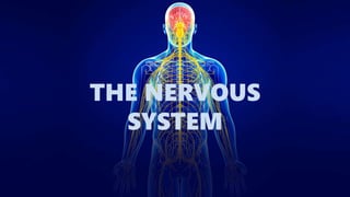
The Nervous System: Anatomy and Functions
- 2. CELLULAR ARCHITECTURE NEURONS (the structural and functional unit of nervous tissue) NEUROGLIAL CELLS (supporting tissue)
- 3. DIVISONS OF NERVOUS SYSTEM It is divided into : Central Nervous System (CNS) which comprises brain and spinal cord. It is responsible for integrating, coordinating the sensory information and ordering appropriate motor actions. Peripheral Nervous System (PNS) includes 12 pairs of cranial nerves and 31 pairs of spinal nerves. These provides afferent impulses to CNS and carries efferent impulses to muscles, glands and blood vessels.
- 4. THE BRAIN Human Brain is divided into three main parts on the basis of their functions and placements. The three main parts of the human brain are : CEREBRUM BRAIN STEM CEREBELLUM MIDBRAIN PONS MEDULLA OBLONGATA
- 5. DEVELOPMENT OF : THE BRAIN
- 7. CEREBRUM • Largest part of the brain • Outer grey matter and inner white matter • Made up of two cerebral hemispheres; incompletely separated by median Longitudinal Fissure. • Each hemisphere contains a cavity – Lateral Ventricle. • There are four lobes : Frontal Lobe Parietal Lobe Temporal Lobe Occipital Lobe Insula (deep within the Lateral sulcus)
- 8. FUNCTIONAL AREAS OF CEREBRAL CORTEX
- 9. THALAMUS
- 10. HYPOTHALAMUS The Hypothalamus is part of the diencephalon. It lies in the floor and lateral wall of the third ventricle. It’s functions are : • Endocrine control • Neurosecretion • General autonomic effect • Temperature regulation • Regulation of food and water intake • Sexual behavior and reproduction • Biological clocks • Emotion, fear, rage, aversion, pleasure and reward
- 11. LIMBIC SYSTEM • The Limbic System is a complex network of cortical areas and subcortical structures interconnected by directional pathways. Major limbic centers includes: • The Cortical Areas : cingulate gyrus, orbitotofrontal, insular and medial prefrontal cortices and parahippocampal gyrus. • Subcortical Structures including : The thalamus, septal area ,nucleus accumbens, hippocampus, hypothalamus and the amygdala. Connecting pathways of limbic system: • The alveus, the fimbria, the fornix, mammilllothalamic tract and the stria terminalis
- 12. BRAIN STEM • Located between the cerebrum and the spinal cord – provides a pathway for tracts running between higher and lower neural centers. • Consists of the Midbrain, Pons and Medulla Oblongata. • It consists of deep gray matter surrounded by white matter fiber tracts. • Produce automatic behaviors necessary for survival. • Each part of brainstem is connected to cerebellum by Cerebellar Peduncles(superior, middle and inferior) • Contains groups of nuclei and related fibers known as Reticulur Formation. • It is responsible for : control of level of consciousness, perception of pain, regulation of cardiovascular and respiratory systems. • Site of origin of Cranial Nerves ( from 3rd to 12th )
- 13. MIDBRAIN • The Midbrain develops from mesencephalon. • Connects the pons and cerebellum with the forebrain. • It comprises two lateral halves called cerebral peduncles which is again divided into: Anterior part – Crus Cerebri and Posterior part – Tegmentum, by a pigmented band of gray matter - Substantia Nigra • The central narrow cavity is called the Cerebral Aqueduct which connects 3rd and 4th ventricles. • The Tectum is the part of the midbrain posterior to the cerebral aqueduct; it has four small surface swellings reffered as two Superior Colliculi and two Inferior Colliculi. • The Tegmentum forms the floor of mid brain.
- 14. PONS • Located in the rostral to the medulla oblongata, caudal to the mid brain and ventral to the cerebellum Functions: • Relays sensory information between the cerebrum and cerebellum • Some theories say that it has a role in dreaming. • Control of respiration: The Apneustic Center- lower pons and The Pneumotaxic Center - upper pons • A number of cranial nerve nuclei are present in it: the trigeminal nerve, abducens nucleus, vestibulicochlear nuclei and facial nerve nucleus.
- 15. MEDULLA OBLONGATA The Medulla Oblongata lies between the Pons and the Spinal Cord. It contains centers which control key, autonomic body functions and it relays nerve signals between the brain and spinal cord. Important control centers include: • The Respiratory Center - controls the rate, rhythm and depth of breathing • The Cardiac Center - regulates heart beat • The Vasomotor Center - controls blood pressure • The Reflex Centers - reflex the centers for vomiting, coughing, sneezing, hiccupping and swallowing
- 16. • Cerebellum consists of three lobes- Anterior, Posterior and Flocculonodular. • The cerebellum compares the motor plan created in the cortex with motor performances and functions to smoothens and coordinate the movements. This is accompanied by making synaptic contacts with the brain stem ‘motor’ centers and the cerebral hemispheres. Functions : • Muscle tone • Coordination goal directed and spontaneous movements • Posture and balance • Eye movements • Motor learning • Some cognitive functions- language processing,selective attention CEREBELLUM (LITTLE BRAIN)
- 17. MENINGES • The Meninges are the membranes covering the brain and spinal cord for protection. • The meninges consists of the three membranes: DURAMATER (hard mother) ARACHNOID MATER (web like) PIAMATER (soft mother) • The space between the duramater and the vertebral wall -EPIDURAL SPACE • The space between the duramater and the arachnoid mater –SUBDURAL SPACE • The space between arachnoid mater and piamater –SUBARACHNOID SPACE
- 18. • It stretches from the upper border of the Foramen Magnum to the invertebral disc between the 1st and 2nd lumbar vertebrae. • In the newborns ,it extends to the level of the 3rd lumbar vertebrae. • Due to differential growth of the vertebral column relative to the spinal cord, the spinal cord segment do not always correspond to the vertebral levels. • Has two enlargements, Cervical and Lumbar due to cells and fibers of the limbs. • Ends inferiorly in a tapering Conus Medullaris. • Anchored to the coccyx by meningeal extension - Filum Terminale. SPINAL CORD
- 19. CROSS SECTION OF SPINAL CORD • Anterior median fissure and Posterior median sulcus - deep clefts partially separating left and right halves • Gray matter : neuron cell bodies, dendrites, axons - divided into horns Posterior (dorsal) horn Anterior (ventral) horn Lateral horn • White matter : myelinated axons - divided into three columns Ventral Dorsal Lateral - Each of these divided into Sensory or Motor tracts.
- 20. THANK YOU