1. NERVOUS SYSTEM
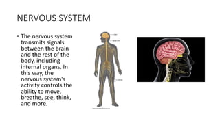
• The nervous system
transmits signals
between the brain
and the rest of the
body, including
internal organs. In
this way, the
nervous system's
activity controls the
ability to move,
breathe, see, think,
and more.
2. CELLS OF THE NERVOUS SYSTEM
There are two cells of the nervous system.
These are;
• Neuron
• Neuroglia
The functional unit of the nervous system is the nerve cell, or neuron
Neuroglia or glial are supportive cells in the nervous system that aid
the function of neurons.
3. NEURONS (Nerve Cells)
Neurons have three fundamental physiological properties:
1. Excitability
2. Conductivity
3. Secretion
A typical neuron is divided into three parts;
1. Soma or cell body
2. Dendrites
3. Axon
4. Cell body
Within the cell body is a nucleus, which controls the cell's activities and
contains the cell's genetic material.
Dendrites
Dendrites are short processes and have a large surface area for receiving
signals from other neurons and pass it to the cell body.
Axon
axon is the conducting region of the neuron and is responsible for
generating and transmitting impulses typically away from the cell body.
5.
6. NEUROGLIA (Glia)
There are six types of supportive cells
Four occur in the CNS
1. Oligodendrocytes
2. Ependymal cells
3. Microglia
4. Astrocytes
Two occur in the PNS
1. Schwann cells
2. Satellite cells
7.
8. Neuroglia in the CNS
There are four types of neuroglia found within the central nervous system:
• Astrocytes – maintain the blood brain barrier and preserve the chemical
environment by recycling ions and neurotransmitters
• Oligodendrocytes – myelinate axons in the central nervous system and
provide an overall structural framework
• Ependymal cells – line ventricles (brain) and central canal (spine) and are
involved in the production of cerebrospinal fluid
• Microglia – remove cell debris, wastes and pathogens via phagocytosis
Neuroglia in the PNS
There are two types of neuroglia found within the peripheral nervous system:
• Schwann cells – myelinate axons in the peripheral nervous system
• Satellite cells – regulate nutrient and neurotransmitter levels around neurons
in ganglia
9. Classification of nervous syestem
A)anatomical classfication
1. The central nervous system (CNS)
2. The peripheral nervous system (PNS)
B)Physiological classification
1. Somatic
2. Visceral or splanchnic or autonomic
C) Embryological Classification
13. CENTRAL NERVOUS SYSTEM
• The CNS consists of the brain and spinal cord
• CNS protected by a cranium surrounding the brain vertebral column
surrounding the spinal cord The CNS is bathed in cerebrospinal fluid
The CNS is composed of gray and white matter.
14. FOREBRAIN
Forebrain consist of the following regions;
1. Telencephalon (cerebrum) and
2. Diencephalon (Thalamus, hypothalamus, epithalamus and pituitary gland)
CEREBRUM
• The cerebrum is the largest part of the brain.
• It is divided into two hemispheres separated by the longitudinal fissure.
• The hemispheres are prominently marked with gyri and sulci.
• Portions of the two hemispheres are connected internally by the corpus
callosum
Each cerebral hemisphere is subdivided by deep sulci, or fissures, into the five
lobes: Frontal, Parietal, Occipital, Temporal and Insula
15.
16.
17.
18. DIENCEPHALON
• it is the median part of forebrain
A major autonomic region of the brain that consists of vital structures such as;
1. Thalamus,
2. Hypothalamus ,
3. Epithalamus ,
4. Metathalmus
5. subthalmus
• The thalamus is a large ovoid mass of gray matter
• The hypothalamus is a small portion of the diencephalon inferior to the
thalamus
• The epithalamus is the dorsal portion of the diencephalon that includes a
thin roof over the third ventricle
19.
20. MIDBRAIN
• The midbrain is a short section of the brain stem between the
diencephalon and the pons
• A short segment of the brainstem that connects the hindbrain and
forebrain
• It includes important centers for vision, hearing, pain, and motor
control
21.
22.
23. HINDBRAIN
• The embryonic hindbrain differentiates into two subdivisions the
• metencephalon and
• myelencephalon
• The metencephalon is the most superior portion of the hindbrain.
• The pons which measures about 2.5 cm long, forms a broad anterior bulge in the
brainstem just rostral to the medulla.
• It conducts signals up and down the brainstem and between the brainstem and
cerebellum
• The cerebellum is the largest part of the hindbrain and receives most of its input
by way of the pons.
• Occupies the inferior and posterior aspect of the cranial cavity
24.
25.
26. MYELENCEPHALON
• Myelencephalon contain the medulla oblongata
• The medulla oblongata is a bulbous structure about 3 cm (1In) long
Externally, the medulla resembles the spinal cord
27. SPINAL CORD
• It is an elongated cylindrical structure that is a ropelike bundle of nervous
tissue In adults, it averages about 1.8 cm thick and 45 cm long
• It begins as a continuation of the medulla oblongata at the level of the
foramen magnum
• The spinal cord serves three principal functions:
• Conduction, Locomotion and Reflexes
• The cord gives rise to 31 pairs of spinal nerves. the part supplied by each
pair of spinal nerves is called a segment.
• The spinal cord is divided into cervical, thoracic, lumbar, and sacral regions.
28. CROSS-SECTIONAL ANATOMY
• The spinal cord consists of two kinds of nervous tissue called gray and white matter.
• Gray matter has a relatively dull color because it contains little myelin. It has butterfly- or
H-shaped in cross sections
• It contains the somas, dendrites, and proximal parts of the axons of neurons.
• It is the site of synaptic contact between neurons (information processing)
GRAY AND WHITE MATTER
• White matter contains an abundance of myelinated axons, which give it a bright, pearly
white appearance.
• It is composed of bundles of axons called tracts or fascicule
• It carry signals from one part of the CNS to another.
• The spinal cord has two tracts; Ascending and descending tract
• Ascending tracts carry sensory information up the cord and descending tracts conduct
motor impulses down
29.
30. PERIPHERAL NERVOUS SYSTEM
• The (PNS) is that portion of the nervous system outside the central
nervous system
• Sensory receptors within the sensory organs, neurons, nerve, ganglia,
and plexuses are all part of the PNS
• The nerves of the PNS are classified as;
1. cranial nerves or
2. spinal nerves,
31. CRANIAL NERVES
• Cranial nerves are nerves that emerge directly from the brain and the
brainstem.
• There are 12 pairs of cranial nerves, numbered I to XII
• they relay information between the brain and parts of the body.
Considers to be parts of both CNS and PNS
• They are traditionally classified as sensory, motor or mixed base on
their functions.
32.
33. SPINAL NERVES
• There are 31 pairs of spinal nerves: 8 cervical (C1–C8), 12 thoracic
(T1–T12), 5 lumbar (L1–L5), 5 sacral (S1–S5), and 1 coccygeal (Co).
• The first cervical nerve emerges between the skull and atlas
• The others emerge through intervertebral foramina, including the
anterior and posterior foramina of the sacrum and the sacral hiatus.
34.
35. AUTONOMIC NERVOUS SYSTEM (ANS)
• ANS is motor nervous system that controls glands, cardiac muscle,
and smooth muscle
• The primary target organs of the ANS are the viscera of the thoracic
and abdominal cavities
• Its job is to regulate such fundamental states and life processes as
heart rate, blood pressure, body temperature, respiratory airflow,
pupillary diameter, digestion, energy metabolism, defecation, and
urination.
36. DIVISION OF THE ANS
• The ANS has two subsystems Sympathetic parasympathetic divisions
Sympathetic (Thoracolumbar) Division The sympathetic division is also called
the thoracolumbar division
• It has relatively short preganglionic and long postganglionic fibers
PARASYMPATHETIC DIVISION
The parasympathetic division is also called the craniosacral
Somas of the preganglionic neurons are located in the pons, medulla
oblongata, and segments S2 to S4 of the spinal cord
The parasympathetic division has long preganglionic fibers reaching almost
all the way to the target cells
short postganglionic fibers that cover the rest of the distance
37. Reflex arc
• A receptor in the skin detects a stimulus (the change in temperature).
• Sensory neurones send electrical impulses to relay neurones, which
are located in the spinal cord. ...
• Motor neurones send electrical impulses to an effector.
• The effector produces a response (muscle contracts to move hand
away).
38.
39.
40. Meninges
• Meninges are three layers of membranes that cover and protect your
brain and spinal cord (your central nervous system [CNS]). They're
known as:
1. Dura mater: This is the outer layer, closest to your skull.
2. Arachnoid mater: This is the middle layer.
3. Pia mater: This is the inner layer, closest to your brain tissue.
41.
42.
43.
44.
45.
46.
47.
48. • The conus medullaris is the terminal end of the spinal cord
• The cauda equina (CE) is a bundle of intradural nerve roots at the end
of the spinal cord, in the subarachnoid space distal to the conus
medullaris. Cauda is Latin for tail, and equina is Latin for horse (ie, the
"horse's tail").
• The filum terminale (FT) is a fibrous band that extends from the conus
medullaris to the periosteum of the coccyx
49.
50.
51.
52.
53.
54. Cerebellum
The cerebellum lies within the posterior cranial fossa of the skull (Figs.
1-8, 1-9, 1-9 and 1-10), posterior to the pons and the medulla
oblongata. It consists of two laterally placed hemispheres connected by
a median portion, the vermis. The cerebellum is connected to
the midbrain by the superior cerebellar peduncles, to the pons by the
middle cerebellar peduncles, and to the medulla by the inferior
cerebellar peduncles (see Fig. 6-9). The peduncles are composed of
large bundles of nerve fibers connecting the cerebellum to the
remainder of the nervous system
55.
56. Cerebellar peduncles connect the cerebellum to the brain stem.There
are six cerebellar peduncles in total, three on each side:
Superior cerebellar peduncle is a paired structure of white matter that
connects the cerebellum to the mid-brain.
Middle cerebellar peduncles connect the cerebellum to the pons and
are composed entirely of centripetal fibers.
Inferior cerebellar peduncle is a thick rope-like strand that occupies the
upper part of the posterior district of the medulla oblongata.
57.
58.
59. The cerebral aqueduct is a narrow 15 mm conduit that allows for
cerebrospinal fluid (CSF) to flow between the third ventricle and
the fourth ventricle.
60.
61.
62. • The falx cerebri is a sickle-shaped structure formed from the
invagination of the dura mater into the longitudinal fissure between
the cerebral hemispheres.