SlideShare a Scribd company logo
1 of 56
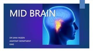
MID BRAIN
DR SANA YASEEN
ANATOMY DEPARTMENT
KIMS
BRAIN STEM REVIEW
BRAIN STEM
 It is a stalk-like part of the brain which connects the spinal cord with the forebrain.
 From below upwards it consists of three parts:
 medulla oblongata,
 pons, and
 midbrain.
 The midbrain is continuous above with the cerebral hemispheres.
 The medulla oblongata is continuous below with spinal cord.
 It is located in the posterior cranial fossa.
 Its ventral surface lies on the clivus.
 Posteriorly, the pons and medulla are separated from the cerebellum by the cavity of the fourth
ventricle.
Anterior view
FUNCTIONS OF
BRAIN STEM
four major functions:
 It provides passage to various
ascending and descending tracts
that connect the spinal cord to the
different parts of the forebrain.
 It contains important autonomic
reflex centres (vital centres)
associated with the control of
respiration heart rate and blood
pressure.
 It contains reticular activating
system which controls
consciousness.
 It contains important nuclei of the
last ten cranial nerves (i.e. IIIrd to
XIIth).
The impairment of reticular activating
system leads to progressive loss of
consciousness.
“
”
MID BRAIN
EXTERNAL FEATURES
 ANTERIOR VENTRAL VIEW
 POSTERIOR DORSAL VIEW
INTERNAL FEATURES
 AT SUPERIOR COLLICULLUS
 AT INFERIOR COLLICULUS
INTRODUCTION TO MID
BRAIN
 It is the upper and shortest part
of the brain-stem.
 It is about 0.8 inches , app 2cm
long and 2.5 cm wide.
 It connects the pons a cerebellum
to diencephalon
 Its cavity, the cerebral aqueduct
(aqueduct of Sylvius) connects the
third ventricle with the fourth
ventricle.
 It passes through the tentorial
notch.
It is related
on each side to the
 optic tract,
 parahippocampal gyrus,
 posterior cerebral artery, and
basal
Anteriorly to
 interpeduncular structures, such
as mammillary bodies, tuber
cinereum, etc.;
.
Posteriorly to
 splenium of corpus callosum,
 great cerebral vein,
 pineal body and
 posterior ends of right and left
thalami.
Cerebral
peduncle
OPTIC NERVE
BASILAR
ARTERY
ANTERIOR TO MIDBRAIN AT INTERPEDUNCULAR FOSSA
POSTERIOR VIEW
OF MIBRAIN
RELATION OF TENTORIUM CEREBELLI
WITH BASILAR ARTERY
ANTERIOR / VENTRAL VIEW
 It presents two crura cerebri which emerges from the cerebral hemispheres and
converge downwards to enter the pons forming the posterolateral boundaries of the
interpeduncular fossa.
 The superficial surface of the crus cerebri is finely corrugated by the underlying
longitudinal fibres.
 It is crossed transversely from above downwards by
 optic tract,
 posterior cerebral artery,
 superior cerebellar artery
 The oculomotor nerve emerges from a groove (occulomotor sulcus) on the medial side
of the crus cerebri ventrally.
 The trochlear nerve emerges on the dorsal aspect of the midbrain and curls around the
lateral aspect of the cerebral peduncle to appear on the ventral aspect of the midbrain
lateral to the oculomotor nerve.
 These two nerves run forward between the posterior cerebral and the superior cerebellar
arteries.
 Within interpeduncular
fossa is the depression
called posterior
perforating
 Its so called as it
posses small openings
for the central
branches of posterior
cerebral arteries to
runs through it.
POSTERIOR / DORSAL VIEW
 It presents four rounded elevations:
two superior and two inferior colliculi (or corpora quadrigemina).
 The colliculi are separated from each other by a cruciform sulcus.
 The vertical limb of sulcus when traced above forms a surface
depression which lodges the pineal body
 Traced below, it becomes continuous with the frenulum veli (a median
ridge on the dorsal surface of the superior medullary velum).
 The trochlear nerves emerges one on each side of the upper part of
frenulum veli after decussation in the superior medullary velum.
POSTERIOR/ DORSAL VIEW
SUPERIOR COLLICULUS
POSTERIOR / DORSAL VIEW
INFERIOR COLLICULUS
 Thick ridges of white matter extending from lateral side of
each colliculus constitute their brachia.
 superior brachium connect the superior colliculus to the lateral
geniculate body and the optic tract, and contributes in visual reflex.
 inferior brachium connect the inferior colliculus to the medial
geniculate body, and contributes in auditory reflex
 Trochlear nerve (IV CN) emerges lateral to frenulum orsal to
inferior colliculus runs in the posterior aspect of midbrain than
curve to run over lateral aspect of cerebral peduncles and
traverses interpeduncular cistern to petrosal end of cavernous
sinus.
INTERNAL STRUCTURE
 Each cerebral
peduncle is further
subdivided into three
parts,
 from dorsal to
ventral these are:
(a) tegmentum,
(b) substantia nigra, and
(c) crus cerebri.
Internal Structure
 The transverse section of midbrain shows a tiny canal, called cerebral aqueduct.
 A coronal plane passing through the aqueduct divides the midbrain into two
parts;
 small posterior part and
 large anterior part
 The small posterior part is called tectum and consists of four colliculi.
 The large anterior part is divided into two equal right and left halves by a vertical
plane, the cerebral peduncle.
Crus cerebri (basis pedunculi)
 It is the part of cerebral peduncle
 It is situated anterolateral to the substantia nigra.
 It contains important descending tracts which connect the cerebral cortex
to the anterior horn cells of the spinal cord, cranial nerve nuclei, and
pontine nuclei.
• corticospinal and corticonuclear fibres (pyramidal tract) occupy the
middle 2/3 of crus
• frontopontine fibres occupy the medial 1/6 of crus.
• temporopontine, parietopontine, and occipitopontine fibres occupy the
lateral 1/6 of the crus.
Substantia nigra
 It is a curved (crescent-shaped) pigmented band of grey matter.
 It is situated between tegmentum and crus cerebri. The concavity is smooth and
directed towards the tegmentum.
 From its convex margin spiky processes project into the substance of the crus
cerebri.
 It is a large motor nucleus that extends throughout the length of midbrain.
 It is divided into two parts:
(a) the dorsal part (pars compacta) containing medium sized cells and
(b) a ventral part (pars reticularis) containing fewer cells.
 The pars reticularis is intermingled with the fibres of crus cerebri.
 The substantia nigra is made up of deeply pigmented nerve cells which
contain melanin & iron.
 These cells synthesize dopamine which is carried through their axons
(nigrostriatal fibres) to the corpus striatum.
Clinical Correlation
 The degeneration or destruction of substantia nigra causes deficiency of
dopamine in the corpus striatum leading to a clinical condition called
Parkinsonism.
Transverse
Section of the
Midbrain at the
Level of the
Inferior Collicul
Grey matter
 It has a central grey matter (grey matter around the cerebral aqueduct)
 It contains two nuclei:
(a) nucleus of trochlear nerve
(b) mesencephalic nucleus of trigeminal nerve.
1. Trochlear nerve nucleus is situated close to the median plane just posterior to the medial longitudinal
fasciculus (MLF).
The emerging fibres of the trochlear nerve pass laterally and posteriorly around the central grey matter
and leave the midbrain just below the inferior colliculi.
The fibres of trochlear nerve now decussate in the superior medullary vellum and
wind round the lateral aspect of the midbrain to enter the lateral wall of cavernous sinus
2. The mesencephalic nucleus of trigeminal nerve lies in the lateral edge
of the central grey matter.
It receives proprioceptive impulses from muscles of mastication, teeth,
ocular and facial muscles.
3. An ovoid mass of grey matter underneath the inferior colliculus forms
the nucleus of inferior colliculus.
 It receives the afferent fibres of lateral lemniscus and gives the efferent
fibres to the medial geniculate body through the inferior brachium.
 4. Substantia nigra.
 5. The reticular formation is smaller than that in the pons and is
situated ventrolaterally between the medial lemniscus and the central
grey matter.
• The decussation of the superior cerebellar peduncles occupies the central part of the
tegmentum.
 This forms the most important feature in the lower part of the midbrain.
• The lemnisci are arranged in the form of a curved compact band of white fibres in the ventrolateral
part of the tegmentum, lateral to cerebellar decussation and dorsal to the substantia nigra.
 From medial to lateral side these are:
 medial lemniscus, trigeminal lemniscus, spinal lemniscus, and lateral lemniscus.
• The medial longitudinal fasciculus lies on the side of median plane ventral to the trochlear nerve
nucleus.
• The tectospinal tracts lie ventral to the medial longitudinal fasciculi.
• The rubrospinal tracts lie ventral to the decussation of the superior cerebellar peduncles.
White matter
Transverse Section of the Midbrain at the
Level of the Superior Colliculi
Grey matter
 The central grey matter in each half contains two nuclei:
 oculomotor nerve nucleus
 mesencephalic nucleus.
1. Oculomotor nucleus , lies in the ventromedial part.
 The nuclei of two sides fuse together forming a single complex having a triangular outline.
 The oculomotor nuclei are bounded laterally by the medial longitudinal fasciculus.
 The Edinger-Westphal nucleus which supplies the sphincter pupillae and ciliary muscle, forms part of
the oculomotor nucleus and is located dorsal to the rostral two-thirds of the main oculomotor
nucleus
 The emerging fibres of oculomotor nerve pass ventrally through the tegmentum intersecting red
nucleus and medial part of the substantia nigra, and emerge in the posterior part of interpeduncular
fossa through the sulcus on the medial aspect of crus cerebri.
2. Mesencephalic nucleus occupies the same position as in the lower part of the
midbrain.
3. Superior colliculus
 It is is a flattened mass formed of seven concentric alternating laminae of white
matter and grey matter.
 Unilateral lesion of the superior colliculus results in inability to track moving objects
in the contralateral field of vision, although the eye movements are normal.
4. Pretectal nucleus
 It is a small group of neurons and lies deep to the superolateral part of the superior
colliculus.
 It receives afferents from the lateral root of the optic tract and
 It gives efferents to the Edinger-Westphal nucleus (the parasympathetic component
of the oculomotor nucleus) of the same as well as of the opposite side.
 The pretectal nucleus is an important part of the pathway for pupillary light reflex
and consensual light reflex.
 Its lesion causes Argyll Robertson pupil in which light reflex is lost but
accommodation reflex remains intact.
Connections of the superior colliculus
The superior colliculus receives
afferent fibres from:
 1. The retinae (mainly the contralateral)
through the lateral geniculate body and
superior brachium,
 2. The spinal cord (pain and tactile fibres)
through spinotectal tract,
 3. The frontal and occipital visual cortex
(conjugate eye movements), and
 4. The inferior colliculus.
The efferent fibres from superior
colliculus form
 tectospinal and tectobulbar tracts, which
are
 probably responsible for the reflex
movements of the eyes, head, and neck in
response to visual stimuli.
 5. Red nucleus
 It is a cigar-shaped mass of grey matter which appears ovoid in cross-section.
 It is about 0.5 cm in diameter
 It is situated dorsomedial to the substantia nigra.
 In the fresh specimen it is red/pink in colour due to its high vascular supply and an iron containing pigment
present in the cytoplasm of its cells.
Connections of red nucleus
Efferents:
 (a) Rubrospinal, rubrobulbar and rubroreticular tracts.
 The fibres from red nucleus before forming these tracts
decussate forming ‘ventral tegmental decussation of Forel’
The fibres of rubrospinal tract end in the anterior horn cells
of the opposite side.
 The rubrobulbar tract ends in the motor nuclei of Vth and
VIIth cranial nerves (also in the nuclei of IIIrd, IVth and VIth
cranial nerves).
 (b) rubroolivary fibres, (c) rubrothalamic fibres, (d)
rubrocerebellar fibres, (e) rubronigral fibres.
 The red nucleus is considered as an integrating and relay
centre on the following pathways:
 (a) cortico-rubro-spinal, (b) cortico-rubro-nuclear, and (c)
cerebello-rubro-spinal
Afferents:
 (a) Cerebellorubral fibres from contralateral
dentate nucleus of the cerebellum through
superior cerebellar peduncle,
 (b) corticorubral fibres, mostly from the
ipsilateral motor area (area 4 & 6 of frontal
cortex),
 (c) pallidorubral fibres from globus pallidus
of the same side, (d) red nucleus also
receives fibres from: subthalamic nucleus
(corpus luysi), hypothalamus, substantia
nigra and tectum.
White matter
 Decussation of fibres (tectospinal and tectobulbar tracts) arising from superior colliculi forming
dorsal tegmental decussation (of Meynert).
 Decussation of fibres (rubrospinal tracts) arising from red nuclei forming ventral tegmental
decussation (of Forel).
 Medial longitudinal fasciculus (MLF) lies ventrolateral to the oculomotor nucleus.
 Tegmentum at this level also contains the same lemnisci (i.e. medial, trigeminal and spinal) as
those at the level of inferior colliculus except for the lateral lemniscus.
 The lateral lemniscus is not seen at this level because it terminates in the nucleus of inferior
colliculus.
 Emerging fibres of oculomotor nerve.
Medial Longitudinal Fasciculus
 It is a heavily myelinated composite tract found in the paramedian plane of the brainstem.
 The MLF extends cranially to the interstitial nucleus of Cajal (accessory oculomotor nucleus)
located at the junction of midbrain and diencephalon near the rostral end of the cerebral
aqueduct,
 caudally it becomes continuous with anterior intersegmental fasciculus of the spinal cord.
 The MLF consists of fibres arising mainly from vestibular nuclei
 some fibres also arise from nucleus of lateral lemniscus and interstitial nucleus of Cajal
 The fibres of MLF interconnect the nuclei of IIIrd, IVth, Vth and VIth cranial nerves and spinal
nucleus of accessory nerve.
 Chief function of MLF is to coordinate the movements of eyes, head and neck in response to
stimulation of the vestibulocochlear nerve.
Chief function of MLF is
to coordinate the
movements of eyes,
head and neck in
response to stimulation
of the vestibulocochlear
nerve.
MLF syndrome (internuclear ophthalmoplegia):
 It occurs due to lesion of in the upper part of the pons in the region between
abducent and oculomotor nuclei.
 The MLF syndrome is mostly seen in multiple sclerosis and presents following
clinical features:
 1. Isolates paralysis of medial rectus muscle of eyeball on the side of lesion on
attempted lateral gaze.
 2. Mononuclear horizontal nystagmus in the adducting eye contralateral to the
side of lesion.
 The convergence and vertical remain unaffected.
MLF SYNDROME
(INTERNUCLEAR
OPHTHALMOPLEGIA):
Blood Supply of the Midbrain
Arterial supply
 The midbrain is supplied by following arteries:
• Basilar artery through its posterior cerebral and superior cerebellar arteries.
Basilar artery also
 supplies mid-brain through direct branches.
• Branches of posterior communicating and anterior choroidal arteries.
Venous drainage
• The veins of midbrain drains into the great cerebral and the basal veins.
Weber’s syndrome
 Weber’s syndrome is produced by a vascular lesion in the basal region of the cerebral peduncle due to
occlusion of a branch of the posterior cerebral artery. This lesion involves the oculomotor nerve and the crus
cerebri
 It produces following important signs and symptoms
 — Ipsilateral lateral squint, due to involvement of third cranial nerve.
 – Contralateral hemiplegia, due to involvement of corticospinal tract in the crus cerebri.
 – Contralateral paralysis of the lower part of the face and tongue, due to involvement of the corticobulbar tract
in the crus cerebri.
 – Drooping of the upper lid (ptosis), due to paralysis of levator palpebrae superioris supplied by oculomotor
nerve.
 – Pupil is dilated and fixed to light and accommodation is lost on the side of lesion due to involvement of
parasympathetic component of oculomotor nerve (Edinger-Westphal nucleus)
Benedikt’s syndrome
 Benedikt’s syndrome occurs due to the vascular ischaemia of the tegmentum of midbrain involving
the medial lemniscus, spinal lemniscus, red nucleus, superior cerebellar peduncle and fibres of
oculomotor nerve
 It is characterised by following signs and symptoms:
 – Ipsilateral lateral squint and ptosis, due to involvement of oculomotor nerve fibres.
 – Contralateral loss of pain and temperature sensation, due to involvement of trigeminal and spinal
lemnisci.
 – Contralateral loss of tactile, muscle, joint and vibration sense, due to involvement of medial
lemniscus.
 – Contralateral tremors and involuntary movements in the limbs, due to involvement of red nucleus
and fibres of superior cerebellar peduncle entering into it
Parinaud’s syndrome
 Parinaud’s syndrome results from a lesion of the superior colliculi as occurs when
this area becomes compressed by the tumours of the pineal gland.
 It is characterised by the loss of upward gaze without affecting the other eye
movements.
Argyll Robertson pupil
 The Argyll Robertson’s pupil is a clinical condition in which light reflex is lost but
the accommodation reflex remains intact.
 occurs because of lesion in the vicinity of pretectal nucleus.
MID BRAIN.pptx

More Related Content

What's hot

MIDBRAIN basic anatomy and applied aspects.
MIDBRAIN basic anatomy and applied aspects.MIDBRAIN basic anatomy and applied aspects.
MIDBRAIN basic anatomy and applied aspects.Pulak Agrawal
 
Anatomy of midbrain & pons
Anatomy of midbrain & ponsAnatomy of midbrain & pons
Anatomy of midbrain & ponsRushdanZakariah
 
white fibres of cns modified
white fibres of cns modifiedwhite fibres of cns modified
white fibres of cns modifiedMed Study
 
Anatomy of brainstem
Anatomy of brainstemAnatomy of brainstem
Anatomy of brainstemMBBS IMS MSU
 
Diencephalon
DiencephalonDiencephalon
DiencephalonMed Study
 
Sulci,Gyri & Functional areas of cerebrum Dr.N.Mugunthan.M.S
Sulci,Gyri & Functional areas of cerebrum Dr.N.Mugunthan.M.SSulci,Gyri & Functional areas of cerebrum Dr.N.Mugunthan.M.S
Sulci,Gyri & Functional areas of cerebrum Dr.N.Mugunthan.M.Smgmcri1234
 
Mid brain part 1
Mid brain part 1Mid brain part 1
Mid brain part 1DHABHAI
 
Medulla oplongata 22 04-16
Medulla oplongata 22 04-16Medulla oplongata 22 04-16
Medulla oplongata 22 04-16mgmcri1234
 
Anatomy of cerebellum
Anatomy of cerebellumAnatomy of cerebellum
Anatomy of cerebellumMBBS IMS MSU
 
Functional cortical area & white matter of cerebrum
Functional cortical area & white matter of cerebrumFunctional cortical area & white matter of cerebrum
Functional cortical area & white matter of cerebrumPrabhakar Yadav
 
Medulla Oblongata
Medulla OblongataMedulla Oblongata
Medulla Oblongatamunnam37
 

What's hot (20)

MIDBRAIN basic anatomy and applied aspects.
MIDBRAIN basic anatomy and applied aspects.MIDBRAIN basic anatomy and applied aspects.
MIDBRAIN basic anatomy and applied aspects.
 
Anatomy of medulla oblongata
Anatomy of medulla oblongataAnatomy of medulla oblongata
Anatomy of medulla oblongata
 
Anatomy of pons
Anatomy of ponsAnatomy of pons
Anatomy of pons
 
Anatomy of midbrain & pons
Anatomy of midbrain & ponsAnatomy of midbrain & pons
Anatomy of midbrain & pons
 
Medulla Oblongata
Medulla OblongataMedulla Oblongata
Medulla Oblongata
 
Ventricles
VentriclesVentricles
Ventricles
 
Thalamus
ThalamusThalamus
Thalamus
 
white fibres of cns modified
white fibres of cns modifiedwhite fibres of cns modified
white fibres of cns modified
 
Anatomy of brainstem
Anatomy of brainstemAnatomy of brainstem
Anatomy of brainstem
 
Diencephalon
DiencephalonDiencephalon
Diencephalon
 
Anatomy of midbrain
Anatomy of midbrainAnatomy of midbrain
Anatomy of midbrain
 
Fourth ventricle
Fourth ventricleFourth ventricle
Fourth ventricle
 
Sulci,Gyri & Functional areas of cerebrum Dr.N.Mugunthan.M.S
Sulci,Gyri & Functional areas of cerebrum Dr.N.Mugunthan.M.SSulci,Gyri & Functional areas of cerebrum Dr.N.Mugunthan.M.S
Sulci,Gyri & Functional areas of cerebrum Dr.N.Mugunthan.M.S
 
Mid brain part 1
Mid brain part 1Mid brain part 1
Mid brain part 1
 
Medulla oplongata 22 04-16
Medulla oplongata 22 04-16Medulla oplongata 22 04-16
Medulla oplongata 22 04-16
 
Anatomy of cerebellum
Anatomy of cerebellumAnatomy of cerebellum
Anatomy of cerebellum
 
4th ventricle
4th ventricle4th ventricle
4th ventricle
 
Lateral Ventricle
Lateral VentricleLateral Ventricle
Lateral Ventricle
 
Functional cortical area & white matter of cerebrum
Functional cortical area & white matter of cerebrumFunctional cortical area & white matter of cerebrum
Functional cortical area & white matter of cerebrum
 
Medulla Oblongata
Medulla OblongataMedulla Oblongata
Medulla Oblongata
 

Similar to MID BRAIN.pptx

anatomy of brainstem and related syndromes .pptx
anatomy of brainstem and  related syndromes .pptxanatomy of brainstem and  related syndromes .pptx
anatomy of brainstem and related syndromes .pptxshaitansingh8
 
BRAINSTEM-ANATOMICAL DETAILS WITH EXPLANATIOON.pptx
BRAINSTEM-ANATOMICAL DETAILS WITH EXPLANATIOON.pptxBRAINSTEM-ANATOMICAL DETAILS WITH EXPLANATIOON.pptx
BRAINSTEM-ANATOMICAL DETAILS WITH EXPLANATIOON.pptxDrRashidKaziMahbubur
 
Abstract-Squares-PowerPoint-Templates.pptx
Abstract-Squares-PowerPoint-Templates.pptxAbstract-Squares-PowerPoint-Templates.pptx
Abstract-Squares-PowerPoint-Templates.pptxExtra74Extra
 
Central nervous system
Central nervous system Central nervous system
Central nervous system Sunil Pahari
 
14. Less Dienceph,mesenceph.pptx
14. Less Dienceph,mesenceph.pptx14. Less Dienceph,mesenceph.pptx
14. Less Dienceph,mesenceph.pptxSachinSaharan9
 
Embryology development of central nervous system
Embryology   development of central nervous systemEmbryology   development of central nervous system
Embryology development of central nervous systemMBBS IMS MSU
 
Spinal cord Gross anatomy with Clinical Anatomy.pptx
Spinal cord Gross anatomy with Clinical Anatomy.pptxSpinal cord Gross anatomy with Clinical Anatomy.pptx
Spinal cord Gross anatomy with Clinical Anatomy.pptxsiddharthroy26587
 
Embryology developmentofcentralnervoussystem-100708132545-phpapp02
Embryology developmentofcentralnervoussystem-100708132545-phpapp02Embryology developmentofcentralnervoussystem-100708132545-phpapp02
Embryology developmentofcentralnervoussystem-100708132545-phpapp02PTCHANDAN221
 
embryology_developmentofcentralnervoussystem_100708132545_phpapp02.pdf
embryology_developmentofcentralnervoussystem_100708132545_phpapp02.pdfembryology_developmentofcentralnervoussystem_100708132545_phpapp02.pdf
embryology_developmentofcentralnervoussystem_100708132545_phpapp02.pdfJoharseid1
 
Neuroanatomy Cerebellum-5 (Muhadharaty).pdf
Neuroanatomy Cerebellum-5 (Muhadharaty).pdfNeuroanatomy Cerebellum-5 (Muhadharaty).pdf
Neuroanatomy Cerebellum-5 (Muhadharaty).pdfmaktabaalarbaeen
 
Overview of the anatomy of the brain and its physiology
Overview of the anatomy of the brain and its physiologyOverview of the anatomy of the brain and its physiology
Overview of the anatomy of the brain and its physiologyssuser1d880f
 
Presentation1.pptx, radiological anatomy of the brain and pituitary gland
Presentation1.pptx, radiological anatomy of the brain and pituitary glandPresentation1.pptx, radiological anatomy of the brain and pituitary gland
Presentation1.pptx, radiological anatomy of the brain and pituitary glandAbdellah Nazeer
 

Similar to MID BRAIN.pptx (20)

anatomy of brainstem and related syndromes .pptx
anatomy of brainstem and  related syndromes .pptxanatomy of brainstem and  related syndromes .pptx
anatomy of brainstem and related syndromes .pptx
 
Brain stem
Brain stemBrain stem
Brain stem
 
BRAINSTEM-ANATOMICAL DETAILS WITH EXPLANATIOON.pptx
BRAINSTEM-ANATOMICAL DETAILS WITH EXPLANATIOON.pptxBRAINSTEM-ANATOMICAL DETAILS WITH EXPLANATIOON.pptx
BRAINSTEM-ANATOMICAL DETAILS WITH EXPLANATIOON.pptx
 
Abstract-Squares-PowerPoint-Templates.pptx
Abstract-Squares-PowerPoint-Templates.pptxAbstract-Squares-PowerPoint-Templates.pptx
Abstract-Squares-PowerPoint-Templates.pptx
 
Central nervous system
Central nervous system Central nervous system
Central nervous system
 
Brain stem.pptx
Brain stem.pptxBrain stem.pptx
Brain stem.pptx
 
Midbrain
MidbrainMidbrain
Midbrain
 
14. Less Dienceph,mesenceph.pptx
14. Less Dienceph,mesenceph.pptx14. Less Dienceph,mesenceph.pptx
14. Less Dienceph,mesenceph.pptx
 
Embryology development of central nervous system
Embryology   development of central nervous systemEmbryology   development of central nervous system
Embryology development of central nervous system
 
Occulo motor nerve dnm
Occulo motor nerve dnmOcculo motor nerve dnm
Occulo motor nerve dnm
 
Spinal cord Gross anatomy with Clinical Anatomy.pptx
Spinal cord Gross anatomy with Clinical Anatomy.pptxSpinal cord Gross anatomy with Clinical Anatomy.pptx
Spinal cord Gross anatomy with Clinical Anatomy.pptx
 
Embryology developmentofcentralnervoussystem-100708132545-phpapp02
Embryology developmentofcentralnervoussystem-100708132545-phpapp02Embryology developmentofcentralnervoussystem-100708132545-phpapp02
Embryology developmentofcentralnervoussystem-100708132545-phpapp02
 
embryology_developmentofcentralnervoussystem_100708132545_phpapp02.pdf
embryology_developmentofcentralnervoussystem_100708132545_phpapp02.pdfembryology_developmentofcentralnervoussystem_100708132545_phpapp02.pdf
embryology_developmentofcentralnervoussystem_100708132545_phpapp02.pdf
 
CNS of Goat
CNS of GoatCNS of Goat
CNS of Goat
 
BRAINSTEM-DETAILED.pptx
BRAINSTEM-DETAILED.pptxBRAINSTEM-DETAILED.pptx
BRAINSTEM-DETAILED.pptx
 
Neuroanatomy Cerebellum-5 (Muhadharaty).pdf
Neuroanatomy Cerebellum-5 (Muhadharaty).pdfNeuroanatomy Cerebellum-5 (Muhadharaty).pdf
Neuroanatomy Cerebellum-5 (Muhadharaty).pdf
 
Overview of the anatomy of the brain and its physiology
Overview of the anatomy of the brain and its physiologyOverview of the anatomy of the brain and its physiology
Overview of the anatomy of the brain and its physiology
 
Presentation1.pptx, radiological anatomy of the brain and pituitary gland
Presentation1.pptx, radiological anatomy of the brain and pituitary glandPresentation1.pptx, radiological anatomy of the brain and pituitary gland
Presentation1.pptx, radiological anatomy of the brain and pituitary gland
 
thebrainstem-170219034215.pdf
thebrainstem-170219034215.pdfthebrainstem-170219034215.pdf
thebrainstem-170219034215.pdf
 
The brainstem
The brainstemThe brainstem
The brainstem
 

More from Dr. sana yaseen

More from Dr. sana yaseen (20)

Abdominal Quadrants of Abdominal cavity.pptx
Abdominal Quadrants of Abdominal cavity.pptxAbdominal Quadrants of Abdominal cavity.pptx
Abdominal Quadrants of Abdominal cavity.pptx
 
PERITONEUM AND THE COMPONENTS OF PERITONEUM.pptx
PERITONEUM AND THE COMPONENTS OF PERITONEUM.pptxPERITONEUM AND THE COMPONENTS OF PERITONEUM.pptx
PERITONEUM AND THE COMPONENTS OF PERITONEUM.pptx
 
mandible.pptx
mandible.pptxmandible.pptx
mandible.pptx
 
NOSE & para nasal sinus.pptx
NOSE & para nasal sinus.pptxNOSE & para nasal sinus.pptx
NOSE & para nasal sinus.pptx
 
FACIAL ARTERY.pptx
FACIAL ARTERY.pptxFACIAL ARTERY.pptx
FACIAL ARTERY.pptx
 
Larynx.ppt
Larynx.pptLarynx.ppt
Larynx.ppt
 
TRIANGLES OF NEK.pptx
TRIANGLES OF NEK.pptxTRIANGLES OF NEK.pptx
TRIANGLES OF NEK.pptx
 
STERNUM.pptx
STERNUM.pptxSTERNUM.pptx
STERNUM.pptx
 
DURAL VENOUS SINUS.pptx
DURAL VENOUS SINUS.pptxDURAL VENOUS SINUS.pptx
DURAL VENOUS SINUS.pptx
 
urinary Bladder.ppt
urinary Bladder.ppturinary Bladder.ppt
urinary Bladder.ppt
 
kidney.pptx
kidney.pptxkidney.pptx
kidney.pptx
 
suboccipital triangle & cervical plexus.pptx
suboccipital triangle & cervical plexus.pptxsuboccipital triangle & cervical plexus.pptx
suboccipital triangle & cervical plexus.pptx
 
PALATE.pptx
PALATE.pptxPALATE.pptx
PALATE.pptx
 
DEEP CERVICAL FASCIA.pptx
DEEP CERVICAL FASCIA.pptxDEEP CERVICAL FASCIA.pptx
DEEP CERVICAL FASCIA.pptx
 
VESTIBULOCOCHLEAR NERVE.pptx
VESTIBULOCOCHLEAR NERVE.pptxVESTIBULOCOCHLEAR NERVE.pptx
VESTIBULOCOCHLEAR NERVE.pptx
 
PTERYGOPALATINE FOSSA.pptx
PTERYGOPALATINE FOSSA.pptxPTERYGOPALATINE FOSSA.pptx
PTERYGOPALATINE FOSSA.pptx
 
MUSCLES OF MASTICATION.pptx
MUSCLES OF MASTICATION.pptxMUSCLES OF MASTICATION.pptx
MUSCLES OF MASTICATION.pptx
 
NEURAL TUBE DEFECTS.pptx
NEURAL TUBE DEFECTS.pptxNEURAL TUBE DEFECTS.pptx
NEURAL TUBE DEFECTS.pptx
 
cerebellum.pptx
cerebellum.pptxcerebellum.pptx
cerebellum.pptx
 
SPINAL CORD LESSIONS.pptx
SPINAL CORD LESSIONS.pptxSPINAL CORD LESSIONS.pptx
SPINAL CORD LESSIONS.pptx
 

Recently uploaded

Low Rate Call Girls Pune Esha 9907093804 Short 1500 Night 6000 Best call girl...
Low Rate Call Girls Pune Esha 9907093804 Short 1500 Night 6000 Best call girl...Low Rate Call Girls Pune Esha 9907093804 Short 1500 Night 6000 Best call girl...
Low Rate Call Girls Pune Esha 9907093804 Short 1500 Night 6000 Best call girl...Miss joya
 
Russian Call Girl Brookfield - 7001305949 Escorts Service 50% Off with Cash O...
Russian Call Girl Brookfield - 7001305949 Escorts Service 50% Off with Cash O...Russian Call Girl Brookfield - 7001305949 Escorts Service 50% Off with Cash O...
Russian Call Girl Brookfield - 7001305949 Escorts Service 50% Off with Cash O...narwatsonia7
 
High Profile Call Girls Jaipur Vani 8445551418 Independent Escort Service Jaipur
High Profile Call Girls Jaipur Vani 8445551418 Independent Escort Service JaipurHigh Profile Call Girls Jaipur Vani 8445551418 Independent Escort Service Jaipur
High Profile Call Girls Jaipur Vani 8445551418 Independent Escort Service Jaipurparulsinha
 
VIP Call Girls Pune Vrinda 9907093804 Short 1500 Night 6000 Best call girls S...
VIP Call Girls Pune Vrinda 9907093804 Short 1500 Night 6000 Best call girls S...VIP Call Girls Pune Vrinda 9907093804 Short 1500 Night 6000 Best call girls S...
VIP Call Girls Pune Vrinda 9907093804 Short 1500 Night 6000 Best call girls S...Miss joya
 
Call Girls Whitefield Just Call 7001305949 Top Class Call Girl Service Available
Call Girls Whitefield Just Call 7001305949 Top Class Call Girl Service AvailableCall Girls Whitefield Just Call 7001305949 Top Class Call Girl Service Available
Call Girls Whitefield Just Call 7001305949 Top Class Call Girl Service Availablenarwatsonia7
 
Bangalore Call Girls Majestic 📞 9907093804 High Profile Service 100% Safe
Bangalore Call Girls Majestic 📞 9907093804 High Profile Service 100% SafeBangalore Call Girls Majestic 📞 9907093804 High Profile Service 100% Safe
Bangalore Call Girls Majestic 📞 9907093804 High Profile Service 100% Safenarwatsonia7
 
Ahmedabad Call Girls CG Road 🔝9907093804 Short 1500 💋 Night 6000
Ahmedabad Call Girls CG Road 🔝9907093804  Short 1500  💋 Night 6000Ahmedabad Call Girls CG Road 🔝9907093804  Short 1500  💋 Night 6000
Ahmedabad Call Girls CG Road 🔝9907093804 Short 1500 💋 Night 6000aliya bhat
 
Housewife Call Girls Hoskote | 7001305949 At Low Cost Cash Payment Booking
Housewife Call Girls Hoskote | 7001305949 At Low Cost Cash Payment BookingHousewife Call Girls Hoskote | 7001305949 At Low Cost Cash Payment Booking
Housewife Call Girls Hoskote | 7001305949 At Low Cost Cash Payment Bookingnarwatsonia7
 
College Call Girls Pune Mira 9907093804 Short 1500 Night 6000 Best call girls...
College Call Girls Pune Mira 9907093804 Short 1500 Night 6000 Best call girls...College Call Girls Pune Mira 9907093804 Short 1500 Night 6000 Best call girls...
College Call Girls Pune Mira 9907093804 Short 1500 Night 6000 Best call girls...Miss joya
 
Housewife Call Girls Bangalore - Call 7001305949 Rs-3500 with A/C Room Cash o...
Housewife Call Girls Bangalore - Call 7001305949 Rs-3500 with A/C Room Cash o...Housewife Call Girls Bangalore - Call 7001305949 Rs-3500 with A/C Room Cash o...
Housewife Call Girls Bangalore - Call 7001305949 Rs-3500 with A/C Room Cash o...narwatsonia7
 
Mumbai Call Girls Service 9910780858 Real Russian Girls Looking Models
Mumbai Call Girls Service 9910780858 Real Russian Girls Looking ModelsMumbai Call Girls Service 9910780858 Real Russian Girls Looking Models
Mumbai Call Girls Service 9910780858 Real Russian Girls Looking Modelssonalikaur4
 
Call Girls Jayanagar Just Call 7001305949 Top Class Call Girl Service Available
Call Girls Jayanagar Just Call 7001305949 Top Class Call Girl Service AvailableCall Girls Jayanagar Just Call 7001305949 Top Class Call Girl Service Available
Call Girls Jayanagar Just Call 7001305949 Top Class Call Girl Service Availablenarwatsonia7
 
Call Girls Frazer Town Just Call 7001305949 Top Class Call Girl Service Avail...
Call Girls Frazer Town Just Call 7001305949 Top Class Call Girl Service Avail...Call Girls Frazer Town Just Call 7001305949 Top Class Call Girl Service Avail...
Call Girls Frazer Town Just Call 7001305949 Top Class Call Girl Service Avail...narwatsonia7
 
Call Girls Budhwar Peth 7001305949 All Area Service COD available Any Time
Call Girls Budhwar Peth 7001305949 All Area Service COD available Any TimeCall Girls Budhwar Peth 7001305949 All Area Service COD available Any Time
Call Girls Budhwar Peth 7001305949 All Area Service COD available Any Timevijaych2041
 
VIP Call Girls Mumbai Arpita 9910780858 Independent Escort Service Mumbai
VIP Call Girls Mumbai Arpita 9910780858 Independent Escort Service MumbaiVIP Call Girls Mumbai Arpita 9910780858 Independent Escort Service Mumbai
VIP Call Girls Mumbai Arpita 9910780858 Independent Escort Service Mumbaisonalikaur4
 
Hemostasis Physiology and Clinical correlations by Dr Faiza.pdf
Hemostasis Physiology and Clinical correlations by Dr Faiza.pdfHemostasis Physiology and Clinical correlations by Dr Faiza.pdf
Hemostasis Physiology and Clinical correlations by Dr Faiza.pdfMedicoseAcademics
 
Call Girl Surat Madhuri 7001305949 Independent Escort Service Surat
Call Girl Surat Madhuri 7001305949 Independent Escort Service SuratCall Girl Surat Madhuri 7001305949 Independent Escort Service Surat
Call Girl Surat Madhuri 7001305949 Independent Escort Service Suratnarwatsonia7
 
Call Girls Service Nandiambakkam | 7001305949 At Low Cost Cash Payment Booking
Call Girls Service Nandiambakkam | 7001305949 At Low Cost Cash Payment BookingCall Girls Service Nandiambakkam | 7001305949 At Low Cost Cash Payment Booking
Call Girls Service Nandiambakkam | 7001305949 At Low Cost Cash Payment BookingNehru place Escorts
 

Recently uploaded (20)

Low Rate Call Girls Pune Esha 9907093804 Short 1500 Night 6000 Best call girl...
Low Rate Call Girls Pune Esha 9907093804 Short 1500 Night 6000 Best call girl...Low Rate Call Girls Pune Esha 9907093804 Short 1500 Night 6000 Best call girl...
Low Rate Call Girls Pune Esha 9907093804 Short 1500 Night 6000 Best call girl...
 
Russian Call Girl Brookfield - 7001305949 Escorts Service 50% Off with Cash O...
Russian Call Girl Brookfield - 7001305949 Escorts Service 50% Off with Cash O...Russian Call Girl Brookfield - 7001305949 Escorts Service 50% Off with Cash O...
Russian Call Girl Brookfield - 7001305949 Escorts Service 50% Off with Cash O...
 
High Profile Call Girls Jaipur Vani 8445551418 Independent Escort Service Jaipur
High Profile Call Girls Jaipur Vani 8445551418 Independent Escort Service JaipurHigh Profile Call Girls Jaipur Vani 8445551418 Independent Escort Service Jaipur
High Profile Call Girls Jaipur Vani 8445551418 Independent Escort Service Jaipur
 
VIP Call Girls Pune Vrinda 9907093804 Short 1500 Night 6000 Best call girls S...
VIP Call Girls Pune Vrinda 9907093804 Short 1500 Night 6000 Best call girls S...VIP Call Girls Pune Vrinda 9907093804 Short 1500 Night 6000 Best call girls S...
VIP Call Girls Pune Vrinda 9907093804 Short 1500 Night 6000 Best call girls S...
 
Call Girls Whitefield Just Call 7001305949 Top Class Call Girl Service Available
Call Girls Whitefield Just Call 7001305949 Top Class Call Girl Service AvailableCall Girls Whitefield Just Call 7001305949 Top Class Call Girl Service Available
Call Girls Whitefield Just Call 7001305949 Top Class Call Girl Service Available
 
Bangalore Call Girls Majestic 📞 9907093804 High Profile Service 100% Safe
Bangalore Call Girls Majestic 📞 9907093804 High Profile Service 100% SafeBangalore Call Girls Majestic 📞 9907093804 High Profile Service 100% Safe
Bangalore Call Girls Majestic 📞 9907093804 High Profile Service 100% Safe
 
Ahmedabad Call Girls CG Road 🔝9907093804 Short 1500 💋 Night 6000
Ahmedabad Call Girls CG Road 🔝9907093804  Short 1500  💋 Night 6000Ahmedabad Call Girls CG Road 🔝9907093804  Short 1500  💋 Night 6000
Ahmedabad Call Girls CG Road 🔝9907093804 Short 1500 💋 Night 6000
 
Housewife Call Girls Hoskote | 7001305949 At Low Cost Cash Payment Booking
Housewife Call Girls Hoskote | 7001305949 At Low Cost Cash Payment BookingHousewife Call Girls Hoskote | 7001305949 At Low Cost Cash Payment Booking
Housewife Call Girls Hoskote | 7001305949 At Low Cost Cash Payment Booking
 
College Call Girls Pune Mira 9907093804 Short 1500 Night 6000 Best call girls...
College Call Girls Pune Mira 9907093804 Short 1500 Night 6000 Best call girls...College Call Girls Pune Mira 9907093804 Short 1500 Night 6000 Best call girls...
College Call Girls Pune Mira 9907093804 Short 1500 Night 6000 Best call girls...
 
Housewife Call Girls Bangalore - Call 7001305949 Rs-3500 with A/C Room Cash o...
Housewife Call Girls Bangalore - Call 7001305949 Rs-3500 with A/C Room Cash o...Housewife Call Girls Bangalore - Call 7001305949 Rs-3500 with A/C Room Cash o...
Housewife Call Girls Bangalore - Call 7001305949 Rs-3500 with A/C Room Cash o...
 
Mumbai Call Girls Service 9910780858 Real Russian Girls Looking Models
Mumbai Call Girls Service 9910780858 Real Russian Girls Looking ModelsMumbai Call Girls Service 9910780858 Real Russian Girls Looking Models
Mumbai Call Girls Service 9910780858 Real Russian Girls Looking Models
 
Call Girls Jayanagar Just Call 7001305949 Top Class Call Girl Service Available
Call Girls Jayanagar Just Call 7001305949 Top Class Call Girl Service AvailableCall Girls Jayanagar Just Call 7001305949 Top Class Call Girl Service Available
Call Girls Jayanagar Just Call 7001305949 Top Class Call Girl Service Available
 
sauth delhi call girls in Bhajanpura 🔝 9953056974 🔝 escort Service
sauth delhi call girls in Bhajanpura 🔝 9953056974 🔝 escort Servicesauth delhi call girls in Bhajanpura 🔝 9953056974 🔝 escort Service
sauth delhi call girls in Bhajanpura 🔝 9953056974 🔝 escort Service
 
Call Girls Frazer Town Just Call 7001305949 Top Class Call Girl Service Avail...
Call Girls Frazer Town Just Call 7001305949 Top Class Call Girl Service Avail...Call Girls Frazer Town Just Call 7001305949 Top Class Call Girl Service Avail...
Call Girls Frazer Town Just Call 7001305949 Top Class Call Girl Service Avail...
 
Call Girls Budhwar Peth 7001305949 All Area Service COD available Any Time
Call Girls Budhwar Peth 7001305949 All Area Service COD available Any TimeCall Girls Budhwar Peth 7001305949 All Area Service COD available Any Time
Call Girls Budhwar Peth 7001305949 All Area Service COD available Any Time
 
Russian Call Girls in Delhi Tanvi ➡️ 9711199012 💋📞 Independent Escort Service...
Russian Call Girls in Delhi Tanvi ➡️ 9711199012 💋📞 Independent Escort Service...Russian Call Girls in Delhi Tanvi ➡️ 9711199012 💋📞 Independent Escort Service...
Russian Call Girls in Delhi Tanvi ➡️ 9711199012 💋📞 Independent Escort Service...
 
VIP Call Girls Mumbai Arpita 9910780858 Independent Escort Service Mumbai
VIP Call Girls Mumbai Arpita 9910780858 Independent Escort Service MumbaiVIP Call Girls Mumbai Arpita 9910780858 Independent Escort Service Mumbai
VIP Call Girls Mumbai Arpita 9910780858 Independent Escort Service Mumbai
 
Hemostasis Physiology and Clinical correlations by Dr Faiza.pdf
Hemostasis Physiology and Clinical correlations by Dr Faiza.pdfHemostasis Physiology and Clinical correlations by Dr Faiza.pdf
Hemostasis Physiology and Clinical correlations by Dr Faiza.pdf
 
Call Girl Surat Madhuri 7001305949 Independent Escort Service Surat
Call Girl Surat Madhuri 7001305949 Independent Escort Service SuratCall Girl Surat Madhuri 7001305949 Independent Escort Service Surat
Call Girl Surat Madhuri 7001305949 Independent Escort Service Surat
 
Call Girls Service Nandiambakkam | 7001305949 At Low Cost Cash Payment Booking
Call Girls Service Nandiambakkam | 7001305949 At Low Cost Cash Payment BookingCall Girls Service Nandiambakkam | 7001305949 At Low Cost Cash Payment Booking
Call Girls Service Nandiambakkam | 7001305949 At Low Cost Cash Payment Booking
 

MID BRAIN.pptx

  • 1. MID BRAIN DR SANA YASEEN ANATOMY DEPARTMENT KIMS
  • 3. BRAIN STEM  It is a stalk-like part of the brain which connects the spinal cord with the forebrain.  From below upwards it consists of three parts:  medulla oblongata,  pons, and  midbrain.  The midbrain is continuous above with the cerebral hemispheres.  The medulla oblongata is continuous below with spinal cord.  It is located in the posterior cranial fossa.  Its ventral surface lies on the clivus.  Posteriorly, the pons and medulla are separated from the cerebellum by the cavity of the fourth ventricle.
  • 5. FUNCTIONS OF BRAIN STEM four major functions:  It provides passage to various ascending and descending tracts that connect the spinal cord to the different parts of the forebrain.  It contains important autonomic reflex centres (vital centres) associated with the control of respiration heart rate and blood pressure.  It contains reticular activating system which controls consciousness.  It contains important nuclei of the last ten cranial nerves (i.e. IIIrd to XIIth). The impairment of reticular activating system leads to progressive loss of consciousness.
  • 6. “ ” MID BRAIN EXTERNAL FEATURES  ANTERIOR VENTRAL VIEW  POSTERIOR DORSAL VIEW INTERNAL FEATURES  AT SUPERIOR COLLICULLUS  AT INFERIOR COLLICULUS
  • 7. INTRODUCTION TO MID BRAIN  It is the upper and shortest part of the brain-stem.  It is about 0.8 inches , app 2cm long and 2.5 cm wide.  It connects the pons a cerebellum to diencephalon  Its cavity, the cerebral aqueduct (aqueduct of Sylvius) connects the third ventricle with the fourth ventricle.  It passes through the tentorial notch.
  • 8.
  • 9. It is related on each side to the  optic tract,  parahippocampal gyrus,  posterior cerebral artery, and basal Anteriorly to  interpeduncular structures, such as mammillary bodies, tuber cinereum, etc.; . Posteriorly to  splenium of corpus callosum,  great cerebral vein,  pineal body and  posterior ends of right and left thalami.
  • 10.
  • 11.
  • 12. Cerebral peduncle OPTIC NERVE BASILAR ARTERY ANTERIOR TO MIDBRAIN AT INTERPEDUNCULAR FOSSA
  • 13. POSTERIOR VIEW OF MIBRAIN RELATION OF TENTORIUM CEREBELLI WITH BASILAR ARTERY
  • 15.
  • 16.
  • 17.  It presents two crura cerebri which emerges from the cerebral hemispheres and converge downwards to enter the pons forming the posterolateral boundaries of the interpeduncular fossa.  The superficial surface of the crus cerebri is finely corrugated by the underlying longitudinal fibres.  It is crossed transversely from above downwards by  optic tract,  posterior cerebral artery,  superior cerebellar artery  The oculomotor nerve emerges from a groove (occulomotor sulcus) on the medial side of the crus cerebri ventrally.  The trochlear nerve emerges on the dorsal aspect of the midbrain and curls around the lateral aspect of the cerebral peduncle to appear on the ventral aspect of the midbrain lateral to the oculomotor nerve.  These two nerves run forward between the posterior cerebral and the superior cerebellar arteries.
  • 18.  Within interpeduncular fossa is the depression called posterior perforating  Its so called as it posses small openings for the central branches of posterior cerebral arteries to runs through it.
  • 20.  It presents four rounded elevations: two superior and two inferior colliculi (or corpora quadrigemina).  The colliculi are separated from each other by a cruciform sulcus.  The vertical limb of sulcus when traced above forms a surface depression which lodges the pineal body  Traced below, it becomes continuous with the frenulum veli (a median ridge on the dorsal surface of the superior medullary velum).  The trochlear nerves emerges one on each side of the upper part of frenulum veli after decussation in the superior medullary velum.
  • 22. POSTERIOR / DORSAL VIEW INFERIOR COLLICULUS
  • 23.  Thick ridges of white matter extending from lateral side of each colliculus constitute their brachia.  superior brachium connect the superior colliculus to the lateral geniculate body and the optic tract, and contributes in visual reflex.  inferior brachium connect the inferior colliculus to the medial geniculate body, and contributes in auditory reflex  Trochlear nerve (IV CN) emerges lateral to frenulum orsal to inferior colliculus runs in the posterior aspect of midbrain than curve to run over lateral aspect of cerebral peduncles and traverses interpeduncular cistern to petrosal end of cavernous sinus.
  • 24.
  • 25.
  • 27.  Each cerebral peduncle is further subdivided into three parts,  from dorsal to ventral these are: (a) tegmentum, (b) substantia nigra, and (c) crus cerebri.
  • 28. Internal Structure  The transverse section of midbrain shows a tiny canal, called cerebral aqueduct.  A coronal plane passing through the aqueduct divides the midbrain into two parts;  small posterior part and  large anterior part  The small posterior part is called tectum and consists of four colliculi.  The large anterior part is divided into two equal right and left halves by a vertical plane, the cerebral peduncle.
  • 29. Crus cerebri (basis pedunculi)  It is the part of cerebral peduncle  It is situated anterolateral to the substantia nigra.  It contains important descending tracts which connect the cerebral cortex to the anterior horn cells of the spinal cord, cranial nerve nuclei, and pontine nuclei. • corticospinal and corticonuclear fibres (pyramidal tract) occupy the middle 2/3 of crus • frontopontine fibres occupy the medial 1/6 of crus. • temporopontine, parietopontine, and occipitopontine fibres occupy the lateral 1/6 of the crus.
  • 30.
  • 31. Substantia nigra  It is a curved (crescent-shaped) pigmented band of grey matter.  It is situated between tegmentum and crus cerebri. The concavity is smooth and directed towards the tegmentum.  From its convex margin spiky processes project into the substance of the crus cerebri.  It is a large motor nucleus that extends throughout the length of midbrain.  It is divided into two parts: (a) the dorsal part (pars compacta) containing medium sized cells and (b) a ventral part (pars reticularis) containing fewer cells.  The pars reticularis is intermingled with the fibres of crus cerebri.
  • 32.  The substantia nigra is made up of deeply pigmented nerve cells which contain melanin & iron.  These cells synthesize dopamine which is carried through their axons (nigrostriatal fibres) to the corpus striatum. Clinical Correlation  The degeneration or destruction of substantia nigra causes deficiency of dopamine in the corpus striatum leading to a clinical condition called Parkinsonism.
  • 33. Transverse Section of the Midbrain at the Level of the Inferior Collicul
  • 34. Grey matter  It has a central grey matter (grey matter around the cerebral aqueduct)  It contains two nuclei: (a) nucleus of trochlear nerve (b) mesencephalic nucleus of trigeminal nerve. 1. Trochlear nerve nucleus is situated close to the median plane just posterior to the medial longitudinal fasciculus (MLF). The emerging fibres of the trochlear nerve pass laterally and posteriorly around the central grey matter and leave the midbrain just below the inferior colliculi. The fibres of trochlear nerve now decussate in the superior medullary vellum and wind round the lateral aspect of the midbrain to enter the lateral wall of cavernous sinus
  • 35.
  • 36. 2. The mesencephalic nucleus of trigeminal nerve lies in the lateral edge of the central grey matter. It receives proprioceptive impulses from muscles of mastication, teeth, ocular and facial muscles. 3. An ovoid mass of grey matter underneath the inferior colliculus forms the nucleus of inferior colliculus.  It receives the afferent fibres of lateral lemniscus and gives the efferent fibres to the medial geniculate body through the inferior brachium.  4. Substantia nigra.  5. The reticular formation is smaller than that in the pons and is situated ventrolaterally between the medial lemniscus and the central grey matter.
  • 37.
  • 38. • The decussation of the superior cerebellar peduncles occupies the central part of the tegmentum.  This forms the most important feature in the lower part of the midbrain. • The lemnisci are arranged in the form of a curved compact band of white fibres in the ventrolateral part of the tegmentum, lateral to cerebellar decussation and dorsal to the substantia nigra.  From medial to lateral side these are:  medial lemniscus, trigeminal lemniscus, spinal lemniscus, and lateral lemniscus. • The medial longitudinal fasciculus lies on the side of median plane ventral to the trochlear nerve nucleus. • The tectospinal tracts lie ventral to the medial longitudinal fasciculi. • The rubrospinal tracts lie ventral to the decussation of the superior cerebellar peduncles. White matter
  • 39. Transverse Section of the Midbrain at the Level of the Superior Colliculi
  • 40. Grey matter  The central grey matter in each half contains two nuclei:  oculomotor nerve nucleus  mesencephalic nucleus. 1. Oculomotor nucleus , lies in the ventromedial part.  The nuclei of two sides fuse together forming a single complex having a triangular outline.  The oculomotor nuclei are bounded laterally by the medial longitudinal fasciculus.  The Edinger-Westphal nucleus which supplies the sphincter pupillae and ciliary muscle, forms part of the oculomotor nucleus and is located dorsal to the rostral two-thirds of the main oculomotor nucleus  The emerging fibres of oculomotor nerve pass ventrally through the tegmentum intersecting red nucleus and medial part of the substantia nigra, and emerge in the posterior part of interpeduncular fossa through the sulcus on the medial aspect of crus cerebri.
  • 41. 2. Mesencephalic nucleus occupies the same position as in the lower part of the midbrain. 3. Superior colliculus  It is is a flattened mass formed of seven concentric alternating laminae of white matter and grey matter.  Unilateral lesion of the superior colliculus results in inability to track moving objects in the contralateral field of vision, although the eye movements are normal. 4. Pretectal nucleus  It is a small group of neurons and lies deep to the superolateral part of the superior colliculus.  It receives afferents from the lateral root of the optic tract and  It gives efferents to the Edinger-Westphal nucleus (the parasympathetic component of the oculomotor nucleus) of the same as well as of the opposite side.  The pretectal nucleus is an important part of the pathway for pupillary light reflex and consensual light reflex.  Its lesion causes Argyll Robertson pupil in which light reflex is lost but accommodation reflex remains intact.
  • 42. Connections of the superior colliculus The superior colliculus receives afferent fibres from:  1. The retinae (mainly the contralateral) through the lateral geniculate body and superior brachium,  2. The spinal cord (pain and tactile fibres) through spinotectal tract,  3. The frontal and occipital visual cortex (conjugate eye movements), and  4. The inferior colliculus. The efferent fibres from superior colliculus form  tectospinal and tectobulbar tracts, which are  probably responsible for the reflex movements of the eyes, head, and neck in response to visual stimuli.
  • 43.
  • 44.  5. Red nucleus  It is a cigar-shaped mass of grey matter which appears ovoid in cross-section.  It is about 0.5 cm in diameter  It is situated dorsomedial to the substantia nigra.  In the fresh specimen it is red/pink in colour due to its high vascular supply and an iron containing pigment present in the cytoplasm of its cells.
  • 45. Connections of red nucleus Efferents:  (a) Rubrospinal, rubrobulbar and rubroreticular tracts.  The fibres from red nucleus before forming these tracts decussate forming ‘ventral tegmental decussation of Forel’ The fibres of rubrospinal tract end in the anterior horn cells of the opposite side.  The rubrobulbar tract ends in the motor nuclei of Vth and VIIth cranial nerves (also in the nuclei of IIIrd, IVth and VIth cranial nerves).  (b) rubroolivary fibres, (c) rubrothalamic fibres, (d) rubrocerebellar fibres, (e) rubronigral fibres.  The red nucleus is considered as an integrating and relay centre on the following pathways:  (a) cortico-rubro-spinal, (b) cortico-rubro-nuclear, and (c) cerebello-rubro-spinal Afferents:  (a) Cerebellorubral fibres from contralateral dentate nucleus of the cerebellum through superior cerebellar peduncle,  (b) corticorubral fibres, mostly from the ipsilateral motor area (area 4 & 6 of frontal cortex),  (c) pallidorubral fibres from globus pallidus of the same side, (d) red nucleus also receives fibres from: subthalamic nucleus (corpus luysi), hypothalamus, substantia nigra and tectum.
  • 46. White matter  Decussation of fibres (tectospinal and tectobulbar tracts) arising from superior colliculi forming dorsal tegmental decussation (of Meynert).  Decussation of fibres (rubrospinal tracts) arising from red nuclei forming ventral tegmental decussation (of Forel).  Medial longitudinal fasciculus (MLF) lies ventrolateral to the oculomotor nucleus.  Tegmentum at this level also contains the same lemnisci (i.e. medial, trigeminal and spinal) as those at the level of inferior colliculus except for the lateral lemniscus.  The lateral lemniscus is not seen at this level because it terminates in the nucleus of inferior colliculus.  Emerging fibres of oculomotor nerve.
  • 47. Medial Longitudinal Fasciculus  It is a heavily myelinated composite tract found in the paramedian plane of the brainstem.  The MLF extends cranially to the interstitial nucleus of Cajal (accessory oculomotor nucleus) located at the junction of midbrain and diencephalon near the rostral end of the cerebral aqueduct,  caudally it becomes continuous with anterior intersegmental fasciculus of the spinal cord.  The MLF consists of fibres arising mainly from vestibular nuclei  some fibres also arise from nucleus of lateral lemniscus and interstitial nucleus of Cajal  The fibres of MLF interconnect the nuclei of IIIrd, IVth, Vth and VIth cranial nerves and spinal nucleus of accessory nerve.  Chief function of MLF is to coordinate the movements of eyes, head and neck in response to stimulation of the vestibulocochlear nerve.
  • 48. Chief function of MLF is to coordinate the movements of eyes, head and neck in response to stimulation of the vestibulocochlear nerve.
  • 49. MLF syndrome (internuclear ophthalmoplegia):  It occurs due to lesion of in the upper part of the pons in the region between abducent and oculomotor nuclei.  The MLF syndrome is mostly seen in multiple sclerosis and presents following clinical features:  1. Isolates paralysis of medial rectus muscle of eyeball on the side of lesion on attempted lateral gaze.  2. Mononuclear horizontal nystagmus in the adducting eye contralateral to the side of lesion.  The convergence and vertical remain unaffected.
  • 51. Blood Supply of the Midbrain Arterial supply  The midbrain is supplied by following arteries: • Basilar artery through its posterior cerebral and superior cerebellar arteries. Basilar artery also  supplies mid-brain through direct branches. • Branches of posterior communicating and anterior choroidal arteries. Venous drainage • The veins of midbrain drains into the great cerebral and the basal veins.
  • 52. Weber’s syndrome  Weber’s syndrome is produced by a vascular lesion in the basal region of the cerebral peduncle due to occlusion of a branch of the posterior cerebral artery. This lesion involves the oculomotor nerve and the crus cerebri  It produces following important signs and symptoms  — Ipsilateral lateral squint, due to involvement of third cranial nerve.  – Contralateral hemiplegia, due to involvement of corticospinal tract in the crus cerebri.  – Contralateral paralysis of the lower part of the face and tongue, due to involvement of the corticobulbar tract in the crus cerebri.  – Drooping of the upper lid (ptosis), due to paralysis of levator palpebrae superioris supplied by oculomotor nerve.  – Pupil is dilated and fixed to light and accommodation is lost on the side of lesion due to involvement of parasympathetic component of oculomotor nerve (Edinger-Westphal nucleus)
  • 53. Benedikt’s syndrome  Benedikt’s syndrome occurs due to the vascular ischaemia of the tegmentum of midbrain involving the medial lemniscus, spinal lemniscus, red nucleus, superior cerebellar peduncle and fibres of oculomotor nerve  It is characterised by following signs and symptoms:  – Ipsilateral lateral squint and ptosis, due to involvement of oculomotor nerve fibres.  – Contralateral loss of pain and temperature sensation, due to involvement of trigeminal and spinal lemnisci.  – Contralateral loss of tactile, muscle, joint and vibration sense, due to involvement of medial lemniscus.  – Contralateral tremors and involuntary movements in the limbs, due to involvement of red nucleus and fibres of superior cerebellar peduncle entering into it
  • 54. Parinaud’s syndrome  Parinaud’s syndrome results from a lesion of the superior colliculi as occurs when this area becomes compressed by the tumours of the pineal gland.  It is characterised by the loss of upward gaze without affecting the other eye movements.
  • 55. Argyll Robertson pupil  The Argyll Robertson’s pupil is a clinical condition in which light reflex is lost but the accommodation reflex remains intact.  occurs because of lesion in the vicinity of pretectal nucleus.