Downloaded 103 times


![Diagram to illustrate division of kidney biopsy cores in the absence of a dissecting
microscope for laboratories using immunofluorescence
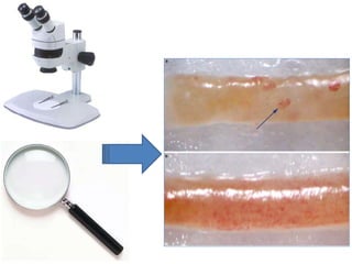
The standard approach is to first procure tissue
for electron microscopy (EM) from each core
by removing 1 mm cubes from the ends and
placing them in cooled glutaraldehyde or other
fixative suitable for EM [Figure 4]. Some
clinicians prefer that the pathology laboratory
obtain tissue for EM from the ends of the
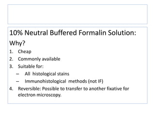
formalin-fixed tissue. If the specimen is to be
sent to a laboratory that uses
immunofluorescence (IF), the first core can be
cut in half by cross-sectioning and the larger
piece placed in formalin or another fixative
suitable for light microscopy (LM); the smaller
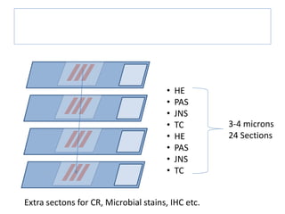
portion is saved for IF evaluation. If a second
core is obtained, the ends should be taken for
EM and the specimen again divided almost in
half, with the larger tissue core now kept for IF
and the smaller for LM.](https://image.slidesharecdn.com/doc-20170121-wa00031-170121171628/85/renal-biopsy-14-320.jpg)
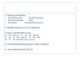


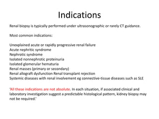
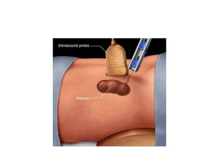
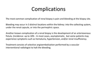
A renal biopsy is a procedure used to obtain renal tissue samples through a needle. The tissue is analyzed to diagnose underlying renal conditions ranging from infections to tumors. Indications for biopsy include unexplained kidney failure or dysfunction, nephrotic syndrome, and kidney masses. Complications can include bleeding or the formation of abnormal vessel connections. The biopsy samples are prepared and examined under the microscope using stains to identify structures and diagnose kidney diseases.


