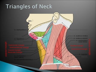
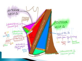
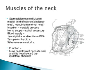
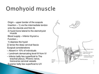
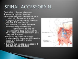
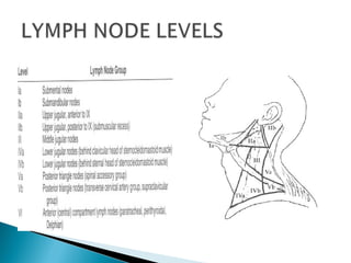
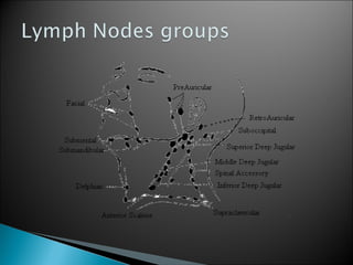
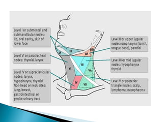
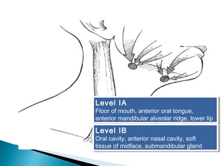
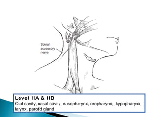
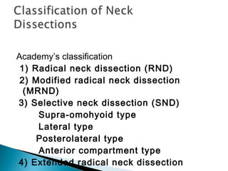
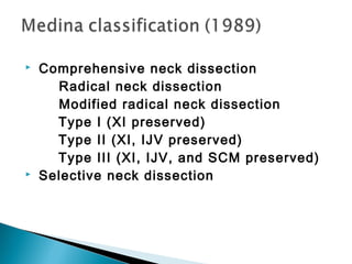
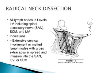
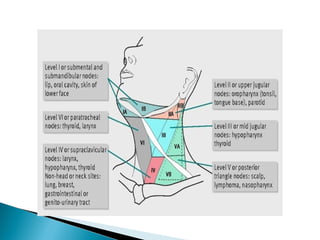
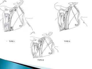
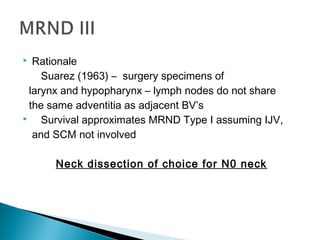
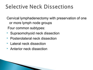
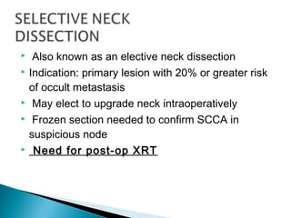
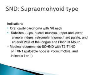
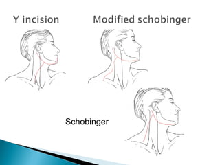
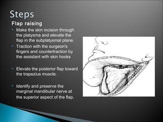
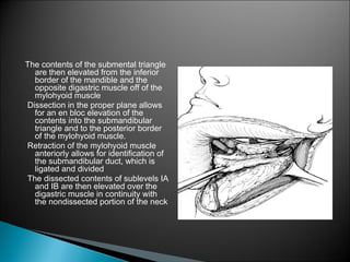
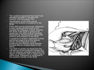
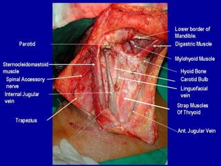
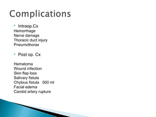
Neck Dissections:Neck Dissections:
Classifications,Classifications,
Indications, andIndications, and
TechniquesTechniques
The neck dissection is a surgical procedure
for control of neck lymph node metastasis.
This can be done for clinically or radiologically
evident lymph nodes or as part of curative
surgery where risk of occult nodal metastasis
is deemed sufficiently high.
 Status of the cervical lymph nodes
is important prognostic factor in SCCA of the
upper aerodigestive tract
 Cure rates drop in half when there is
regional lymph node involvement
Emil Theodor Kocher
Earned Nobel Prize in 1909 for
his work in thyroid and neck
surgery — the first ever
awarded to a surgeon.
1880 – Kocher proposed
removing nodal
metastases
1906 – George Crile
described the classic radical
neck dissection (RND)
1967 - Bocca and Pignataro
described the “functional neck dissection”
(FND)
Fascial layers of the neck
Superficial cervical fascia
Deep cervical fascia
– Superficial layer
SCM, strap muscles,
trapezius
– Middle or Visceral Layer
Thyroid,Trachea, esophagus
– Deep layer (also prevertebral fascia)
Vertebral muscles
Phrenic nerve
 Sternocleidomastoid Muscle
medial third of clavicle(clavicular
head), manubrium (sternal head)
Insertion – mastoid process
Nerve supply – spinal accessory
Blood supply –
1) occipital a. or direct from ECA
2) superior thyroid a.
3) transverse cervical a.
 Function –
 turns head toward opposite side
and tilts head toward the
ipsilateral shoulder
Origin – upper border of the scapula
Insertion – 1) via the intermediate tendon
onto the clavicle and first rib
2) hyoid bone lateral to the sternohyoid
muscle
Blood supply – Inferior thyroid a.
Function –
1) depress the hyoid
2) tense the deep cervical fascia
Surgical considerations
– Absent in 10% of individuals
– Landmark demarcating level III from IV
– Inferior belly lies superficial to The
brachial plexus, Phrenic nerve,
transverse cervical vessels
Superior belly lies superficial to IJV
Origin –
1) medial 1/3 of the sup. Nuchal line
2) external occipital protuberance
3) ligamentum nuchae
4) spinous process of C7 and T1-T12
Insertion –
1) lateral 1/3 of the clavicle
2) acromion process
3) spine of the scapula
Function – elevate and rotate the scapula and
stabilize the shoulder
 Surgical considerations
 – Posterior limit of Level V neck dissection
 – Denervation results in shoulder drop and winged scapula
Origin – digastric fossa of the mandible
Insertion –
1) hyoid bone via the intermediate
tendon
2) mastoid process
Function – 1) elevate the hyoid bone
2) depress the mandible (assists
lateral pterygoid)
Surgical considerations
“Residents friend”
Posterior belly is superficial to:
ECA, Hypoglossal nerve, ICA, IJV
– Anterior belly
Landmark for identification of mylohyoid
for dissection of the submandibular
triangle
Should be preserved in
neck dissections
Most commonly injury
dissection level Ib
Can be found:
– 1cm anterior and inferior
to angle of mandible
 Deep to fascia of the
submandibular gland
 Superficial to adventitia of
the facial vein
Originates in the spinal nucleus
Passes through two foramen
– Foramen Magnum – enters the skull
posterior to the vertebral artery
 – Jugular Foramen – exits the skull
with CN IX,X and the IJV
Occipital artery crosses the nerve
Descends obliquely in level II
(forms Level IIa and IIb)
 Penetrates the deep surface of the
SCM Exits posterior surface of SCM
deep to Erb’s point
Traverses the posterior triangle
ensheathed by the superficial cervical
fascia and lies on the levator
scapulae
● Enters the trapezius approx. 5
cm above the clavicle
Sole nerve supply to the
diaphragm
Supplied by nerve roots
C3-5
Runs obliquely toward
midline on the anterior
surface of anterior
scalene
Covered by prevertebral
fascia
Lies posterior and lateral
to the carotid sheath
Motor nerve to the tongue
Cell bodies are in the Hypoglossal
nucleus of the Medulla oblongata
Exits the skull via the hypoglossal
canal
Lies deep to the IJV, ICA, CN IX, X,
and XI
Curves 90 degrees and passes
between the IJV and ICA
Extends upward along hyoglossus
muscle and into the genioglossus
to the tip of the tongue
 Iatrogenic injury Most common site
- floor of the submandibular
triangle, just deep to the duct
“N” classification AJCC (1997)
Consistent for all mucosal sites except the
nasopharynx
Thyroid and nasopharynx have different
staging based on tumor behavior and
prognosis
NX: Regional lymph nodes cannot
be assessed
N0: No regional lymph node
metastasis
N1: Metastasis in a single ipsilateral
lymph node, < 3
N2a: Metastasis in a single
ipsilateral
lymph node 3 to 6 cm
N2b: Metastasis in multiple
ipsilateral
lymph nodes, none more than 6 cm
N2c: Metastasis in bilateral or
contralateral nodes < 6cm
N3: Metastasis in a lymph node
more than 6 cm in greatest
dimension
Level IA
Floor of mouth, anterior oral tongue,
anterior mandibular alveolar ridge, lower lip
Level IA
Floor of mouth, anterior oral tongue,
anterior mandibular alveolar ridge, lower lip
Level IB
Oral cavity, anterior nasal cavity, soft
tissue of midface, submandibular gland
Level IB
Oral cavity, anterior nasal cavity, soft
tissue of midface, submandibular gland
Level IIA & IIB
Oral cavity, nasal cavity, nasopharynx, oropharynx,, hypopharynx,
larynx, parotid gland
 With oral, tongue, retromolar trigone, and
tonsillar fossa subsites, the
jugulodigastric, submandibular, and
midjugular lymph node stations are
involved.
 With the floor of the mouth as the subsite,
the submandibular and
jugulodigastric lymph node stations are
involved.
 With the soft palate, base of the tongue,
oropharynx, supraglottis, and
hypopharynx subsites, the
jugulodigastric, midjugular, and
contralateral lymph node stations are
involved.
 With the nasopharynx as the subsite,
lymph node stations of the widest nodal
distribution are involved.
 Contralateral metastasis is found in the
supraglottis, the base of the tongue, and the
posterior pharyngeal wall palate.
 Bilateral metastasis is found in the
nasopharynx, the base of the tongue, the soft
palate, the floor of mouth, and the supraglottis.
 Multiple cervical metastases
(adenocarcinoma) occur with thyroid carcinoma,
breast carcinoma, and nasopharyngeal
carcinoma.
Academy’s classification
1) Radical neck dissection (RND)
2) Modified radical neck dissection
(MRND)
3) Selective neck dissection (SND)
Supra-omohyoid type
Lateral type
Posterolateral type
Anterior compartment type
4) Extended radical neck dissection
 Comprehensive neck dissection
Radical neck dissection
Modified radical neck dissection
Type I (XI preserved)
Type II (XI, IJV preserved)
Type III (XI, IJV, and SCM preserved)
 Selective neck dissection
 All lymph nodes in Levels
I-V including spinal
accessory nerve (SAN),
SCM, and IJV
 Indications
 – Extensive cervical
involvement or matted
lymph nodes with gross
extracapsular spread and
invasion into the SAN,
IJV, or SCM
 Excision of same lymph node bearing regions as
RND with preservation of one or more
nonlymphatic structures (SAN, SCM, IJV)
 Type I: Preservation of SAN
 Type II: Preservation of SAN and IJV
 Type III: Preservation of SAN, IJV, and
SCM
 TYPE I TYPE II
 TYPE III
Indications
 – Clinically obvious lymph node metastases
 – SAN not involved by tumor
 Rationale
RND vs MRND Type I:
 5-year survival and neck failure rates for RND
(63% and 12%) not statistically different from
MRND (71% and 12%).
 No difference in pattern of neck failure
Indications
 Rarely planned
 Intraoperative tumor found adherent to the
SCM, but not IJV and SAN
 Rationale
Suarez (1963) – surgery specimens of
larynx and hypopharynx – lymph nodes do not share
the same adventitia as adjacent BV’s
 Survival approximates MRND Type I assuming IJV,
and SCM not involved
Neck dissection of choice for N0 neck
Cervical lymphadenectomy with preservation of one
or more lymph node groups
Four common subtypes:
 Supraomohyoid neck dissection
 Posterolateral neck dissection
 Lateral neck dissection
 Anterior neck dissection
 Also known as an elective neck dissection
 Indication: primary lesion with 20% or greater risk
of occult metastasis
 May elect to upgrade neck intraoperatively
 Frozen section needed to confirm SCCA in
suspicious node
 Need for post-op XRT
Indications
 Oral cavity carcinoma with N0 neck
 Subsites - Lips, buccal mucosa, upper and lower
alveolar ridges, retromolar trigone, hard palate, and
anterior 2/3s of the tongue and Floor Of Mouth.
– Medina recommends SOHND with T2-T4NO
or TXN1 (palpable node is <3cm, mobile, and
in levels I or II)
Indications
 Anterior tongue
 Oral tongue and
FOM that approach
the midline
 Adjuvant XRT given
to patients with
> 2positive nodes +/-
En bloc removal of the
jugular lymph nodes
including Levels II-IV
 Indications
 N0 neck in
carcinomas of the
oropharynx,hypophar
ynx, supraglottis, and
larynx
En bloc excision of lymph bearing tissues in
Levels II-V
Indications
 Cutaneous malignancies
 Melanoma
 Squamous cell carcinoma
 Merkel cell carcinoma
 Soft tissue sarcomas of the scalp and neck
En bloc removal of lymph structures in Level VI
 Perithyroidal nodes
 Pretracheal nodes
 Precricoid nodes (Delphian)
 Paratracheal nodes along recurrent nerves
 – Limits of the dissection are the hyoid
bone,suprasternal notch and carotid sheaths
 Indications
 – Selected cases of thyroid carcinoma
 – Parathyroid carcinoma
 – Subglottic carcinoma
 – Laryngeal carcinoma with subglottic extension
 – CA of the cervical esophagus
Any previous dissection which includes removal of one or
more additional lymph node groups and/or non-
lymphatic structures.
 Usually performed with N+ necks in MRND or RND
when metastases invade structures usually preserved
 Examples:
Resection of the hypoglossal nerve, resection of
digastric muscle, Carotid artery resection,
dissection of mediastinal nodes and central
compartment for subglottic involvement, and removal of
retropharyngeal lymph nodes for tumors originating in
the pharyngeal walls.
Schobinger
Flap raising
 Make the skin incision through
the platysma and elevate the
flap in the subplatysmal plane.
 Traction with the surgeon's
fingers and countertraction by
the assistant with skin hooks
 Elevate the posterior flap toward
the trapezius muscle.
 Identify and preserve the
marginal mandibular nerve at
the superior aspect of the flap.
The contents of the submental triangle
are then elevated from the inferior
border of the mandible and the
opposite digastric muscle off of the
mylohyoid muscle
Dissection in the proper plane allows
for an en bloc elevation of the
contents into the submandibular
triangle and to the posterior border
of the mylohyoid muscle.
Retraction of the mylohyoid muscle
anteriorly allows for identification of
the submandibular duct, which is
ligated and divided
The dissected contents of sublevels IA
and IB are then elevated over the
digastric muscle in continuity with
the nondissected portion of the neck
 The contents dissected from level I are
elevated caudally to visualize the
superior internal jugular vein..
 Identification of the SAN can be
performed anterior or posterior to the
SCM.
 If the SAN can be preserved, dissection
is then continued from near to IJV
towards the trapezius muscle, dividing
the SCM. If the SCM is going to be
preserved, the SAN must be carefully
dissected by identifying the nerve both
anterior and posterior to the SCM.
 A posterior to anterior dissection is then
performed beginning at the anterior
border of the trapezius muscle,
preserving the phrenic nerve and the
brachial plexus, located deep to this
fascia.
 The SAN must then be freed from the
soft tissues of the posterior triangle and
can be carefully retracted away from the
region of dissection with a vessel loop or
nerve hook. Dissection is continued to
the posterior border of the SCM.
.
 At this point, the posterior triangle contents,
with or without the SAN and SCM, have been
elevated to the lateral aspect of the IJV. If the
SCM is being resected, transection is
performed below the mastoid tip and above
the clavicle as in a RND.
 The nodes along IJV can usually be removed
en bloc with the remainder of the dissection
in a posteroanterior fashion, sharply incising
the fascia of the jugular vein with a scalpel
blade using a feather-light touch. If the IJV
requires sacrifice due to metastatic nodal
involvement or tumor thrombosis, the vein is
ligated and divided superiorly and inferiorly
following identification and preservation of
the vagus nerve.
 Dissection is continued anteriorly, elevating
the fascia and soft tissues up to the
infrahyoid strap muscles and the hyoid-
digastric junction.
 Preservation of a fascial layer superficial to
the carotid artery is usually possible, and
exposure of the carotid artery should be
discouraged unless necessary.
 Suction drains are strategically placed and a
layered closure is performed.
 Intraop.Cx
Hemorrhage
Nerve damage
Thoracic duct injury
Pneumothorax
 Post op. Cx
Hematoma
Wound infection
Skin flap loss
Salivary fistula
Chylous fistula 500 ml
Facial edema
Carotid artery rupture
THANK YOU

Neck dissection

  • 2.
  • 3.
    The neck dissectionis a surgical procedure for control of neck lymph node metastasis. This can be done for clinically or radiologically evident lymph nodes or as part of curative surgery where risk of occult nodal metastasis is deemed sufficiently high.
  • 4.
     Status ofthe cervical lymph nodes is important prognostic factor in SCCA of the upper aerodigestive tract  Cure rates drop in half when there is regional lymph node involvement
  • 5.
    Emil Theodor Kocher EarnedNobel Prize in 1909 for his work in thyroid and neck surgery — the first ever awarded to a surgeon. 1880 – Kocher proposed removing nodal metastases
  • 6.
    1906 – GeorgeCrile described the classic radical neck dissection (RND)
  • 7.
    1967 - Boccaand Pignataro described the “functional neck dissection” (FND)
  • 9.
    Fascial layers ofthe neck Superficial cervical fascia Deep cervical fascia – Superficial layer SCM, strap muscles, trapezius – Middle or Visceral Layer Thyroid,Trachea, esophagus – Deep layer (also prevertebral fascia) Vertebral muscles Phrenic nerve
  • 13.
     Sternocleidomastoid Muscle medialthird of clavicle(clavicular head), manubrium (sternal head) Insertion – mastoid process Nerve supply – spinal accessory Blood supply – 1) occipital a. or direct from ECA 2) superior thyroid a. 3) transverse cervical a.  Function –  turns head toward opposite side and tilts head toward the ipsilateral shoulder
  • 14.
    Origin – upperborder of the scapula Insertion – 1) via the intermediate tendon onto the clavicle and first rib 2) hyoid bone lateral to the sternohyoid muscle Blood supply – Inferior thyroid a. Function – 1) depress the hyoid 2) tense the deep cervical fascia Surgical considerations – Absent in 10% of individuals – Landmark demarcating level III from IV – Inferior belly lies superficial to The brachial plexus, Phrenic nerve, transverse cervical vessels Superior belly lies superficial to IJV
  • 15.
    Origin – 1) medial1/3 of the sup. Nuchal line 2) external occipital protuberance 3) ligamentum nuchae 4) spinous process of C7 and T1-T12 Insertion – 1) lateral 1/3 of the clavicle 2) acromion process 3) spine of the scapula Function – elevate and rotate the scapula and stabilize the shoulder  Surgical considerations  – Posterior limit of Level V neck dissection  – Denervation results in shoulder drop and winged scapula
  • 16.
    Origin – digastricfossa of the mandible Insertion – 1) hyoid bone via the intermediate tendon 2) mastoid process Function – 1) elevate the hyoid bone 2) depress the mandible (assists lateral pterygoid) Surgical considerations “Residents friend” Posterior belly is superficial to: ECA, Hypoglossal nerve, ICA, IJV – Anterior belly Landmark for identification of mylohyoid for dissection of the submandibular triangle
  • 17.
    Should be preservedin neck dissections Most commonly injury dissection level Ib Can be found: – 1cm anterior and inferior to angle of mandible  Deep to fascia of the submandibular gland  Superficial to adventitia of the facial vein
  • 18.
    Originates in thespinal nucleus Passes through two foramen – Foramen Magnum – enters the skull posterior to the vertebral artery  – Jugular Foramen – exits the skull with CN IX,X and the IJV Occipital artery crosses the nerve Descends obliquely in level II (forms Level IIa and IIb)  Penetrates the deep surface of the SCM Exits posterior surface of SCM deep to Erb’s point Traverses the posterior triangle ensheathed by the superficial cervical fascia and lies on the levator scapulae ● Enters the trapezius approx. 5 cm above the clavicle
  • 19.
    Sole nerve supplyto the diaphragm Supplied by nerve roots C3-5 Runs obliquely toward midline on the anterior surface of anterior scalene Covered by prevertebral fascia Lies posterior and lateral to the carotid sheath
  • 21.
    Motor nerve tothe tongue Cell bodies are in the Hypoglossal nucleus of the Medulla oblongata Exits the skull via the hypoglossal canal Lies deep to the IJV, ICA, CN IX, X, and XI Curves 90 degrees and passes between the IJV and ICA Extends upward along hyoglossus muscle and into the genioglossus to the tip of the tongue  Iatrogenic injury Most common site - floor of the submandibular triangle, just deep to the duct
  • 22.
    “N” classification AJCC(1997) Consistent for all mucosal sites except the nasopharynx Thyroid and nasopharynx have different staging based on tumor behavior and prognosis
  • 23.
    NX: Regional lymphnodes cannot be assessed N0: No regional lymph node metastasis N1: Metastasis in a single ipsilateral lymph node, < 3 N2a: Metastasis in a single ipsilateral lymph node 3 to 6 cm N2b: Metastasis in multiple ipsilateral lymph nodes, none more than 6 cm N2c: Metastasis in bilateral or contralateral nodes < 6cm N3: Metastasis in a lymph node more than 6 cm in greatest dimension
  • 27.
    Level IA Floor ofmouth, anterior oral tongue, anterior mandibular alveolar ridge, lower lip Level IA Floor of mouth, anterior oral tongue, anterior mandibular alveolar ridge, lower lip Level IB Oral cavity, anterior nasal cavity, soft tissue of midface, submandibular gland Level IB Oral cavity, anterior nasal cavity, soft tissue of midface, submandibular gland
  • 28.
    Level IIA &IIB Oral cavity, nasal cavity, nasopharynx, oropharynx,, hypopharynx, larynx, parotid gland
  • 29.
     With oral,tongue, retromolar trigone, and tonsillar fossa subsites, the jugulodigastric, submandibular, and midjugular lymph node stations are involved.  With the floor of the mouth as the subsite, the submandibular and jugulodigastric lymph node stations are involved.  With the soft palate, base of the tongue, oropharynx, supraglottis, and hypopharynx subsites, the jugulodigastric, midjugular, and contralateral lymph node stations are involved.  With the nasopharynx as the subsite, lymph node stations of the widest nodal distribution are involved.
  • 30.
     Contralateral metastasisis found in the supraglottis, the base of the tongue, and the posterior pharyngeal wall palate.  Bilateral metastasis is found in the nasopharynx, the base of the tongue, the soft palate, the floor of mouth, and the supraglottis.  Multiple cervical metastases (adenocarcinoma) occur with thyroid carcinoma, breast carcinoma, and nasopharyngeal carcinoma.
  • 32.
    Academy’s classification 1) Radicalneck dissection (RND) 2) Modified radical neck dissection (MRND) 3) Selective neck dissection (SND) Supra-omohyoid type Lateral type Posterolateral type Anterior compartment type 4) Extended radical neck dissection
  • 33.
     Comprehensive neckdissection Radical neck dissection Modified radical neck dissection Type I (XI preserved) Type II (XI, IJV preserved) Type III (XI, IJV, and SCM preserved)  Selective neck dissection
  • 34.
     All lymphnodes in Levels I-V including spinal accessory nerve (SAN), SCM, and IJV  Indications  – Extensive cervical involvement or matted lymph nodes with gross extracapsular spread and invasion into the SAN, IJV, or SCM
  • 36.
     Excision ofsame lymph node bearing regions as RND with preservation of one or more nonlymphatic structures (SAN, SCM, IJV)  Type I: Preservation of SAN  Type II: Preservation of SAN and IJV  Type III: Preservation of SAN, IJV, and SCM
  • 37.
     TYPE ITYPE II  TYPE III
  • 38.
    Indications  – Clinicallyobvious lymph node metastases  – SAN not involved by tumor  Rationale RND vs MRND Type I:  5-year survival and neck failure rates for RND (63% and 12%) not statistically different from MRND (71% and 12%).  No difference in pattern of neck failure
  • 39.
    Indications  Rarely planned Intraoperative tumor found adherent to the SCM, but not IJV and SAN
  • 40.
     Rationale Suarez (1963)– surgery specimens of larynx and hypopharynx – lymph nodes do not share the same adventitia as adjacent BV’s  Survival approximates MRND Type I assuming IJV, and SCM not involved Neck dissection of choice for N0 neck
  • 41.
    Cervical lymphadenectomy withpreservation of one or more lymph node groups Four common subtypes:  Supraomohyoid neck dissection  Posterolateral neck dissection  Lateral neck dissection  Anterior neck dissection
  • 42.
     Also knownas an elective neck dissection  Indication: primary lesion with 20% or greater risk of occult metastasis  May elect to upgrade neck intraoperatively  Frozen section needed to confirm SCCA in suspicious node  Need for post-op XRT
  • 43.
    Indications  Oral cavitycarcinoma with N0 neck  Subsites - Lips, buccal mucosa, upper and lower alveolar ridges, retromolar trigone, hard palate, and anterior 2/3s of the tongue and Floor Of Mouth. – Medina recommends SOHND with T2-T4NO or TXN1 (palpable node is <3cm, mobile, and in levels I or II)
  • 44.
    Indications  Anterior tongue Oral tongue and FOM that approach the midline  Adjuvant XRT given to patients with > 2positive nodes +/-
  • 45.
    En bloc removalof the jugular lymph nodes including Levels II-IV  Indications  N0 neck in carcinomas of the oropharynx,hypophar ynx, supraglottis, and larynx
  • 46.
    En bloc excisionof lymph bearing tissues in Levels II-V Indications  Cutaneous malignancies  Melanoma  Squamous cell carcinoma  Merkel cell carcinoma  Soft tissue sarcomas of the scalp and neck
  • 47.
    En bloc removalof lymph structures in Level VI  Perithyroidal nodes  Pretracheal nodes  Precricoid nodes (Delphian)  Paratracheal nodes along recurrent nerves  – Limits of the dissection are the hyoid bone,suprasternal notch and carotid sheaths  Indications  – Selected cases of thyroid carcinoma  – Parathyroid carcinoma  – Subglottic carcinoma  – Laryngeal carcinoma with subglottic extension  – CA of the cervical esophagus
  • 48.
    Any previous dissectionwhich includes removal of one or more additional lymph node groups and/or non- lymphatic structures.  Usually performed with N+ necks in MRND or RND when metastases invade structures usually preserved  Examples: Resection of the hypoglossal nerve, resection of digastric muscle, Carotid artery resection, dissection of mediastinal nodes and central compartment for subglottic involvement, and removal of retropharyngeal lymph nodes for tumors originating in the pharyngeal walls.
  • 53.
  • 54.
    Flap raising  Makethe skin incision through the platysma and elevate the flap in the subplatysmal plane.  Traction with the surgeon's fingers and countertraction by the assistant with skin hooks  Elevate the posterior flap toward the trapezius muscle.  Identify and preserve the marginal mandibular nerve at the superior aspect of the flap.
  • 55.
    The contents ofthe submental triangle are then elevated from the inferior border of the mandible and the opposite digastric muscle off of the mylohyoid muscle Dissection in the proper plane allows for an en bloc elevation of the contents into the submandibular triangle and to the posterior border of the mylohyoid muscle. Retraction of the mylohyoid muscle anteriorly allows for identification of the submandibular duct, which is ligated and divided The dissected contents of sublevels IA and IB are then elevated over the digastric muscle in continuity with the nondissected portion of the neck
  • 56.
     The contentsdissected from level I are elevated caudally to visualize the superior internal jugular vein..  Identification of the SAN can be performed anterior or posterior to the SCM.  If the SAN can be preserved, dissection is then continued from near to IJV towards the trapezius muscle, dividing the SCM. If the SCM is going to be preserved, the SAN must be carefully dissected by identifying the nerve both anterior and posterior to the SCM.  A posterior to anterior dissection is then performed beginning at the anterior border of the trapezius muscle, preserving the phrenic nerve and the brachial plexus, located deep to this fascia.  The SAN must then be freed from the soft tissues of the posterior triangle and can be carefully retracted away from the region of dissection with a vessel loop or nerve hook. Dissection is continued to the posterior border of the SCM.
  • 57.
    .  At thispoint, the posterior triangle contents, with or without the SAN and SCM, have been elevated to the lateral aspect of the IJV. If the SCM is being resected, transection is performed below the mastoid tip and above the clavicle as in a RND.  The nodes along IJV can usually be removed en bloc with the remainder of the dissection in a posteroanterior fashion, sharply incising the fascia of the jugular vein with a scalpel blade using a feather-light touch. If the IJV requires sacrifice due to metastatic nodal involvement or tumor thrombosis, the vein is ligated and divided superiorly and inferiorly following identification and preservation of the vagus nerve.  Dissection is continued anteriorly, elevating the fascia and soft tissues up to the infrahyoid strap muscles and the hyoid- digastric junction.  Preservation of a fascial layer superficial to the carotid artery is usually possible, and exposure of the carotid artery should be discouraged unless necessary.  Suction drains are strategically placed and a layered closure is performed.
  • 59.
     Intraop.Cx Hemorrhage Nerve damage Thoracicduct injury Pneumothorax  Post op. Cx Hematoma Wound infection Skin flap loss Salivary fistula Chylous fistula 500 ml Facial edema Carotid artery rupture
  • 60.