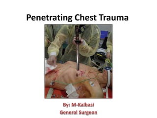
Penetrating chest trauma
•Download as PPTX, PDF•
6 likes•437 views

thoracic injury during trauma is one of most important life threaten that maybe occurred. so all of medical practitioner must learn and must do some primary survey

Recommended
More Related Content
What's hot
What's hot (20)
Similar to Penetrating chest trauma
Similar to Penetrating chest trauma (20)
Recently uploaded
Recently uploaded (20)
Penetrating chest trauma
- 2. Anatomy of the Chest
- 3. Specific organ chest injury • Great Vessels Injury • Heart Injury • Trachea and Bronchi Injury • Lung Parenchyma Injury • Esophagus Injury • Chest Wall and Diaphragm Injury
- 4. History of the Procedure • One of the earliest writings of thoracic injury was noted in the Edwin Smith Surgical Papyrus, written in 3000 BCE. Galen reported attempts to treat gladiators with chest injuries with open packing. • Hill performed the first cardiorrhaphy in the United States in 1902 and initiated the modern treatment of the wounded heart.
- 6. Problem • Any penetrating wound below the nipples (front) and the inferior scapular angles (dorsum) should be considered an entry point for a course that may have carried into the abdominal cavity
- 7. • Low-velocity injuries include impalement (eg, knife wounds), which disrupts only the structures penetrated. • Medium-velocity injuries include bullet wounds characterized by much less primary tissue destruction than wounds caused by high- velocity forces. • High-velocity injuries include bullet wounds caused by rifles and wounds resulting from military weapons. Mechanism of injury The mechanism of injury may be categorized as:
- 8. • The initial management of seriously injured patients consists of performing the primary survey (the “ABCs”—Airway with cervical spine protection, Breathing, and Circulation); • the goals of the primary survey are to identify and treat conditions that constitute an immediate threat to life. • For penetrating thoracic trauma, physical examination, plain posteroanterior and lateral chest radiographs with metallic markings of wounds, pericardial ultrasound, and CVP measurement will identify the majority of injuries. • Bronchoscopy should be performed to evaluate the trachea in patients with a persistent air leak from the chest tube or mediastinal air.
- 9. • As with neck injuries, hemodynamically stable patients with transmediastinal gunshot wounds should undergo CT scanning to determine the path of the bullet. • CT scanning identifies the vascular or visceral structures at risk for injury and directs angiography or endoscopy as appropriate. • Therefore, CTA should be performed based on injury proximity to intrathoracic vasculature.
- 10. • The most common injuries from penetrating thoracic trauma are hemothorax and pneumothorax. • The majority of pulmonary parenchymal injuries are suspected based upon identification of a pneumothorax. • the vast majority is managed by tube thoracostomy.
- 11. • Pneumothorax • The alveoli can rupture, causing a pneumothorax • In traumatic pneumothorax, chest tube drainage is recommended, even for small collection of air. • When large air leak is present or reexpansion of the lung is incomplete, a tracheobronchial injury should be suspected and prompt flexible bronchoscopy performed
- 12. • Very large pneumothoraxes produce tension by shifting the mediastinal structures toward the contralateral side. • In these situations, the anatomic distortion combined with the increase in intrathoracic pressure decreases cardiac output. • If untreated, this can cause cardiac arrest. • Immediate release of a tension pneumothorax may be accomplished by placing a needle into pleural space to allow pressure in the pleura to equilibrate with the outside air.
- 13. • Tension pneumothorax should be suspected in any patient with chest wall trauma receiving general anesthesia who suddenly develops deterioration associated with a markedly increased requirement o inspiratory ventilator pressure.
- 14. • Hemothorax Sources of intrathoracic bleeding include: - Intercostal vessels - Internal mammary arteries - Pulmonary parenchyma - Major pulmonary vessels - Cardiac and great vessels injuries Intrathoracic bleeding recognized in the emergency department requires a closed-tube thoracostomy. Massive hemorrhage defines the need for exploration. The generally accepted criteria are: - initial drainage of 1,500 mL at the time of chest tube placement - continued drainage of 250 mL/hr for next 4 hours.
- 15. • Residual hemothorax must be evacuated as soon as the patient is stable to reduce the risk for empyema or prevent the formation of a fibrous peel(resulting in lung entrapment). • It is not emergency situation if bleeding has stopped. However, thoracoscopic drainage is the best perfomed within 1 to 3 days after injury to reduce the risk for empyema or lung entrapment.
- 17. • Peripheral lacerations with persistent bleeding can be managed with stapled wedge resection using a stapler. • central injuries, the current treatment is pulmonary tractotomy, which permits selective ligation of individual bronchioles and bleeders, prevents the development of an intraparenchymal hematoma or air embolism, and reduces the need for formal lobar resection. • The injury track is thus filleted open, which allows direct access to the bleeding vessels and leaking bronchi. The majority of injuries are definitively managed with selective ligation, and the defect is left open.
- 18. • Less than 1% of all injured patients sustain intrathoracic tracheobronchial injuries and penetrating injuries may occur throughout the tracheobronchial system. • For patients with a massive air leak requiring emergent exploration, initial control of the injury to provide effective ventilation is obtained by passing an endotracheal tube either beyond the injury or into the contralateral mainstem bronchus. • For patients with a massive air leak requiring emergent exploration, initial control of the injury to provide effective ventilation is obtained by passing an endotracheal tube either beyond the injury or into the contralateral mainstem bronchus.
- 19. • Principles of repair are devitalized tissue is debrided, and primary end-to-end anastomosis with 3-0 PDS suture is performed. • Suture lines should be encircled with vascularized tissue, either pericardium, intercostal muscle, or pleura. • Expectant management is employed for bronchial injuries that are less than one- third the circumference of the airway and have no evidence of a persistent major air leak • In patients with peripheral bronchial injuries, indicated by persistent air leaks from the chest tube and documented by endoscopy, bronchoscopically directed fibrin glue sealing may be useful.
- 20. • Traumatic cardiac penetration is highly lethal, with case fatality rates of 70-80%. • Ventricular injuries are more common than atrial injuries, and the right side is involved more often than the left side. • The Beck triad (ie, high venous pressure, low arterial pressure, muffled heart sounds) is documented in only 10-30% of patients who have proven tamponade. • Pericardiocentesis can be both diagnostic and therapeutic. This procedure is reserved for patients with significant hemodynamic compromise without another likely etiology. • Echocardiography is a rapid, noninvasive, and accurate test for pericardial fluid.
- 21. • The management algorithm is based on the patient's hemodynamic status, with patients who are in extremis or who are profoundly unstable benefiting from emergency thoracotomy with ongoing aggressive resuscitation. • Stable patients with cardiac wounds may be diagnosed using a subxiphoid pericardial window.
- 22. • Over 90% of thoracic great vessel injuries are due to penetrating trauma. • Simple lacerations of the ascending or transverse aortic arch can be repaired with lateral aortorrhaphy. • Repair of posterior injuries, or those requiring interposition grafting of the arch, require full cardiopulmonary bypass, and repair of complex injuries may require circulatory arrest.
- 23. • Innominate artery injuries are repaired using the bypass exclusion technique, which avoids the need for cardiopulmonary bypass. • Bypass grafting from the proximal aorta to the distal innominate with a prosthetic tube graft is performed before the postinjury hematoma is entered. The PTFE graft is anastomosed end to side from the proximal undamaged aorta and anastomosed end-to-end to the innominate artery. • The origin of the innominate is then oversewn at its base to exclude the pseudoaneurysm or other injury
- 25. • Subclavian artery injuries can be repaired using lateral arteriorrhaphy or PTFE graft interposition; due to its multiple branches and tethering of the artery, end-to-end anastomosis is not advocated if there is a significant segmental loss.
- 27. • Descending thoracic aortic injuries may require urgent if not emergent intervention. However, operative intervention for intracranial or intra- abdominal hemorrhage or unstable pelvic fractures takes precedence. • To prevent aortic rupture, pharmacologic therapy with a selective β1 antagonist, esmolol, should be instituted in the trauma bay, with a target SBP of <100 mm Hg and heart rate of <100/min. • Endovascular stenting is now the mainstay of treatment, but open operative reconstruction is warranted, or necessary, in select patients. • Open repair of the descending aorta is accomplished using partial left heart bypass. • In most patients a short PTFE graft (usually 18 mm in diameter) is placed using a running 3-0 polypropylene suture.
- 28. • Esophageal injuries often occur with tracheobronchial injuries, particularly in cases of penetrating trauma. • Operative options are based on the extent and location of esophageal injury. • With sufficient mobilization, a primary single-layer end-to-end anastomosis may be performed after appropriate debridement.
- 29. • Perforations at the gastroesophageal junction may be treated with repair and Nissan fundoplication or, for destructive injuries, segmental resection and gastric pull-up. • With large destructive injuries or delayed presentation of injuries, esophageal exclusion with wide drainage, diverting loop esophagostomy, and placement of a gastrostomy tube should be considered. As with cervical repairs, if there are two suture lines in close approximation (trachea or bronchi and esophagus) interposition of a vascularized pedicle is warranted to prevent fistula formation.
- 30. • Open Wounds of the Chest Wall • These injuries, also known as “sucking chest wounds”, are the result of full-thickness loss of portion of the chest wall. • The injury presents as a life- threatening emergency. • The loss of negative intrathoracic pressure results in an open pneumothorax leading to collapse of ipsilateral lung.
- 31. • A temporary dressing should be applied to have wound at the scene of accident in an attempt to improve ventilation. The “three-way dreaaing” involves placing a plastic sheet taped shut on three sides, leaving the fourth one(at least a few inches) unsealed, thus creating a one-way valve dressing.
- 32. • In the emergency department, a chest tube is placed to reexpand the lung and the wound is covered with an impermeable dressing.. • Most open wounds require operative debridment with removal of devitalized tissue and foreign bodies. • Closure of the wounds may be accomplish by mobilizing the surrounding tissues. • However, large defects may require rotational or free musculocutaneous flaps.
- 33. • Injured intercostal arteries • Persistent hemorrhage from a chest tube after blunt trauma most often is due to injured intercostal arteries; • for unusual persistent bleeding, thoracotomy with direct ligation or angioembolization may be required to arrest hemorrhage.
- 34. • Diaphragmatic Injuries • Penetrating diaphragmatic injuries are variable in size and location depending on the agent of injury. • Penetrating injuries also have high rates of associated injuries, especially to intra-abdominal viscera, but including a hemothorax or pneumothorax, as well. Specific intra-abdominal organs typically injured with penetrating diaphragmatic injury include the upper abdominal organs. • In general, acute injuries are approached with laparoscopy or laparotomy because of associated injuries and chronic injuries are approached with thoracoscopy because of dense adhesions that arise between the abdominal contents and the lung.
- 35. • Most injuries require repair with heavy, nonabsorbable sutures
- 36. • some large tears may require mesh closure
- 38. • Only a low percentage of trauma patients require an emergency thoracotomy(EDT) • Indication for EDT : – Acute pericardial tamponade unresponsive to cardiac massage. – Exsanguinating intrathoracic hemorrhage. – Need for internal cardiac massage