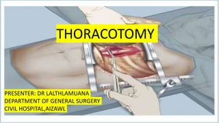
2. Thoracotomy
• A thoracotomy is a major surgery in which a cut is made between the ribs to view
and access organs in the chest cavity.
• The most frequent indication for thoracotomy is lung cancer.
• The standard route into the thoracic cavity is through a posterolateral thoracotomy
3. The incision is used for access to the:
● lung and major bronchi;
● pleura;
● thoracic aorta;
● esophagus;
● posterior mediastinum.
4. Reasons for a Thoracotomy
• Thoracotomy allows to study the conditions of the lungs such as removal of lung
or part of lung or removal of rib.
• It can be used to diagnose various conditions, as it allows surgeons to directly see
inside the body and, if needed, perform a biopsy to obtain a sample of tissue that
can be evaluated in a lab.
5. • It is also a traditional approach used as part of surgeries to treat several diseases
and conditions, including:
• Lung cancer (accounts for at least 90% of the activity of thoracic surgery
departments)
• Esophageal cancer
• Issues involving the heart and aorta
• Chest trauma
• Persistent pneumothorax (collapsed lung)
6. • Chronic obstructive pulmonary disease (COPD)
• Tuberculosis
• An unknown mediastinal mass
• Emergencies requiring resuscitation, such as a chest haemorrhage
7. Preparing for a Thoracotomy
• A thoracotomy is often done as an emergency procedure. But when one is planned,
a careful medical history and physical examination is done in advance of the
procedure.
• Tests, such as a pulmonary function test, to assess lung health.
• Various other blood test, imaging, and other tests to evaluate heart function.
8. • Smoking, should be stop ahead of surgery.
• Exercise, such as walking, will help the patient to be fit ahead of the
surgery.
9. Subtypes of thoracotomy
• Posterolateral thoracotomy
• Anterolateral thoracotomy
• Trans axillary lateral thoracotomy
10. Posterolateral Thoracotomy
• It is a gold standard for access to the thorax. It provides access to all thoracic
viscera, & is mainly used for pulmonary resections.
• It is very common approach for operations on lungs. When performed on 5th
intercostal space , it allows optimal access to pulmonary hilum( pulmonary artery
and pulmonary vein)
• Incision of choice for pulmonary resections i.e pneumonectomy & lobectomy.
11. • Incision is made in patient in lateral decubitus position.
• It starts from between scapula and mid-spinal line and extends laterally to anterior
axillary line.
• Before reaching thoracic cavity, incision is passed through latissimus dorsi &
serratus anterior muscle then transects the rhomboids & trapezius.
12.
13.
14. • A double-lumen endotracheal tube is used to allow ventilation of one lung while the other is collapsed, to
facilitate surgery and to protect the non-operated lung and retain control of ventilation
15. • The ribs are spread carefully using a self retaining retractor, but overspreading
should be avoided as this can also damage the intercostal neurovascular bundle,
with resultant postoperative neuralgia.
• One or two chest drains are commonly placed in the pleural cavity at the end of
surgery before closure, and are brought out through separate stab incisions.
• Rib apposition can be held by four peri costal sutures spaced along the incision,
followed by a continuous suture.
• The chest wall muscles are then repaired with absorbable sutures.
16.
17. Anterolateral and Clamshell Thoracotomy
• A left anterolateral thoracotomy is often the incision of choice for emergency access to
the heart and may be the prelude to a bilateral anterior thoracotomy or ‘clamshell’.
• Access to the posterolateral aspect of the left ventricle is superior to that obtained with a
median sternotomy but, more importantly, it is a faster and safer approach
• The patient is laid obliquely supine with the ipsilateral hip and shoulder raised and the
arm abducted.
18.
19. • A left 5th space anterolateral thoracotomy can be extended by a transverse or oblique division of
the sternal body to a 5th or 4th interspace right anterolateral thoracotomy if greater access is
required.
• In this clamshell thoracotomy both internal mammary arteries must be ligated and divided.
• A clamshell thoracotomy is the preferred approach to the heart and chest cavity in an extreme
emergency.
• For blunt chest trauma the incision is started as a left anterolateral thoracotomy to gain rapid
access to the pericardium and heart and is then extended as required.
• For penetrating chest trauma the side of the initial incision is guided by the site of injury.
20.
21. Trans axillary Lateral Thoracotomy
• This is a limited lateral thoracotomy, performed through the medial wall of the axilla,
which affords restricted access to the apex of the lung.
• It was a standard approach for a thoracic sympathectomy but it has now generally been
superseded by a thoracoscopic technique
22.
23. Median Sternotomy
• This is the incision that is performed most frequently for elective cardiac surgery
and provides excellent access to the heart and anterior mediastinum.
• It may also be the most appropriate incision in an emergency when damage to the
heart or to the great vessels of the superior mediastinum is suspected.
• The incision can be extended up into the neck along the anterior border of
sternocleidomastoid for injuries of the carotid root, or laterally above the clavicle
for access to an injury to the subclavian root.
24.
25. • After surgery within the pericardium, drains should be left both inside the
pericardium and in the anterior mediastinum.
• The two halves of the sternum are then drawn together.
• It is important that the sides of the sternum are very firmly pulled together by
the sternal wires.
• Poor closure at this point will result in chronic pain, non-union and dehiscence.
26. Thoracotomy vs. VATS
• With VATS, several small incisions are made in the chest and surgery is performed
by using an inserted scope with a camera.
• The VATS procedure is associated with shorter recovery times and fewer pain-
related complications than traditional thoracotomy. This may lead to improved
outcomes, because VATS may limit the effects on breathing that lead
to atelectasis (collapsed lung) or pneumonia when compared with thoracotomy.
27.
28. ED Thorocotomy
• 25% – 50% all traumatic injuries involve thorax
• Thoracotomy is an integral part of resuscitation in selected patients
• Need to decide quickly if thoracotomy is indicated to increase chance of survival
• Patients may deteriorate prehospital or in the ED and this may be the only option
to restore life
29. Aims Of ED Thoracotomy
• Release cardiac tamponade
• Control haemorrhage
• Perform open cardiac massage
• Cross clamp the descending thoracic aorta
• Control air embolism
31. Indications
• Blunt thoracic injury
◦ Cardiac tamponade diagnosed rapidly on US with no obvious non survivable injury.
◦ Unresponsive hypotension (SPB < 70mmHg)
◦ >1500ml from chest tube immediately after insertion
• Continuous Bleeding >200ml per hour over 3 hours
• Brisk Bleeding >100 ml every 15mins for 1 hour
32. • A posterolateral thoracotomy is done.
• The bleeding vessel identified and secured by ligation.
• Chest closed by placing an intercoastal tube drainage.
33. Contraindications
• Non traumatic cardiac arrest
• Severe head or multisystem injury
• Improperly trained team
• Insufficient equipment
34. • Penetrating cardiac injury
◦ Direct digital pressure
◦ Cardiac defect closed
◦ Suture closure of injury
◦ Pass Foleys catheter through defect, inflate balloon, apply traction
35. • Abdominal Haemorrhage / Hypoperfusion
◦ Cross clamp thoracic aorta to redistribute blood to myocardium and brain
• Haemorrhage from pulmonary parenchyma or major pulmonary vasculature
◦ Clamp pulmonary Hilum / Injured tissue / Bleeding vessel
36. Pericardiotomy
◦ Identify phrenic nerve in the anterolateral surface of pericardium
◦ Evacuate blood clots / Pericardial fluid
◦ Deliver heart from pericardial sac to inspect or fix defects
37.
38. Air Embolism
◦ Air in coronary vessels, heart or aorta is diagnostic
◦ Clamp hilum of affected lung
◦ Ventilate unaffected lung only
39. Complications
• Depends on the type of procedure and technique, whether VATS or open
thoracotomy.
• Prolonged need for ventilatory assistance after surgery.
• Persistent air leak resulting in a prolonged need for a chest tube after surgery
• Infection.
• Bleeding.
40. • Blood clots: Deep vein thrombosis and pulmonary emboli are common and
serious complications of chest surgery.
• Complications of general anesthesia
• Heart attack or arrhythmias
• Vocal cord dysfunction or paralysis
• Bronchopleural fistula
41. RATS
• In this approach, the thoracoscopy is done using a robotic system with three-dimensional vision.
• The surgeon sits at a control panel in the operating room and moves robotic arms to operate
through several small incisions in the patient’s chest.
• RATS is similar to VATS in terms of less pain, less blood loss and a shorter recovery time.
• For the surgeon, the robotic system may provide more maneuverability and more precision when
moving the instruments than standard VATS.
• It may have advantages when performing more complex lung resections such as segmentectomies
or mediastinal tumors (thymectomy).