SlideShare a Scribd company logo
1 of 76
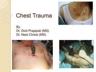
Chest Trauma
By
Dr. Dixit Prajapati (MS)
Dr. Neet Chotai (MS)
Introduction
ī‚— Thoracic injuries are directly
responsible for 25% of all trauma
deaths and are a major contributory
factor to mortality in a further 25%.
ī‚— Although many of these deaths occur
almost immediately, there is a
significant group of patients that may
be salvaged with early effective
management.
ī‚— The majority (approximately 90%) of
all patients who sustain thoracic
trauma can be managed
conservatively, with no more than a
chest drain, monitoring and
analgesia.
ī‚— Few patients require surgery, and an
emergency department thoracotomy
is indicated in only a very small
minority.
ī‚— A reproducible and safe approach to
the diagnosis and management of
chest injury is taught by the ATLS
course of the American College of
Surgeons.
Anatomy of thoracic cavity
â€ĸ 12 pair of ribs with intercostal muscles.
â€ĸ The lungs occupy the majority of the
thoracic volume.
â€ĸ Mediastinum - heart and great
vessels.
â€ĸ Diaphragm
PATHOPHYSIOLOGY OF
CHEST TRAUMA
Blunt Trauma- Blunt force to chest. E.g.
automobile crashes and falls.
Penetrating Trauma- Projectile that enters chest
causing small or large hole. E.g. gun shot and
stabbing.
ī‚— Anatomical Injuries
â—Ļ Thoracic Cage (Skeletal)
â—Ļ Cardiovascular
â—Ļ Pleural and Pulmonary
â—Ļ Mediastinal
â—Ļ Diaphragmatic
â—Ļ Esophageal
â—Ļ Penetrating Cardiac
ī‚— The main consequences of chest
trauma occur as a result of its
combined effects on respiratory and
haemodynamic function.
ī‚— The commonest manifestation of
thoracic trauma is hypoxia
ī‚— Mechanism of injury is important in so
far as blunt and penetrating injuries
have different pathophysiologies and
clinical courses.
ī‚— Most blunt injuries are managed non-
operatively or with simple
interventions such as intubation and
ventilation and chest tube insertion.
ī‚— Diagnosis of blunt injuries may be
more difficult and require additional
investigations such as CT scanning
(when the patient is STABLE).
ī‚— In contrast, penetrating injuries are
more likely to require operation, and
complex investigations are required
infrequently.
â€ĸ Impairments in ventilatory efficiency
ī‚– Chest movement compromise due to
ī‚–pain
ī‚–air in pleural space
ī‚–asymmetrical movement
ī‚– Bleeding in pleural space
ī‚– Ineffective diaphragm contraction
â—Ļ Impairments in gas exchange
ī‚– Atelectasis
ī‚– Pulmonary contusion
ī‚– Respiratory tract disruption
Causes of hypoxia in chest
trauma
ī‚— Haemorrhage;
ī‚— Lung collapse and compression;
ī‚— Ventilatory or cardiac failure;
ī‚— Pulmonary contusion;
ī‚— Changes in intrathoracic pressure;
and
ī‚— Mediastinal displacement.
ī‚— Profound hypovolaemia in chest
trauma due to
Great vessel damage,
Pulmonary hilar injury
Cardiac or pericardial
laceration without tamponade.
Hypovolaemia produces a low cardiac
output state, which further contributes
to the pathophysiological
consequences of chest injury.
ī‚— Hypovolaemia produces a low cardiac
output state, which further contributes
to the pathophysiological
consequences of chest injury.
ī‚— Pulmonary contusion is one of the
main factors responsible for the
increased morbidity and mortality
associated with chest trauma.
ī‚— It is a progressive condition,
Alveolar haemorrhage and oedema
Interstitial fluid accumulation
Decreased alveolar membrane
diffusion.
ī‚— These changes produce
īƒ˜ Relative hypoxaemia,
īƒ˜ Increased pulmonary vascular
resistance,
īƒ˜ Decreased pulmonary vascular flow
īƒ˜ Reduced lung compliance.
ī‚— Importantly, there is a Ventilation-
perfusion mismatch' (alveoli are
perfused, but are unavailable for gas
exchange because they are full of
blood).
ī‚— This contributes significantly to the
hypoxaemia, especially in the early
stages following trauma.
ī‚— Later, hypoxia-induced pulmonary
vasoconstriction will divert the blood
away from the non-ventilated alveoli
ī‚— A loss of mechanical function of the
chest wall will also result in hypoxia
ī‚— If the chest wall is sufficiently
disrupted, the patient may be unable
spontaneously to generate sufficient
movement of air to allow adequate
gas transfer.
ī‚— Cardiac output may be directly
reduced by
īƒ˜ Decreased myocardial contractility (e.g.
myocardial contusion),
īƒ˜ Cardiac disruption (e.g. a tear in a cardiac valve),
īƒ˜ Reduced venous filling (e.g. in cardiac
tamponade),
īƒ˜ With changes in intrathoracic pressure (tension
pneumothorax).
ASSESSMENT
ī‚— On arrival in the emergency
department, decisions and action
need to be taken without delay.
ī‚— Important information may be obtained
from the ambulance service relating to
the patient's history and mechanism of
injury.
The sequence of questions as follow
ī‚— Mechanism of injury
ī‚— Injuries found and suspected
ī‚— Signs (respiratory rate, SpO2, pulse,
blood pressure)
ī‚— Treatment given pre-hospital.
ī‚— In every case, the system of a primary
survey with simultaneous resuscitation
is followed.
ī‚— In the stable patient, once this has
been completed, a secondary survey
can be performed.
ī‚— Certain wounds or bruising patterns
highlight the likelihood of underlying
injury
ī‚— for example a seat-belt mark on the
chest wall may arouse suspicion of
fractured ribs, lung contusion, or
solid organ injury in the abdomen,
ī‚— Penetrating wound medial to the
nipple or the scapula suggests
possible damage to the heart (with
potential cardiac tamponade), the
great vessels, or the hilar
structures.
ī‚— However, major intrathoracic injuries
may occur without obvious external
damage
ī‚— Additionally, some injuries point to
possible associated more serious
pathology
ī‚— for example, fractures of the first
and second ribs are associated
with major vessel injury.
ī‚— The Advanced Trauma Life Support
(ATLS) course of the American
College of Surgeons Committee on
Trauma was developed in the late
1970s,
ī‚— based on the premise that appropriate
and timely care can significantly
improve the outcome for the injured
patient.
ī‚— ATLS provides a structured approach
to the trauma patient with standard
algorithms of care
ī‚— It emphasizes the “golden hour”
concept that timely, prioritized
interventions are necessary to prevent
death and disability.
ī‚— The initial management of seriously
injured patients consists of phases
that include
īƒ˜ Primary survey/concurrent
Resuscitation,
īƒ˜ Secondary survey/diagnostic
evaluation,
īƒ˜ Tertiary survey.
ī‚— The first step in patient management is
performing the primary survey, the goal
of which is to identify and treat
conditions that constitute an
immediate threat to life.
ī‚— The ATLS course refers to the primary
survey as assessment of the “ABCs”
(Airway with cervical spine protection,
Breathing, and Circulation).
ī‚— Although the concepts within the
primary survey are presented in a
sequential fashion, in reality they are
pursued simultaneously in coordinated
team resuscitation.
ī‚— Life-threatening injuries must be
identified and treated before being
distracted by the secondary survey.
AIRWAY
ī‚— It is necessary to recognize and
address major injuries affecting the
airway during the primary survey.
ī‚— Airway patency and air exchange
should be assessed by listening for air
movement at the patient’s nose,
mouth, and lung fields; inspecting the
oropharynx for foreign-body
obstruction.
ī‚— Laryngeal injury can accompany major
thoracic trauma.
ī‚— Patients who have an abnormal
voice, abnormal breathing sounds,
tachypnea, or altered mental status
require further airway evaluation
ī‚— Endotracheal intubation is indicated in
īƒ˜Patients with apnea
īƒ˜Inability to protect the airway due to
altered mental status
īƒ˜Impending airway compromise due to
inhalation injury, hematoma, facial
bleeding, soft tissue swelling, or
aspiration;
īƒ˜Inability to maintain oxygenation.
ī‚— Altered mental status is the most
common indication for intubation.
ī‚— Options for endotracheal intubation
include nasotracheal, orotracheal, or
operative routes.
ī‚— Patients in whom attempts at intubation
have failed or who are precluded from
intubation due to extensive facial injuries
require operative establishment
(cricothyroidotmy/tracheostomy) of an
airway.
ī‚— In patients under the age of 11,
cricothyroidotomy is relatively
contraindicated due to the risk of
subglottic stenosis, and tracheostomy
should be performed.
ī‚— Emergent tracheostomy is indicated in
patients with laryngotracheal
separation or laryngeal fractures, in
whom cricothyroidotomy may cause
further damage or result in complete
loss
BREATHING
ī‚— Before examining the chest, the neck
should be carefully examined for
īƒ˜wounds,
īƒ˜bleeding,
īƒ˜tracheal deviation,
īƒ˜laryngeal crepitus,
īƒ˜jugular vein engorgement
ī‚— If a cervical collar is already in place, it
should ideally be removed temporarily
to allow examination, Bt
ī‚— Do not forget to examine the neck
ī‚— The chest must be completely
exposed so that respiratory movement
and quality of ventilation can be
assessed.
ī‚— The mechanics of breathing can be
disrupted by major airway obstruction,
haemothorax or pneumothorax, pain
or pulmonary contusion.
ī‚— Impending hypoxia is sometimes
indicated by subtle changes in the
breathing pattern, which may become
shallow and rapid.
ī‚— Visual inspection and palpation of the
chest wall may reveal
īƒ˜deformity,
īƒ˜contusion,
īƒ˜abrasion,
īƒ˜penetrating injury,
īƒ˜paradoxical breathing,
īƒ˜tenderness,
īƒ˜crepitus.
ī‚— All are markers suggestive of
underlying injury.
CIRCULATION
ī‚— The pulse should be assessed for
quality, rate and regularity.
ī‚— The peripheral circulation is assessed
by skin colour, temperature and
capillary return.
ī‚— Venous distension in the neck may not
always be present in a patient with
cardiac tamponade who has
hypovolaemia.
Circulation maintained by
ī‚— IV fluids (crystalloids,PCV )
ī‚— External control of any visible
hemorrhage should be achieved
promptly while circulating volume is
restored.
ī‚— Manual compression of open wounds
with ongoing bleeding should be done
with a single 4 × 4 gauze and a gloved
hand.
ī‚— During the circulation section of the
primary survey, two life-threatening
injuries must be identified promptly:
(a) massive hemothorax, (b) cardiac
tamponade.
ī‚— Two critical tools used to differentiate
these in trauma patient are chest
radiograph and focused abdominal
sonography for trauma (FAST)
ī‚— A massive hemothorax is defined as
>1500 mL of blood or, in the pediatric
population, >25% of the patient’s
blood volume in the pleural space
ī‚— Although it may be estimated on
chest radiograph, tube thoracostomy
is the only reliable means to quantify
the amount of hemothorax.
ī‚— After blunt trauma, a major
hemothorax usually is due to
īƒ˜Multiple rib fractures with severed
intercostal arteries
īƒ˜Lacerated lung parenchyma
ī‚— Cardiac tamponade occurs most
commonly after penetrating thoracic
wounds, although occasionally blunt
rupture of the heart, particularly the
atrial appendage, is seen.
ī‚— Acutely, <100 mL of pericardial blood
may cause pericardial tamponade.
ī‚— The classic Beck’s triad—
īƒ˜dilated neck veins,
īƒ˜muffled heart tones, and a
īƒ˜decline in arterial pressure
ī‚— is usually not appreciated in the
trauma bay because of the noisy
environment and associated
hypovolemia.
DISABILITY
ī‚— The hypoxic patient will initially be
confused. Primary head injury will also
cause an altered mental state, which
will be compounded by hypoxia or
hypercarbia.
Immediately life-threatening injuries to be
identified during the primary survey
ī‚— Airway
īƒ˜Airway obstruction
īƒ˜Airway injury
ī‚— Breathing
īƒ˜Tension pneumothorax
īƒ˜Open pneumothorax
īƒ˜Flail chest
ī‚— Circulation
īƒ˜Hemorrhagic shock
īƒ˜Massive hemothorax
īƒ˜Cardiac tamponade
ī‚— Disability
īƒ˜spine injury
Secondary Survey
ī‚— Once the immediate threats to life
have been addressed, a thorough
history is obtained and the patient is
examined in a systematic fashion
ī‚— Adjuncts to the physical examination
include
īƒ˜vital sign and CVP monitoring,
īƒ˜ECG monitoring
īƒ˜nasogastric tube placement,
īƒ˜Foley catheter placement,
īƒ˜Xray chest
īƒ˜hemoglobin
īƒ˜USG chest.
Assessment Findings
â—Ļ Mental Status (decreased)
â—Ļ Pulse (absent, tachy or brady)
â—Ļ BP (hypotension)
â—Ļ Ventilatory rate & effort (tachy- or
bradypnea, labored, retractions)
â—Ļ Skin (pallor, cyanosis, open injury,
ecchymosis)
â—Ļ Neck (tracheal position, subcutaneous
emphysema, JVD, open injury)
â—Ļ Chest (contusions, tenderness,
asymmetry, absent or decreased lung
sounds, abnormal percussion, open injury,
impaled object, crepitus, hemoptysis)
â—Ļ Heart Sounds (muffled)
â—Ļ Upper abdomen (contusion, open injury)
Red flag signs of chest injury
ī‚§ Hemoptysis.
ī‚§ Chest wall contusion.
ī‚§ Flail chest.
ī‚§ Open wounds.
ī‚§ Jugular vein distention (JVD).
ī‚§ Subcutaneous empysema.
ī‚§ Tracheal deviation.
ī‚— Lung sounds:
â—Ļ Absent or decreased
ī‚– Unilateral
ī‚– Bilateral
â—Ļ Bowel sounds in chest
ī‚— Percussion.
â—Ļ Hyperresonance
ī‚– Pneumothorax
ī‚– Tension pneumothorax
â—Ļ Hyporesonance (hemothorax)
DEADLY DOZEN Threats to life from chest
injury
ī‚— Immediately life threatening – primary
survey
â—Ļ Airway obstruction
â—Ļ Tension pneumothorax
â—Ļ Pericardial tamponade
â—Ļ Open pneumothorax
â—Ļ Massive hemothorax
â—Ļ Flail chest
ī‚— Potentially life threatening –
secondary survey
â—Ļ Aortic injuries
â—Ļ Tracheobronchial injuries
â—Ļ Myocardial contusion
â—Ļ Rupture of diaphragm
â—Ļ Oesophageal injury
â—Ļ Pulmonary contusion
Evaluation
ī‚— Most thoracic injuries can be identified
with a physical examination and plain
chest radiography.
ī‚— Physical examination will reveal
superficial injuries, including chest wall
defects and penetrating wounds.
ī‚— Chest radiography is performed on all
significantly injured patients at risk for
thoracic injuries.
ī‚— The chest radiograph easily identifies
the presence of a pneumothorax or
hemothorax, as well as rib and sternal
fractures.
ī‚— The appearance of the mediastinum
may suggest a thoracic aortic injury.
ī‚— An ultrasound of the pericardium is a
component of the FAST examination,
which may reveal pericardial blood.
ī‚— In recent years, thoracic CT
angiography has emerged as a
valuable tool in the evaluation of blunt
thoracic trauma.
ī‚— CT provides visualization of the chest
wall and hemithoraces, allowing
determination of rib fractures,
pneumothoraces and hemothoraces,
and pulmonary contusion.
ī‚— Chest CT angiography is able to
identify transection of the aortic wall,
as well as lower grade injuries that
involve only the aortic intima.
Chest trauma seminar

More Related Content

What's hot

chest trauma management
 chest trauma management chest trauma management
chest trauma managementSumer Yadav
 
Thoracic trauma
Thoracic traumaThoracic trauma
Thoracic traumaVASS Yukon
 
Thoracic trauma presentation
Thoracic trauma presentationThoracic trauma presentation
Thoracic trauma presentationMazin Eragat
 
Chest trauma PTT
Chest trauma PTTChest trauma PTT
Chest trauma PTTAnsam Azzam
 
Thoracictrauma
ThoracictraumaThoracictrauma
ThoracictraumaFatma Elbadry
 
Chest Trauma
Chest Trauma Chest Trauma
Chest Trauma SCGH ED CME
 
Chest trauma presentation
Chest trauma  presentationChest trauma  presentation
Chest trauma presentationOM VERMA
 
Chest trauma m ibrahim copy
Chest trauma  m ibrahim   copyChest trauma  m ibrahim   copy
Chest trauma m ibrahim copyMohamed ELSAYED
 
Initial Management of the Trauma Patient
Initial Management of the Trauma Patient Initial Management of the Trauma Patient
Initial Management of the Trauma Patient Hadi Munib
 
Chest injuries and hemothorax.pptx
Chest injuries and hemothorax.pptxChest injuries and hemothorax.pptx
Chest injuries and hemothorax.pptxPradeep Pande
 
Chest trauma
Chest trauma Chest trauma
Chest trauma Ela Maran
 

What's hot (20)

Chest Trauma
Chest Trauma Chest Trauma
Chest Trauma
 
chest trauma management
 chest trauma management chest trauma management
chest trauma management
 
Chest trauma
Chest traumaChest trauma
Chest trauma
 
Thoracic trauma
Thoracic traumaThoracic trauma
Thoracic trauma
 
Thoracic trauma presentation
Thoracic trauma presentationThoracic trauma presentation
Thoracic trauma presentation
 
Chest trauma PTT
Chest trauma PTTChest trauma PTT
Chest trauma PTT
 
Thoracic Trauma
Thoracic TraumaThoracic Trauma
Thoracic Trauma
 
Lung contusion
Lung contusionLung contusion
Lung contusion
 
Chest Trauma
Chest TraumaChest Trauma
Chest Trauma
 
Thoracictrauma
ThoracictraumaThoracictrauma
Thoracictrauma
 
Chest Trauma
Chest Trauma Chest Trauma
Chest Trauma
 
Chest trauma presentation
Chest trauma  presentationChest trauma  presentation
Chest trauma presentation
 
Chest trauma
Chest traumaChest trauma
Chest trauma
 
Chest injuries
Chest injuriesChest injuries
Chest injuries
 
Chest trauma m ibrahim copy
Chest trauma  m ibrahim   copyChest trauma  m ibrahim   copy
Chest trauma m ibrahim copy
 
Chest trauma
Chest traumaChest trauma
Chest trauma
 
Initial Management of the Trauma Patient
Initial Management of the Trauma Patient Initial Management of the Trauma Patient
Initial Management of the Trauma Patient
 
Chest injuries and hemothorax.pptx
Chest injuries and hemothorax.pptxChest injuries and hemothorax.pptx
Chest injuries and hemothorax.pptx
 
Chest trauma
Chest trauma Chest trauma
Chest trauma
 
Thoracic trauma
Thoracic traumaThoracic trauma
Thoracic trauma
 

Similar to Chest trauma seminar

chest injury.pptx, hemothorax# pneumothorax # management
chest injury.pptx, hemothorax# pneumothorax # managementchest injury.pptx, hemothorax# pneumothorax # management
chest injury.pptx, hemothorax# pneumothorax # managementkhadeejakhurshid
 
CHEST INJURIES AND TUMORS.pptx
CHEST INJURIES AND TUMORS.pptxCHEST INJURIES AND TUMORS.pptx
CHEST INJURIES AND TUMORS.pptxEDWINjose43
 
Chest injury
Chest injuryChest injury
Chest injuryGAMANDEEP
 
Chest trauma
Chest traumaChest trauma
Chest traumapateldipz
 
A discription of chest wall trauma in a clinical setting
A discription of chest wall trauma in a clinical settingA discription of chest wall trauma in a clinical setting
A discription of chest wall trauma in a clinical settingAbdulelahMurshid
 
Mediastinal injury (PNEUMOMEdIASTINUM)- Bhadra.pptx
Mediastinal injury (PNEUMOMEdIASTINUM)- Bhadra.pptxMediastinal injury (PNEUMOMEdIASTINUM)- Bhadra.pptx
Mediastinal injury (PNEUMOMEdIASTINUM)- Bhadra.pptxVigneshSNair3
 
Chest 12. Chest Trauma.pptx
Chest 12. Chest Trauma.pptxChest 12. Chest Trauma.pptx
Chest 12. Chest Trauma.pptxTsegayeChebo
 
Chest injuries and related medical conditions.pptx
Chest injuries and related medical conditions.pptxChest injuries and related medical conditions.pptx
Chest injuries and related medical conditions.pptxcolmanny
 
Chest injury
Chest injuryChest injury
Chest injuryEkta Patel
 
Thoracic Injury and Trauma
Thoracic Injury and TraumaThoracic Injury and Trauma
Thoracic Injury and Traumaalyaa akma
 
Thoracic trauma
Thoracic traumaThoracic trauma
Thoracic traumaDr Vaziri
 
Penetrating chest trauma.pptx
Penetrating chest  trauma.pptxPenetrating chest  trauma.pptx
Penetrating chest trauma.pptxTsholanang2
 
Presentation on TRAUMA(chest injury) PPT. pptx
Presentation on TRAUMA(chest injury)  PPT. pptxPresentation on TRAUMA(chest injury)  PPT. pptx
Presentation on TRAUMA(chest injury) PPT. pptxMonalika6
 
Chest Trauma.pptx
Chest Trauma.pptxChest Trauma.pptx
Chest Trauma.pptxThlamuana Knox
 

Similar to Chest trauma seminar (20)

chest injury.pptx, hemothorax# pneumothorax # management
chest injury.pptx, hemothorax# pneumothorax # managementchest injury.pptx, hemothorax# pneumothorax # management
chest injury.pptx, hemothorax# pneumothorax # management
 
17.9.pptx
17.9.pptx17.9.pptx
17.9.pptx
 
CHEST INJURIES AND TUMORS.pptx
CHEST INJURIES AND TUMORS.pptxCHEST INJURIES AND TUMORS.pptx
CHEST INJURIES AND TUMORS.pptx
 
Chest injury
Chest injuryChest injury
Chest injury
 
Chest trauma
Chest traumaChest trauma
Chest trauma
 
Chest trauma
Chest traumaChest trauma
Chest trauma
 
A discription of chest wall trauma in a clinical setting
A discription of chest wall trauma in a clinical settingA discription of chest wall trauma in a clinical setting
A discription of chest wall trauma in a clinical setting
 
Chest trauma
Chest traumaChest trauma
Chest trauma
 
Surgery 6th year, Tutorial (Dr. Aram Baram)
Surgery 6th year, Tutorial (Dr. Aram Baram)Surgery 6th year, Tutorial (Dr. Aram Baram)
Surgery 6th year, Tutorial (Dr. Aram Baram)
 
Mediastinal injury (PNEUMOMEdIASTINUM)- Bhadra.pptx
Mediastinal injury (PNEUMOMEdIASTINUM)- Bhadra.pptxMediastinal injury (PNEUMOMEdIASTINUM)- Bhadra.pptx
Mediastinal injury (PNEUMOMEdIASTINUM)- Bhadra.pptx
 
Chest 12. Chest Trauma.pptx
Chest 12. Chest Trauma.pptxChest 12. Chest Trauma.pptx
Chest 12. Chest Trauma.pptx
 
Chest injuries and related medical conditions.pptx
Chest injuries and related medical conditions.pptxChest injuries and related medical conditions.pptx
Chest injuries and related medical conditions.pptx
 
Chest injury
Chest injuryChest injury
Chest injury
 
Thoracic Injury and Trauma
Thoracic Injury and TraumaThoracic Injury and Trauma
Thoracic Injury and Trauma
 
8 trauma
8 trauma8 trauma
8 trauma
 
Thoracic trauma
Thoracic traumaThoracic trauma
Thoracic trauma
 
Penetrating chest trauma.pptx
Penetrating chest  trauma.pptxPenetrating chest  trauma.pptx
Penetrating chest trauma.pptx
 
Presentation on TRAUMA(chest injury) PPT. pptx
Presentation on TRAUMA(chest injury)  PPT. pptxPresentation on TRAUMA(chest injury)  PPT. pptx
Presentation on TRAUMA(chest injury) PPT. pptx
 
Chest trauma review article muhammad saaiq
Chest trauma review article  muhammad  saaiqChest trauma review article  muhammad  saaiq
Chest trauma review article muhammad saaiq
 
Chest Trauma.pptx
Chest Trauma.pptxChest Trauma.pptx
Chest Trauma.pptx
 

Recently uploaded

call girls in aerocity DELHI 🔝 >āŧ’9540349809 🔝 genuine Escort Service 🔝✔ī¸âœ”ī¸
call girls in aerocity DELHI 🔝 >āŧ’9540349809 🔝 genuine Escort Service 🔝✔ī¸âœ”ī¸call girls in aerocity DELHI 🔝 >āŧ’9540349809 🔝 genuine Escort Service 🔝✔ī¸âœ”ī¸
call girls in aerocity DELHI 🔝 >āŧ’9540349809 🔝 genuine Escort Service 🔝✔ī¸âœ”ī¸saminamagar
 
call girls in Dwarka Sector 21 Metro DELHI 🔝 >āŧ’9540349809 🔝 genuine Escort Se...
call girls in Dwarka Sector 21 Metro DELHI 🔝 >āŧ’9540349809 🔝 genuine Escort Se...call girls in Dwarka Sector 21 Metro DELHI 🔝 >āŧ’9540349809 🔝 genuine Escort Se...
call girls in Dwarka Sector 21 Metro DELHI 🔝 >āŧ’9540349809 🔝 genuine Escort Se...saminamagar
 
SWD (Short wave diathermy)- Physiotherapy.ppt
SWD (Short wave diathermy)- Physiotherapy.pptSWD (Short wave diathermy)- Physiotherapy.ppt
SWD (Short wave diathermy)- Physiotherapy.pptMumux Mirani
 
PresentaciÃŗ "Real-Life VR Integration for Mild Cognitive Impairment Rehabilit...
PresentaciÃŗ "Real-Life VR Integration for Mild Cognitive Impairment Rehabilit...PresentaciÃŗ "Real-Life VR Integration for Mild Cognitive Impairment Rehabilit...
PresentaciÃŗ "Real-Life VR Integration for Mild Cognitive Impairment Rehabilit...Badalona Serveis Assistencials
 
POST NATAL EXERCISES AND ITS IMPACT.pptx
POST NATAL EXERCISES AND ITS IMPACT.pptxPOST NATAL EXERCISES AND ITS IMPACT.pptx
POST NATAL EXERCISES AND ITS IMPACT.pptxvirengeeta
 
Presentation on Parasympathetic Nervous System
Presentation on Parasympathetic Nervous SystemPresentation on Parasympathetic Nervous System
Presentation on Parasympathetic Nervous SystemPrerana Jadhav
 
Report Back from SGO: What’s New in Uterine Cancer?.pptx
Report Back from SGO: What’s New in Uterine Cancer?.pptxReport Back from SGO: What’s New in Uterine Cancer?.pptx
Report Back from SGO: What’s New in Uterine Cancer?.pptxbkling
 
call girls in Connaught Place DELHI 🔝 >āŧ’9540349809 🔝 genuine Escort Service ...
call girls in Connaught Place  DELHI 🔝 >āŧ’9540349809 🔝 genuine Escort Service ...call girls in Connaught Place  DELHI 🔝 >āŧ’9540349809 🔝 genuine Escort Service ...
call girls in Connaught Place DELHI 🔝 >āŧ’9540349809 🔝 genuine Escort Service ...saminamagar
 
PULMONARY EMBOLISM AND ITS MANAGEMENTS.pdf
PULMONARY EMBOLISM AND ITS MANAGEMENTS.pdfPULMONARY EMBOLISM AND ITS MANAGEMENTS.pdf
PULMONARY EMBOLISM AND ITS MANAGEMENTS.pdfDolisha Warbi
 
Apiculture Chapter 1. Introduction 2.ppt
Apiculture Chapter 1. Introduction 2.pptApiculture Chapter 1. Introduction 2.ppt
Apiculture Chapter 1. Introduction 2.pptkedirjemalharun
 
SYNDESMOTIC INJURY- ANATOMICAL REPAIR.pptx
SYNDESMOTIC INJURY- ANATOMICAL REPAIR.pptxSYNDESMOTIC INJURY- ANATOMICAL REPAIR.pptx
SYNDESMOTIC INJURY- ANATOMICAL REPAIR.pptxdrashraf369
 
See the 2,456 pharmacies on the National E-Pharmacy Platform
See the 2,456 pharmacies on the National E-Pharmacy PlatformSee the 2,456 pharmacies on the National E-Pharmacy Platform
See the 2,456 pharmacies on the National E-Pharmacy PlatformKweku Zurek
 
Measurement of Radiation and Dosimetric Procedure.pptx
Measurement of Radiation and Dosimetric Procedure.pptxMeasurement of Radiation and Dosimetric Procedure.pptx
Measurement of Radiation and Dosimetric Procedure.pptxDr. Dheeraj Kumar
 
Culture and Health Disorders Social change.pptx
Culture and Health Disorders Social change.pptxCulture and Health Disorders Social change.pptx
Culture and Health Disorders Social change.pptxDr. Dheeraj Kumar
 
97111 47426 Call Girls In Delhi MUNIRKAA
97111 47426 Call Girls In Delhi MUNIRKAA97111 47426 Call Girls In Delhi MUNIRKAA
97111 47426 Call Girls In Delhi MUNIRKAAjennyeacort
 
Pharmaceutical Marketting: Unit-5, Pricing
Pharmaceutical Marketting: Unit-5, PricingPharmaceutical Marketting: Unit-5, Pricing
Pharmaceutical Marketting: Unit-5, PricingArunagarwal328757
 
Big Data Analysis Suggests COVID Vaccination Increases Excess Mortality Of ...
Big Data Analysis Suggests COVID  Vaccination Increases Excess Mortality Of  ...Big Data Analysis Suggests COVID  Vaccination Increases Excess Mortality Of  ...
Big Data Analysis Suggests COVID Vaccination Increases Excess Mortality Of ...sdateam0
 
VarSeq 2.6.0: Advancing Pharmacogenomics and Genomic Analysis
VarSeq 2.6.0: Advancing Pharmacogenomics and Genomic AnalysisVarSeq 2.6.0: Advancing Pharmacogenomics and Genomic Analysis
VarSeq 2.6.0: Advancing Pharmacogenomics and Genomic AnalysisGolden Helix
 
METHODS OF ACQUIRING KNOWLEDGE IN NURSING.pptx by navdeep kaur
METHODS OF ACQUIRING KNOWLEDGE IN NURSING.pptx by navdeep kaurMETHODS OF ACQUIRING KNOWLEDGE IN NURSING.pptx by navdeep kaur
METHODS OF ACQUIRING KNOWLEDGE IN NURSING.pptx by navdeep kaurNavdeep Kaur
 

Recently uploaded (20)

call girls in aerocity DELHI 🔝 >āŧ’9540349809 🔝 genuine Escort Service 🔝✔ī¸âœ”ī¸
call girls in aerocity DELHI 🔝 >āŧ’9540349809 🔝 genuine Escort Service 🔝✔ī¸âœ”ī¸call girls in aerocity DELHI 🔝 >āŧ’9540349809 🔝 genuine Escort Service 🔝✔ī¸âœ”ī¸
call girls in aerocity DELHI 🔝 >āŧ’9540349809 🔝 genuine Escort Service 🔝✔ī¸âœ”ī¸
 
call girls in Dwarka Sector 21 Metro DELHI 🔝 >āŧ’9540349809 🔝 genuine Escort Se...
call girls in Dwarka Sector 21 Metro DELHI 🔝 >āŧ’9540349809 🔝 genuine Escort Se...call girls in Dwarka Sector 21 Metro DELHI 🔝 >āŧ’9540349809 🔝 genuine Escort Se...
call girls in Dwarka Sector 21 Metro DELHI 🔝 >āŧ’9540349809 🔝 genuine Escort Se...
 
SWD (Short wave diathermy)- Physiotherapy.ppt
SWD (Short wave diathermy)- Physiotherapy.pptSWD (Short wave diathermy)- Physiotherapy.ppt
SWD (Short wave diathermy)- Physiotherapy.ppt
 
PresentaciÃŗ "Real-Life VR Integration for Mild Cognitive Impairment Rehabilit...
PresentaciÃŗ "Real-Life VR Integration for Mild Cognitive Impairment Rehabilit...PresentaciÃŗ "Real-Life VR Integration for Mild Cognitive Impairment Rehabilit...
PresentaciÃŗ "Real-Life VR Integration for Mild Cognitive Impairment Rehabilit...
 
POST NATAL EXERCISES AND ITS IMPACT.pptx
POST NATAL EXERCISES AND ITS IMPACT.pptxPOST NATAL EXERCISES AND ITS IMPACT.pptx
POST NATAL EXERCISES AND ITS IMPACT.pptx
 
Presentation on Parasympathetic Nervous System
Presentation on Parasympathetic Nervous SystemPresentation on Parasympathetic Nervous System
Presentation on Parasympathetic Nervous System
 
Report Back from SGO: What’s New in Uterine Cancer?.pptx
Report Back from SGO: What’s New in Uterine Cancer?.pptxReport Back from SGO: What’s New in Uterine Cancer?.pptx
Report Back from SGO: What’s New in Uterine Cancer?.pptx
 
call girls in Connaught Place DELHI 🔝 >āŧ’9540349809 🔝 genuine Escort Service ...
call girls in Connaught Place  DELHI 🔝 >āŧ’9540349809 🔝 genuine Escort Service ...call girls in Connaught Place  DELHI 🔝 >āŧ’9540349809 🔝 genuine Escort Service ...
call girls in Connaught Place DELHI 🔝 >āŧ’9540349809 🔝 genuine Escort Service ...
 
PULMONARY EMBOLISM AND ITS MANAGEMENTS.pdf
PULMONARY EMBOLISM AND ITS MANAGEMENTS.pdfPULMONARY EMBOLISM AND ITS MANAGEMENTS.pdf
PULMONARY EMBOLISM AND ITS MANAGEMENTS.pdf
 
Apiculture Chapter 1. Introduction 2.ppt
Apiculture Chapter 1. Introduction 2.pptApiculture Chapter 1. Introduction 2.ppt
Apiculture Chapter 1. Introduction 2.ppt
 
SYNDESMOTIC INJURY- ANATOMICAL REPAIR.pptx
SYNDESMOTIC INJURY- ANATOMICAL REPAIR.pptxSYNDESMOTIC INJURY- ANATOMICAL REPAIR.pptx
SYNDESMOTIC INJURY- ANATOMICAL REPAIR.pptx
 
See the 2,456 pharmacies on the National E-Pharmacy Platform
See the 2,456 pharmacies on the National E-Pharmacy PlatformSee the 2,456 pharmacies on the National E-Pharmacy Platform
See the 2,456 pharmacies on the National E-Pharmacy Platform
 
Measurement of Radiation and Dosimetric Procedure.pptx
Measurement of Radiation and Dosimetric Procedure.pptxMeasurement of Radiation and Dosimetric Procedure.pptx
Measurement of Radiation and Dosimetric Procedure.pptx
 
Epilepsy
EpilepsyEpilepsy
Epilepsy
 
Culture and Health Disorders Social change.pptx
Culture and Health Disorders Social change.pptxCulture and Health Disorders Social change.pptx
Culture and Health Disorders Social change.pptx
 
97111 47426 Call Girls In Delhi MUNIRKAA
97111 47426 Call Girls In Delhi MUNIRKAA97111 47426 Call Girls In Delhi MUNIRKAA
97111 47426 Call Girls In Delhi MUNIRKAA
 
Pharmaceutical Marketting: Unit-5, Pricing
Pharmaceutical Marketting: Unit-5, PricingPharmaceutical Marketting: Unit-5, Pricing
Pharmaceutical Marketting: Unit-5, Pricing
 
Big Data Analysis Suggests COVID Vaccination Increases Excess Mortality Of ...
Big Data Analysis Suggests COVID  Vaccination Increases Excess Mortality Of  ...Big Data Analysis Suggests COVID  Vaccination Increases Excess Mortality Of  ...
Big Data Analysis Suggests COVID Vaccination Increases Excess Mortality Of ...
 
VarSeq 2.6.0: Advancing Pharmacogenomics and Genomic Analysis
VarSeq 2.6.0: Advancing Pharmacogenomics and Genomic AnalysisVarSeq 2.6.0: Advancing Pharmacogenomics and Genomic Analysis
VarSeq 2.6.0: Advancing Pharmacogenomics and Genomic Analysis
 
METHODS OF ACQUIRING KNOWLEDGE IN NURSING.pptx by navdeep kaur
METHODS OF ACQUIRING KNOWLEDGE IN NURSING.pptx by navdeep kaurMETHODS OF ACQUIRING KNOWLEDGE IN NURSING.pptx by navdeep kaur
METHODS OF ACQUIRING KNOWLEDGE IN NURSING.pptx by navdeep kaur
 

Chest trauma seminar

  • 1. Chest Trauma By Dr. Dixit Prajapati (MS) Dr. Neet Chotai (MS)
  • 2. Introduction ī‚— Thoracic injuries are directly responsible for 25% of all trauma deaths and are a major contributory factor to mortality in a further 25%. ī‚— Although many of these deaths occur almost immediately, there is a significant group of patients that may be salvaged with early effective management.
  • 3. ī‚— The majority (approximately 90%) of all patients who sustain thoracic trauma can be managed conservatively, with no more than a chest drain, monitoring and analgesia. ī‚— Few patients require surgery, and an emergency department thoracotomy is indicated in only a very small minority.
  • 4. ī‚— A reproducible and safe approach to the diagnosis and management of chest injury is taught by the ATLS course of the American College of Surgeons.
  • 5. Anatomy of thoracic cavity â€ĸ 12 pair of ribs with intercostal muscles. â€ĸ The lungs occupy the majority of the thoracic volume. â€ĸ Mediastinum - heart and great vessels. â€ĸ Diaphragm
  • 6.
  • 7.
  • 9. Blunt Trauma- Blunt force to chest. E.g. automobile crashes and falls.
  • 10.
  • 11. Penetrating Trauma- Projectile that enters chest causing small or large hole. E.g. gun shot and stabbing.
  • 12.
  • 13.
  • 14. ī‚— Anatomical Injuries â—Ļ Thoracic Cage (Skeletal) â—Ļ Cardiovascular â—Ļ Pleural and Pulmonary â—Ļ Mediastinal â—Ļ Diaphragmatic â—Ļ Esophageal â—Ļ Penetrating Cardiac
  • 15. ī‚— The main consequences of chest trauma occur as a result of its combined effects on respiratory and haemodynamic function. ī‚— The commonest manifestation of thoracic trauma is hypoxia
  • 16. ī‚— Mechanism of injury is important in so far as blunt and penetrating injuries have different pathophysiologies and clinical courses. ī‚— Most blunt injuries are managed non- operatively or with simple interventions such as intubation and ventilation and chest tube insertion.
  • 17. ī‚— Diagnosis of blunt injuries may be more difficult and require additional investigations such as CT scanning (when the patient is STABLE). ī‚— In contrast, penetrating injuries are more likely to require operation, and complex investigations are required infrequently.
  • 18. â€ĸ Impairments in ventilatory efficiency ī‚– Chest movement compromise due to ī‚–pain ī‚–air in pleural space ī‚–asymmetrical movement ī‚– Bleeding in pleural space ī‚– Ineffective diaphragm contraction
  • 19. â—Ļ Impairments in gas exchange ī‚– Atelectasis ī‚– Pulmonary contusion ī‚– Respiratory tract disruption
  • 20. Causes of hypoxia in chest trauma ī‚— Haemorrhage; ī‚— Lung collapse and compression; ī‚— Ventilatory or cardiac failure; ī‚— Pulmonary contusion; ī‚— Changes in intrathoracic pressure; and ī‚— Mediastinal displacement.
  • 21. ī‚— Profound hypovolaemia in chest trauma due to Great vessel damage, Pulmonary hilar injury Cardiac or pericardial laceration without tamponade. Hypovolaemia produces a low cardiac output state, which further contributes to the pathophysiological consequences of chest injury.
  • 22. ī‚— Hypovolaemia produces a low cardiac output state, which further contributes to the pathophysiological consequences of chest injury. ī‚— Pulmonary contusion is one of the main factors responsible for the increased morbidity and mortality associated with chest trauma.
  • 23. ī‚— It is a progressive condition, Alveolar haemorrhage and oedema Interstitial fluid accumulation Decreased alveolar membrane diffusion.
  • 24. ī‚— These changes produce īƒ˜ Relative hypoxaemia, īƒ˜ Increased pulmonary vascular resistance, īƒ˜ Decreased pulmonary vascular flow īƒ˜ Reduced lung compliance.
  • 25. ī‚— Importantly, there is a Ventilation- perfusion mismatch' (alveoli are perfused, but are unavailable for gas exchange because they are full of blood). ī‚— This contributes significantly to the hypoxaemia, especially in the early stages following trauma.
  • 26. ī‚— Later, hypoxia-induced pulmonary vasoconstriction will divert the blood away from the non-ventilated alveoli ī‚— A loss of mechanical function of the chest wall will also result in hypoxia
  • 27. ī‚— If the chest wall is sufficiently disrupted, the patient may be unable spontaneously to generate sufficient movement of air to allow adequate gas transfer.
  • 28. ī‚— Cardiac output may be directly reduced by īƒ˜ Decreased myocardial contractility (e.g. myocardial contusion), īƒ˜ Cardiac disruption (e.g. a tear in a cardiac valve), īƒ˜ Reduced venous filling (e.g. in cardiac tamponade), īƒ˜ With changes in intrathoracic pressure (tension pneumothorax).
  • 29.
  • 30. ASSESSMENT ī‚— On arrival in the emergency department, decisions and action need to be taken without delay. ī‚— Important information may be obtained from the ambulance service relating to the patient's history and mechanism of injury.
  • 31. The sequence of questions as follow ī‚— Mechanism of injury ī‚— Injuries found and suspected ī‚— Signs (respiratory rate, SpO2, pulse, blood pressure) ī‚— Treatment given pre-hospital.
  • 32. ī‚— In every case, the system of a primary survey with simultaneous resuscitation is followed. ī‚— In the stable patient, once this has been completed, a secondary survey can be performed.
  • 33. ī‚— Certain wounds or bruising patterns highlight the likelihood of underlying injury ī‚— for example a seat-belt mark on the chest wall may arouse suspicion of fractured ribs, lung contusion, or solid organ injury in the abdomen,
  • 34. ī‚— Penetrating wound medial to the nipple or the scapula suggests possible damage to the heart (with potential cardiac tamponade), the great vessels, or the hilar structures. ī‚— However, major intrathoracic injuries may occur without obvious external damage
  • 35. ī‚— Additionally, some injuries point to possible associated more serious pathology ī‚— for example, fractures of the first and second ribs are associated with major vessel injury.
  • 36. ī‚— The Advanced Trauma Life Support (ATLS) course of the American College of Surgeons Committee on Trauma was developed in the late 1970s, ī‚— based on the premise that appropriate and timely care can significantly improve the outcome for the injured patient.
  • 37. ī‚— ATLS provides a structured approach to the trauma patient with standard algorithms of care ī‚— It emphasizes the “golden hour” concept that timely, prioritized interventions are necessary to prevent death and disability.
  • 38. ī‚— The initial management of seriously injured patients consists of phases that include īƒ˜ Primary survey/concurrent Resuscitation, īƒ˜ Secondary survey/diagnostic evaluation, īƒ˜ Tertiary survey.
  • 39. ī‚— The first step in patient management is performing the primary survey, the goal of which is to identify and treat conditions that constitute an immediate threat to life. ī‚— The ATLS course refers to the primary survey as assessment of the “ABCs” (Airway with cervical spine protection, Breathing, and Circulation).
  • 40. ī‚— Although the concepts within the primary survey are presented in a sequential fashion, in reality they are pursued simultaneously in coordinated team resuscitation. ī‚— Life-threatening injuries must be identified and treated before being distracted by the secondary survey.
  • 41. AIRWAY ī‚— It is necessary to recognize and address major injuries affecting the airway during the primary survey. ī‚— Airway patency and air exchange should be assessed by listening for air movement at the patient’s nose, mouth, and lung fields; inspecting the oropharynx for foreign-body obstruction.
  • 42. ī‚— Laryngeal injury can accompany major thoracic trauma. ī‚— Patients who have an abnormal voice, abnormal breathing sounds, tachypnea, or altered mental status require further airway evaluation
  • 43. ī‚— Endotracheal intubation is indicated in īƒ˜Patients with apnea īƒ˜Inability to protect the airway due to altered mental status īƒ˜Impending airway compromise due to inhalation injury, hematoma, facial bleeding, soft tissue swelling, or aspiration; īƒ˜Inability to maintain oxygenation.
  • 44. ī‚— Altered mental status is the most common indication for intubation. ī‚— Options for endotracheal intubation include nasotracheal, orotracheal, or operative routes.
  • 45. ī‚— Patients in whom attempts at intubation have failed or who are precluded from intubation due to extensive facial injuries require operative establishment (cricothyroidotmy/tracheostomy) of an airway. ī‚— In patients under the age of 11, cricothyroidotomy is relatively contraindicated due to the risk of subglottic stenosis, and tracheostomy should be performed.
  • 46. ī‚— Emergent tracheostomy is indicated in patients with laryngotracheal separation or laryngeal fractures, in whom cricothyroidotomy may cause further damage or result in complete loss
  • 47. BREATHING ī‚— Before examining the chest, the neck should be carefully examined for īƒ˜wounds, īƒ˜bleeding, īƒ˜tracheal deviation, īƒ˜laryngeal crepitus, īƒ˜jugular vein engorgement
  • 48. ī‚— If a cervical collar is already in place, it should ideally be removed temporarily to allow examination, Bt ī‚— Do not forget to examine the neck
  • 49. ī‚— The chest must be completely exposed so that respiratory movement and quality of ventilation can be assessed. ī‚— The mechanics of breathing can be disrupted by major airway obstruction, haemothorax or pneumothorax, pain or pulmonary contusion.
  • 50. ī‚— Impending hypoxia is sometimes indicated by subtle changes in the breathing pattern, which may become shallow and rapid. ī‚— Visual inspection and palpation of the chest wall may reveal īƒ˜deformity, īƒ˜contusion, īƒ˜abrasion, īƒ˜penetrating injury,
  • 52. CIRCULATION ī‚— The pulse should be assessed for quality, rate and regularity. ī‚— The peripheral circulation is assessed by skin colour, temperature and capillary return. ī‚— Venous distension in the neck may not always be present in a patient with cardiac tamponade who has hypovolaemia.
  • 53. Circulation maintained by ī‚— IV fluids (crystalloids,PCV ) ī‚— External control of any visible hemorrhage should be achieved promptly while circulating volume is restored. ī‚— Manual compression of open wounds with ongoing bleeding should be done with a single 4 × 4 gauze and a gloved hand.
  • 54. ī‚— During the circulation section of the primary survey, two life-threatening injuries must be identified promptly: (a) massive hemothorax, (b) cardiac tamponade. ī‚— Two critical tools used to differentiate these in trauma patient are chest radiograph and focused abdominal sonography for trauma (FAST)
  • 55. ī‚— A massive hemothorax is defined as >1500 mL of blood or, in the pediatric population, >25% of the patient’s blood volume in the pleural space ī‚— Although it may be estimated on chest radiograph, tube thoracostomy is the only reliable means to quantify the amount of hemothorax.
  • 56. ī‚— After blunt trauma, a major hemothorax usually is due to īƒ˜Multiple rib fractures with severed intercostal arteries īƒ˜Lacerated lung parenchyma
  • 57. ī‚— Cardiac tamponade occurs most commonly after penetrating thoracic wounds, although occasionally blunt rupture of the heart, particularly the atrial appendage, is seen. ī‚— Acutely, <100 mL of pericardial blood may cause pericardial tamponade.
  • 58. ī‚— The classic Beck’s triad— īƒ˜dilated neck veins, īƒ˜muffled heart tones, and a īƒ˜decline in arterial pressure ī‚— is usually not appreciated in the trauma bay because of the noisy environment and associated hypovolemia.
  • 59. DISABILITY ī‚— The hypoxic patient will initially be confused. Primary head injury will also cause an altered mental state, which will be compounded by hypoxia or hypercarbia.
  • 60. Immediately life-threatening injuries to be identified during the primary survey ī‚— Airway īƒ˜Airway obstruction īƒ˜Airway injury ī‚— Breathing īƒ˜Tension pneumothorax īƒ˜Open pneumothorax īƒ˜Flail chest
  • 61. ī‚— Circulation īƒ˜Hemorrhagic shock īƒ˜Massive hemothorax īƒ˜Cardiac tamponade ī‚— Disability īƒ˜spine injury
  • 62. Secondary Survey ī‚— Once the immediate threats to life have been addressed, a thorough history is obtained and the patient is examined in a systematic fashion ī‚— Adjuncts to the physical examination include īƒ˜vital sign and CVP monitoring, īƒ˜ECG monitoring īƒ˜nasogastric tube placement,
  • 63. īƒ˜Foley catheter placement, īƒ˜Xray chest īƒ˜hemoglobin īƒ˜USG chest.
  • 64. Assessment Findings â—Ļ Mental Status (decreased) â—Ļ Pulse (absent, tachy or brady) â—Ļ BP (hypotension) â—Ļ Ventilatory rate & effort (tachy- or bradypnea, labored, retractions) â—Ļ Skin (pallor, cyanosis, open injury, ecchymosis)
  • 65. â—Ļ Neck (tracheal position, subcutaneous emphysema, JVD, open injury) â—Ļ Chest (contusions, tenderness, asymmetry, absent or decreased lung sounds, abnormal percussion, open injury, impaled object, crepitus, hemoptysis) â—Ļ Heart Sounds (muffled) â—Ļ Upper abdomen (contusion, open injury)
  • 66. Red flag signs of chest injury ī‚§ Hemoptysis. ī‚§ Chest wall contusion. ī‚§ Flail chest. ī‚§ Open wounds. ī‚§ Jugular vein distention (JVD). ī‚§ Subcutaneous empysema. ī‚§ Tracheal deviation.
  • 67. ī‚— Lung sounds: â—Ļ Absent or decreased ī‚– Unilateral ī‚– Bilateral â—Ļ Bowel sounds in chest ī‚— Percussion. â—Ļ Hyperresonance ī‚– Pneumothorax ī‚– Tension pneumothorax â—Ļ Hyporesonance (hemothorax)
  • 68. DEADLY DOZEN Threats to life from chest injury ī‚— Immediately life threatening – primary survey â—Ļ Airway obstruction â—Ļ Tension pneumothorax â—Ļ Pericardial tamponade â—Ļ Open pneumothorax â—Ļ Massive hemothorax â—Ļ Flail chest
  • 69. ī‚— Potentially life threatening – secondary survey â—Ļ Aortic injuries â—Ļ Tracheobronchial injuries â—Ļ Myocardial contusion â—Ļ Rupture of diaphragm â—Ļ Oesophageal injury â—Ļ Pulmonary contusion
  • 70. Evaluation ī‚— Most thoracic injuries can be identified with a physical examination and plain chest radiography. ī‚— Physical examination will reveal superficial injuries, including chest wall defects and penetrating wounds. ī‚— Chest radiography is performed on all significantly injured patients at risk for thoracic injuries.
  • 71. ī‚— The chest radiograph easily identifies the presence of a pneumothorax or hemothorax, as well as rib and sternal fractures. ī‚— The appearance of the mediastinum may suggest a thoracic aortic injury. ī‚— An ultrasound of the pericardium is a component of the FAST examination, which may reveal pericardial blood.
  • 72. ī‚— In recent years, thoracic CT angiography has emerged as a valuable tool in the evaluation of blunt thoracic trauma. ī‚— CT provides visualization of the chest wall and hemithoraces, allowing determination of rib fractures, pneumothoraces and hemothoraces, and pulmonary contusion.
  • 73. ī‚— Chest CT angiography is able to identify transection of the aortic wall, as well as lower grade injuries that involve only the aortic intima.