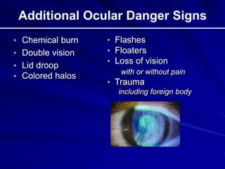
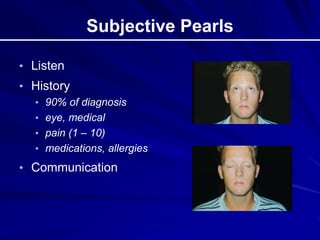
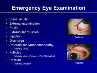
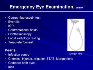
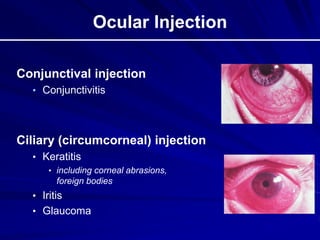
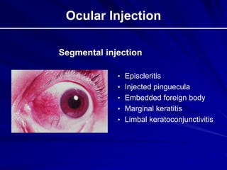
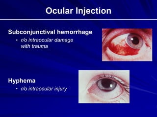
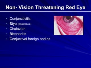
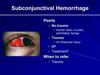
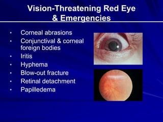
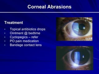
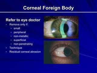
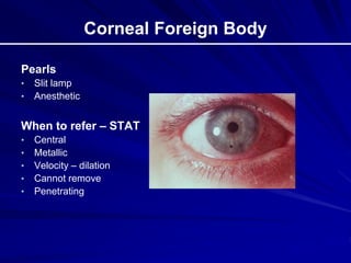
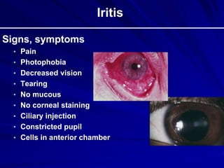
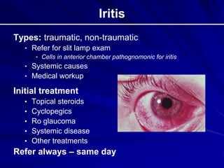
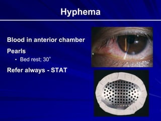
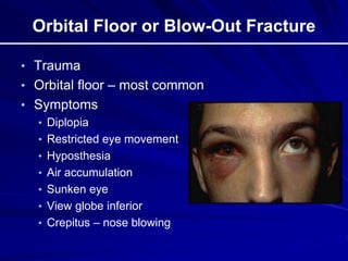
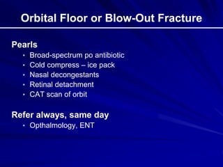
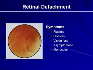
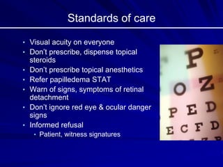
Basic Ocular
Emergencies
www.wordpress.com
Dr. Muhmmad Jawad Awan
PGT Ophthalamology BBH
Learning Objectives
Review:
• Ocular anatomy, danger signs, subjective pearls,
eye examination & pearls, ocular injection,
antibiotics
• Non- vision threatening red eye
• Vision-threatening red eye & emergencies
• Clinical pearls & indications for referral
Ocular Anatomy
Red Eye Danger Signs
• Decreased visual acuity
• Pain
• Ciliary flush
• Pupillary asymmetry
• Irregular corneal light reflex
• Corneal infiltrate
• Photophobia
• Trauma
Additional Ocular Danger Signs
• Chemical burn
• Double vision
• Lid droop
• Colored halos
• Flashes
• Floaters
• Loss of vision
with or without pain
• Trauma
including foreign body
Subjective Pearls
• Listen
• History
• 90% of diagnosis
• eye, medical
• pain (1 – 10)
• medications, allergies
• Communication
Emergency Eye Examination
• Visual acuity
• External examination
• Pupils
• Extraocular muscles
• Injection
• Discharge
• Preauricular lymphadenopathy
• (usually viral)
• Follicles
• (usually viral; chronic – r/o chlamydial)
• Papillae
• (usually allergy)
Follicles
Papillae
Emergency Eye Examination, cont’d.
• Cornea-fluorescein test
• Evert lid
• IOP
• Confrontational fields
• Ophthalmoscopy
• Lab & radiology testing
• Treat/refer/consult
Pearls
• Infection control
• Chemical injuries, irrigation STAT, Morgan lens
• Compare both eyes
• Iritis
Morgan lens
Ocular Injection
Conjunctival injection
• Conjunctivitis
Ciliary (circumcorneal) injection
• Keratitis
• including corneal abrasions,
foreign bodies
• Iritis
• Glaucoma
Ocular Injection
• Episcleritis
• Injected pinguecula
• Embedded foreign body
• Marginal keratitis
• Limbal keratoconjunctivitis
Segmental injection
Ocular Injection
Subconjunctival hemorrhage
• r/o intraocular damage
with trauma
Hyphema
• r/o intraocular injury
Hypopyon
White blood cells (pus) in anterior chamber
“Tells you it’s bad”
Hypopyon
Non- Vision Threatening Red Eye
• Conjunctivitis
• Stye (hordeolum)
• Chalazion
• Blepharitis
• Conjuctival foreign bodies
Chemical Conjunctivitis
• Chemosis • Morgan lens
Subconjunctival Hemorrhage
Pearls
• No trauma
• normal vision, no pain,
self-limited, benign
• Trauma
• r/o intraocular injury
• BP
• Treatment?
When to refer
• Trauma
Vision-Threatening Red Eye
& Emergencies
• Corneal abrasions
• Conjunctival & corneal
foreign bodies
• Iritis
• Hyphema
• Blow-out fracture
• Retinal detachment
• Papilledema
Corneal Abrasions
Treatment
• Topical antibiotics drops
• Ointment @ bedtime
• Cyclopegics – refer
• PO pain medication
• Bandage contact lens
Corneal Abrasions
When to refer
• Large abrasions
• > 3 mm
• Central abrasions
• especially large ones
• Without daily improvement
Scleral Perforation
Eye laceration
Conjunctival Foreign Bodies
Pearls
• Remove w/o anesthetic if
possible (why?)
• Lid inversion
• Treat residual corneal
abrasion
When to refer
• Unable to find, remove fb
• If fb sensation persists
Corneal Foreign Body
Refer to eye doctor
• Remove only if:
• small
• peripheral
• non-metallic
• superficial
• non-penetrating
• Technique
• Residual corneal abrasion
Corneal Foreign Body
Pearls
• Slit lamp
• Anesthetic
When to refer – STAT
• Central
• Metallic
• Velocity – dilation
• Cannot remove
• Penetrating
Iritis
Signs, symptoms
• Pain
• Photophobia
• Decreased vision
• Tearing
• No mucous
• No corneal staining
• Ciliary injection
• Constricted pupil
• Cells in anterior chamber
Iritis
Types: traumatic, non-traumatic
• Refer for slit lamp exam
• Cells in anterior chamber pathognomonic for iritis
• Systemic causes
• Medical workup
Initial treatment
• Topical steroids
• Cyclopegics
• Ro glaucoma
• Systemic disease
• Other treatments
Refer always – same day
Hyphema
Blood in anterior chamber
Pearls
• Bed rest; 30°
Refer always - STAT
Orbital Floor or Blow-Out Fracture
• Trauma
• Orbital floor – most common
• Symptoms
• Diplopia
• Restricted eye movement
• Hyposthesia
• Air accumulation
• Sunken eye
• View globe inferior
• Crepitus – nose blowing
Orbital Floor or Blow-Out Fracture
Pearls
• Broad-spectrum po antibiotic
• Cold compress – ice pack
• Nasal decongestants
• Retinal detachment
• CAT scan of orbit
Refer always, same day
• Opthalmology, ENT
Retinal Detachment
Symptoms
• Flashes
• Floaters
• Vision loss
• Asymptomatic
• Monocular
Retinal Detachment
Risk Factors
• High myopia
• Trauma (5-10%)
• Previous ocular surgery,
• Diabetic retinopathy
• Tumor, inflammation, lesions
• RD in non-involved eye (10 – 20%)
When to refer – STAT
Papilledema
Possibly life-threatening
Optic nerve swelling
• Cause: increased intracranial pressure
• Develops in hours; dissipates over months
Look for
• Bilateral swollen, hyperemic discs
• Blurred disc margins
• Disc hemorrhages
Papilledema
Normal

Swollen, blurred,
disc hemorrhages

Papilledema
Rule out most common
• Primary, metastatic intracranial masses
• Pseudotumor cerebri
• overweight women?
Pearls
• Neuroimaging- head, orbit
• Lumbar puncture
When to refer - STAT
Standards of care
• Visual acuity on everyone
• Don’t prescribe, dispense topical
steroids
• Don’t prescribe topical anesthetics
• Refer papilledema STAT
• Warn of signs, symptoms of retinal
detachment
• Don’t ignore red eye & ocular danger
signs
• Informed refusal
• Patient, witness signatures
Topical Corticosteriods
Don’t prescribe
• Side effects
• Herpes simplex
• Bacterial infection
• Wound healing
• Glaucoma
• Cataract
• Fungal (mycotic)
• Corneal melting, perforation
Thank you!

Ocular emergencies.ppt

  • 1.
  • 2.
    Learning Objectives Review: • Ocularanatomy, danger signs, subjective pearls, eye examination & pearls, ocular injection, antibiotics • Non- vision threatening red eye • Vision-threatening red eye & emergencies • Clinical pearls & indications for referral
  • 3.
  • 4.
    Red Eye DangerSigns • Decreased visual acuity • Pain • Ciliary flush • Pupillary asymmetry • Irregular corneal light reflex • Corneal infiltrate • Photophobia • Trauma
  • 5.
    Additional Ocular DangerSigns • Chemical burn • Double vision • Lid droop • Colored halos • Flashes • Floaters • Loss of vision with or without pain • Trauma including foreign body
  • 6.
    Subjective Pearls • Listen •History • 90% of diagnosis • eye, medical • pain (1 – 10) • medications, allergies • Communication
  • 7.
    Emergency Eye Examination •Visual acuity • External examination • Pupils • Extraocular muscles • Injection • Discharge • Preauricular lymphadenopathy • (usually viral) • Follicles • (usually viral; chronic – r/o chlamydial) • Papillae • (usually allergy) Follicles Papillae
  • 8.
    Emergency Eye Examination,cont’d. • Cornea-fluorescein test • Evert lid • IOP • Confrontational fields • Ophthalmoscopy • Lab & radiology testing • Treat/refer/consult Pearls • Infection control • Chemical injuries, irrigation STAT, Morgan lens • Compare both eyes • Iritis Morgan lens
  • 9.
    Ocular Injection Conjunctival injection •Conjunctivitis Ciliary (circumcorneal) injection • Keratitis • including corneal abrasions, foreign bodies • Iritis • Glaucoma
  • 10.
    Ocular Injection • Episcleritis •Injected pinguecula • Embedded foreign body • Marginal keratitis • Limbal keratoconjunctivitis Segmental injection
  • 11.
    Ocular Injection Subconjunctival hemorrhage •r/o intraocular damage with trauma Hyphema • r/o intraocular injury
  • 12.
    Hypopyon White blood cells(pus) in anterior chamber “Tells you it’s bad” Hypopyon
  • 13.
    Non- Vision ThreateningRed Eye • Conjunctivitis • Stye (hordeolum) • Chalazion • Blepharitis • Conjuctival foreign bodies
  • 14.
  • 15.
    Subconjunctival Hemorrhage Pearls • Notrauma • normal vision, no pain, self-limited, benign • Trauma • r/o intraocular injury • BP • Treatment? When to refer • Trauma
  • 16.
    Vision-Threatening Red Eye &Emergencies • Corneal abrasions • Conjunctival & corneal foreign bodies • Iritis • Hyphema • Blow-out fracture • Retinal detachment • Papilledema
  • 17.
    Corneal Abrasions Treatment • Topicalantibiotics drops • Ointment @ bedtime • Cyclopegics – refer • PO pain medication • Bandage contact lens
  • 18.
    Corneal Abrasions When torefer • Large abrasions • > 3 mm • Central abrasions • especially large ones • Without daily improvement
  • 19.
  • 20.
  • 21.
    Conjunctival Foreign Bodies Pearls •Remove w/o anesthetic if possible (why?) • Lid inversion • Treat residual corneal abrasion When to refer • Unable to find, remove fb • If fb sensation persists
  • 22.
    Corneal Foreign Body Referto eye doctor • Remove only if: • small • peripheral • non-metallic • superficial • non-penetrating • Technique • Residual corneal abrasion
  • 23.
    Corneal Foreign Body Pearls •Slit lamp • Anesthetic When to refer – STAT • Central • Metallic • Velocity – dilation • Cannot remove • Penetrating
  • 24.
    Iritis Signs, symptoms • Pain •Photophobia • Decreased vision • Tearing • No mucous • No corneal staining • Ciliary injection • Constricted pupil • Cells in anterior chamber
  • 25.
    Iritis Types: traumatic, non-traumatic •Refer for slit lamp exam • Cells in anterior chamber pathognomonic for iritis • Systemic causes • Medical workup Initial treatment • Topical steroids • Cyclopegics • Ro glaucoma • Systemic disease • Other treatments Refer always – same day
  • 26.
    Hyphema Blood in anteriorchamber Pearls • Bed rest; 30° Refer always - STAT
  • 27.
    Orbital Floor orBlow-Out Fracture • Trauma • Orbital floor – most common • Symptoms • Diplopia • Restricted eye movement • Hyposthesia • Air accumulation • Sunken eye • View globe inferior • Crepitus – nose blowing
  • 28.
    Orbital Floor orBlow-Out Fracture Pearls • Broad-spectrum po antibiotic • Cold compress – ice pack • Nasal decongestants • Retinal detachment • CAT scan of orbit Refer always, same day • Opthalmology, ENT
  • 29.
    Retinal Detachment Symptoms • Flashes •Floaters • Vision loss • Asymptomatic • Monocular
  • 30.
    Retinal Detachment Risk Factors •High myopia • Trauma (5-10%) • Previous ocular surgery, • Diabetic retinopathy • Tumor, inflammation, lesions • RD in non-involved eye (10 – 20%) When to refer – STAT
  • 31.
    Papilledema Possibly life-threatening Optic nerveswelling • Cause: increased intracranial pressure • Develops in hours; dissipates over months Look for • Bilateral swollen, hyperemic discs • Blurred disc margins • Disc hemorrhages
  • 32.
  • 33.
    Papilledema Rule out mostcommon • Primary, metastatic intracranial masses • Pseudotumor cerebri • overweight women? Pearls • Neuroimaging- head, orbit • Lumbar puncture When to refer - STAT
  • 34.
    Standards of care •Visual acuity on everyone • Don’t prescribe, dispense topical steroids • Don’t prescribe topical anesthetics • Refer papilledema STAT • Warn of signs, symptoms of retinal detachment • Don’t ignore red eye & ocular danger signs • Informed refusal • Patient, witness signatures
  • 35.
    Topical Corticosteriods Don’t prescribe •Side effects • Herpes simplex • Bacterial infection • Wound healing • Glaucoma • Cataract • Fungal (mycotic) • Corneal melting, perforation
  • 36.