Anatomy and Physiology of Digestive System
•
48 likes•18,019 views

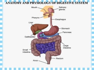
The digestive system includes the organs of the alimentary canal and accessory structures. The alimentary canal forms a continuous tube that is open to the outside environment at both ends. The organs of the alimentary canal are the mouth, pharynx, esophagus, stomach, small intestine, and large intestine.

Recommended
More Related Content
What's hot
What's hot (20)
Similar to Anatomy and Physiology of Digestive System
Similar to Anatomy and Physiology of Digestive System (20)
More from DR .PALLAVI PATHANIA
More from DR .PALLAVI PATHANIA (20)
Recently uploaded
Recently uploaded (20)
Anatomy and Physiology of Digestive System
- 1. ANATOMY AND PHYSIOLOGY OF DIGESTIVE SYSTEM
- 2. ANATOMY AND PHYSIOLOGY OF DIGESTIVE SYSTEM Submitted to: Submitted by: Dr. Pallavi Pathania Nisha Kumari Associate Professor M.Sc. Nursing 1st year (Medical Surgical Nursing) Shimla Nursing College Shimla Nursing College
- 3. INDEX SR. NO. CONTENT 1 Gastrointestinal Tract 2 organs of digestive system 3 Another name of digestive system 4 Primary organs of digestive system 5 Name of Accessory digestive organs 6 Layers of Gastro Intestinal wall 7 Digestion 8 Movements in digestive system 9 Mouth
- 4. SR. NO. CONTENT 10 Tongue 11 Teeth 12 Salivary glands 13 Pharynx 14 Oesophagus 15 Stomach 16 Pancreas 17 Liver 18 Gallbladder
- 5. SR. NO. CONTENT 19 Small intestine 20 Large intestine 21 Rectum 22 Anus 23 Gut flora 24 Physiology of digestion 25 Function of GI Tract 26 Conclusion
- 6. GASTROINTESTINAL TRACT (GIT) The gastrointestinal tract is an organ system within humans and other animals which takes in food, digests it and absorb energy and nutrients, and expels the remaining waste as feces.
- 7. CONTD… The bodily system concerned with the ingestion, digestion, and absorption of food and the discharge of residual wastes and consisting of the digestive tract and accessory glands (such as the salivary glands and the pancreas) that secrete digestive enzymes.
- 8. ORGANS OF DIGESTIVE SYSTEM Mouth Salivary glands Pharynx esophagus Stomach Pancreas
- 11. DIGESTIVE SYSTEM Digestive system also called digestive tract, digestional tract, GI tract, GIT, gut, or alimentary canal.
- 12. PRIMARY ORGANS OF THE DIGESTIVE SYSTEM Mouth. Pharynx. Esophagus. Stomach. Small Intestine. Large Intestine. Rectum. Anus
- 13. ACCESSORY DIGESTIVE ORGANS Liver Gallbladder Pancreas. Salivary gland
- 14. LAYER OF GASTROINTESTINAL WALL Four main layers. From inside (the lumen) to outside the are: 1. Mucosa 2. Sub mucosa 3. Muscularis (external) 4. Serosa ( visceral peritoneum)
- 15. MUCOSA ( VISCERAL PERITONEUM) It is the innermost, moist, epithelial membrane that lines the entire digestive tract. (1) It secretes mucus, digestive enzymes, and hormones. (2) Absorbs digestive end products into the blood. (3) Protects against infectious disease.
- 16. CONTD… Consists of a lining epithelium, a lamina propria, and a Muscularis mucosa. Epithelium - simple columnar epithelium and goblet cells Lamina propria – it is large layer of connective tissue. Which separate innermost layer of epithelial cell from a layer of muscle tissue called muscularis mucosa Muscularis mucosa - thin layer, produces local movements of the mucosa.
- 17. SUB MUCOSA It is a moderately dense connective tissue layer containing blood and lymphatic vessels, lymphoid follicles, and nerve fibers.
- 18. MUSCULARIS EXTERNA It typically consists of smooth muscle and is responsible for peristalsis and segmentation. Contains the myenteric plexus of Auerbach, the other major intrinsic nerve plexus. Located between the two layers of smooth muscle, controls motility of the Gastro Intestinal tract.
- 19. SEROSA It is the protective outer layer of the intraperitoneal organs, is the visceral peritoneum.
- 20. DIGESTION Digestion is the process by which insoluble food, consisting of large molecules is broken down into soluble compounds Digestion starts at the mouth and ends at the anus.
- 21. FOOD IS BROKEN DOWN BY TWO ACTIONS 2. CHEMICAL 1. PHYSICAL /MECHANICAL
- 22. 1. PHYSICAL DIGESTION BY Teeth Peristalsis of the alimentary canal Chewing
- 23. 2. CHEMICAL DIGESTION BY: Enzymes Digestive enzymes are the chemicals that break large insoluble food molecules into smaller soluble molecules.
- 24. PHASES OF DIGESTION 1. Ingestion 2. Digestion 3.Absorption into the bloodstream 4. Egestion
- 25. CONTD… The esophagus, stomach, small intestine, and large intestine are the main regions of the GI tract. They are separated from each other by special muscles, called sphincters, which regulate the movement of ingested material from one part to another. Each part of the GI tract has a unique function to perform in digestion, and each has a distinct type of motility and sensation.
- 26. MOVEMENTS IN ESOPHAGUS The function of the esophagus is to transport the ingested material from the pharynx to the stomach by peristaltic waves.
- 28. PERISTALSIS MOVEMENTS Peristalsis is a series of organized muscle contractions that occur throughout the digestive tract. (that propels foodstuffs distally through the esophagus and intestines). The synchronized contraction of the esophagus, stomach, and intestine is called peristalsis
- 29. CONTD… Peristalsis is a series of wave- like muscle contractions that moves food to different processing stations in the digestive tract. The process of peristalsis begins in the esophagus when a bolus of food is swallowed. It was first described by Bayliss and Starling in 1899 as a type of motility in which there is contraction above and relaxation below a segment being stimulated.
- 30. CONTD… The strong wave like motions of the smooth muscle in the esophagus carry the food to the stomach , where it is churned into a liquid mixture called chyme. Peristalsis continuous in the small intestines where it mixes and shifts the chyme back and forth, allowing nutrients to be absorbed into the bloodstream through the small intestine walls.
- 31. CONTD… Peristalsis concludes in the large intestines where water from the undigested food material is absorbed into the bloodstream. While peristalsis involves one-way motion in the caudal direction, segmentation contractions move chyme in both directions, which allows greater mixing with the secretions of the intestines.
- 32. TYPES OF PERISTALSIS MOVEMENT Primary peristalsis: Triggered by the swallowing center in the brain stem and the contraction wave travel at speed 2cm/s. Secondary peristalsis: Induced by esophageal distension from retained bolus, refluxed material. Its role is to clear the esophagus form retained bolus.
- 33. CONTD… Tertiary peristalsis: Tertiary contractions are non-propulsive and uncoordinated and their non-peristaltic nature means they move the bolus up as well as down the oesophagus.
- 35. SEGMENTATION CONTRACTIONS (OR MOVEMENTS) It is a type of intestinal motility. Unlike peristalsis, which predominates in the esophagus, segmentation contractions occur in the large intestine and small intestine, while predominating in the later.
- 36. CONTD… Segmentation contraction occurs 12time/min in duodenum and 8time/min in the ileum. These contraction last for 5-6sec, they occur through out the digestive period. Segmentation involves contractions of the circular muscles in the digestive tract, while peristalsis involves rhythmic contractions of the longitudinal muscles in the gastrointestinal tract.
- 37. MOVEMENTS OF SMALL INTESTINE Mixing (Segmentation Contractions) Peristalsis (Propulsive Movements) Movements of Muscularis Mucosa Contraction of villi
- 38. MOVEMENTS OF COLON Mixing Movements (Haustrations): Same manner that segmentation movements the circular muscle contracts,
- 39. CONTD… At the same time, the longitudinal muscle of the colon, which is aggregated into three longitudinal strips called the teniae coli, contracts. move slowly toward the anus. The combination of constricted and relaxed portions of the colon create haustrations (bag-like pouches). Haustrations mix the chyme or fecal material and provide slow persistent forward movement.
- 40. CONTD…. Propulsive Movements (Mass Movements): Much of the propulsion in the cecum and ascending colon results from the slow but persistent haustral contractions From the transverse colon to the sigmoid,
- 41. CONTD…. Mass movements one to three times each day, in many people especially for about 15 minutes during the first hour after eating breakfast. Occasionally (1-3 times/day), a modified contraction will propel the contents forward. This occurs by a peristaltic contraction pushing the contents through 20 cm or more of colon in which the haustrations are relaxed.
- 42. ORGANS
- 43. 1. MOUTH The mouth is the first portion of the alimentary canal that receives food and produces saliva.
- 44. CONTD… Mouth or oral cavity is bounded by muscles and bones. The oral cavity is lined throughout with mucous membrane, consisting of stratified squamous epithelium containing small mucus secreting gland. The part of mouth between the gums and the cheeks is the vestibule and the remainder of its interior is the oral cavity
- 45. CONTD… The palate forms the roof of the mouth & is divided into the anterior hard palate & posterior soft palate. The uvula is a curved fold of muscle covered with mucous membrane, hanging down from the middle of the free border of the soft palate.
- 46. CONTD… Relations: Anteriorly- by the lips Posteriorly- continue with the oropharynx Laterally- muscles of cheeks Superiorly- bony hard palate Inferiorly- muscular tongue & the soft tissues of the floor of the mouth
- 47. FUNCTION OF MOUTH The two main functions of the mouth are eating and speaking. The face's trigeminal nerve provides sensation (feeling) and helps us to bite, chew and swallow.
- 48. ORAL CAVITY CONSIST OF: 1. Tongue 2. Teeth 3. Salivary glands
- 49. 1. TONGUE
- 50. TONGUE The tongue is a muscular organ in the mouth and composed of voluntary muscle. It attached by its base to the hyoid bone and by fold of its mucous membrane covering called the frenulum to the floor of the mouth, It is of importance in the digestive system and is the primary organ of taste in the gustatory system.
- 51. CONTD… Length :3 inches Shape: triangular The tongue's upper surface (dorsum) is covered by taste buds housed in numerous lingual papillae The human tongue is divided into two parts, an oral part at the front and a pharyngeal part at the back.
- 52. PAPPILAE Papillae (singular papilla) are nodules on the surface of the tongue that increase the surface area for the taste buds.
- 53. TYPES OF PAPILAE 1. Vallate/ circumvallate (8-12 in number) 2. Filiform (numerous in numbers) 3. Fungiform( larger than filiform pappilae) 4. Foliate
- 54. TASTE BUDS These are sensory receptor for taste. The sensation of taste is called Gustation. Taste buds are located on the surface of papilae except filiform papillae
- 55. BLOOD SUPPLY TO TONGUE The tongue receives its blood supply primarily from the lingual artery, a branch of the external carotid artery. The lingual veins drain into the internal jugular vein. The floor of the mouth also receives its blood supply from the lingual artery.
- 56. FUNCTIONS OF TONGUE Mastication (chewing) Deglutition (swallowing) Taste Speech
- 57. 2. TEETH
- 58. ERUPTION OF TEETH Tooth eruption in humans is a process in tooth development in which the teeth enter the mouth and become visible. Primary teeth erupt into the mouth from around six months until two years of age.
- 59. TEETH The teeth are embedded in the alveoli or sockets of the alveolar ridges of the mandible and the maxilla. Babies are born with two sets, or dentitions, the temporary or deciduous teeth and permanent teeth. At birth the teeth of both dentitions are present, in immature form, in the mandible and maxilla.
- 60. There are 20 temporary teeth, 10 in each jaw. They begin to erupt at about 6 months of age, and should all be present by 24 months. The permanent teeth begin to replace the deciduous teeth in the 6th year of age and this dentition, consisting of 32 teeth, is usually complete by the 21st year. The human teeth function to mechanically break down items of food by cutting and crushing them in preparation for swallowing and digesting. CONTD…
- 61. CONTD… Humans have four types of teeth: incisors, canines, premolars, and molars, each with a specific function.
- 62. PRIMARY TEETH Among deciduous (primary) teeth, ten are found in the maxilla (upper jaw) and ten in the mandible (lower jaw), for a total of 20. The dental formula for primary teeth is 2.1.0.2/2.1.0.2. In the primary set of teeth, two types of incisors – centrals and laterals, one canine & two types of molars – first and second. All primary teeth are normally later replaced with their permanent counterparts.
- 63. PERMANENT TEETH Among permanent teeth, 16 are found in the maxilla and 16 in the mandible, for a total of 32. The dental formula is 2.1.2.3/2.1.2.3. Age 21, all 32 of the permanent teeth have usually erupted. The permanent teeth are the: Two incisor (for cutting)-central incisor, lateral incisor One canine (for tearing) Two premolar(for crushing)-first premolar, second premolar, Three molar (for grinding)-first molar, second molar, and third molar.
- 64. STRUCTURE OF TEETH ENAMEL Enamel is the hardest and most highly mineralized substance of the body. It is one of the four major tissues which make up the tooth, along with dentin, cementum, and dental pulp. 96% of enamel consists of mineral, with water and organic material comprising the rest.
- 65. • The normal color of enamel varies from light yellow to grayish white. DENTIN • Dentin is the substance between enamel or cementum and the pulp chamber. • Dentin is a mineralized connective tissue with an organic matrix of collagenous proteins. • The porous, yellow-hued material is made up of 70% inorganic materials, 20% organic materials, and 10% water by weight CONTD…
- 66. CEMENTUM Cementum is a specialized bone like substance covering the root of a tooth. Its coloration is yellowish and it is softer than dentin and enamel. CONTD…
- 67. DENTAL PULP The dental pulp is the central part of the tooth filled with soft connective tissue. This tissue contains blood vessels and nerves that enter the tooth from a hole at the apex of the root. CONTD…
- 68. BLOOD SUPPLY OF TEETH The blood supply to the upper jaw is provided by the superior alveolar arteries, which arise from the infraorbital and maxillary arteries. Blood to the lower jaw is carried by the inferior alveolar artery, another branch of the maxillary artery.
- 69. FUNCTIONS OF TEETH Two incisor -for cutting One canine -for tearing Two premolar-for crushing Three molar-for grinding
- 70. 3. SALIVARY GLANDS The salivary glands in are exocrine glands that produce saliva through a system of ducts into the mouth. Humans have 3 paired major salivary glands: Parotid submandibular and Sublingual as well hundreds of minor salivary glands.
- 72. a) PAROTID GLANDS The two parotid glands are major salivary glands situated one on each side of the face just below the external meatus. Each gland has a parotid duct opening into the mouth at the level of the second upper molar tooth. The largest of the salivary glands.
- 73. FUNCTION OF PAROTID GLANDS They secrete saliva to facilitate mastication and swallowing, and amylase to begin the digestion of starches.
- 74. b) SUBMANDIBULAR GLANDS The submandibular glands are a pair of major salivary glands located beneath the lower jaws. The secretion produced is a mixture of both serous fluid and mucus, and enters the oral cavity via the submandibular duct. The submandibular ducts open on to the floor of mouth, one on each side of the frenulum of the tongue
- 75. FUNCTIONS OF SUBMANDIBULAR GLANDS They also secrete saliva that are essential for digestion and for maintaining healthy mouth. Saliva that contain enzyme that begin to breakdown food before it passes to the stomach. It moistens food so that it slips easily to the esophagus.
- 76. c) SUBLINGUAL GLANDS The sublingual glands are a pair of major salivary glands located inferior to the tongue, anterior to the submandibular glands. Approximately 5% of saliva entering the oral cavity comes from these glands. The secretion produced is mainly mucous in nature They have numerous small ducts that open into the floor of mouth.
- 77. FUNCTIONS OF SUBLINGUAL GLANDS The main role of sublingual gland is to produce saliva, which has many functions like food moisturizing, carbohydrate digestion, protection against bacteria flora etc.
- 78. d) MINOR SALIVARY GLANDS There are 800 to 1,000 minor salivary glands located throughout the oral cavity within the submucosa of the oral mucosa in the tissue of the buccal, and lingual mucosa.
- 79. COMPOSITION OF SALIVA About 1.5 litres of saliva is produced daily & it consists of: Water Mineral salts An enzyme Mucus Lysozyme Immunoglobulins
- 80. FUNCTION OF SALIVA 1. Saliva contributes to the digestion of food and to the maintenance of oral hygiene. 2. Without normal salivary function the frequency of dental caries, gum disease and other oral problems increases significantly.
- 81. LUBRICANT 3. Saliva, coats the oral mucosa, mechanically protecting it from trauma during eating, swallowing and speaking. 4. In people with little saliva soreness of the mouth is very common, and the food (especially dry food) sticks to the inside of the mouth.
- 82. DIGESTION 5. The digestive functions of saliva include moistening food and helping to create a food bolus. 6. This lubricative function of saliva allows the food bolus to be passed easily from the mouth into the esophagus.
- 83. ROLE IN TASTE Saliva is very important in the sense of taste. It is the liquid medium in which chemicals are carried to taste receptor cells (mostly associated with lingual papillae). Other Function Saliva maintains the pH of the mouth. Saliva is supersaturated with various ions.
- 84. HOW FOOD MOVE STEP 1. Mouth: when we eat food mastication & saliva helps to make the soft bolus of food so the content travel easily and smoothly through pharynx. STEP 2. PHARYNX: The pharynx receives the food from the mouth. When food reaches the pharynx food moves towards the esophagus by the involuntary muscle contractions.
- 85. CONTD… STEP 3. ESOPHAGUS: the bolus move through the esophagus by a movement called peristalsis ( contraction and relaxation of muscles). That pushes the bolus towards the stomach. STEP 4. STOMACH: the stomach is an organ where food stores and further broken down by gastric acid and powerful enzymes. From there, food moves into the small intestine.
- 86. CONTD… STEP 5. SMALL INTESTINE: Most of nutrients are absorbed in the small intestine, where food is broken down even more by enzymes released from the pancreas & bile from the liver. Anything left in the small intestines move into the large intestine by the segmentation contraction. STEP 6. LARGE INTESTINE: the colon is a 5-7 foot long muscular tube that connect the small intestine to the rectum. It is responsible for processing waste. On average, it takes about 36 hours for waste, or stool, to get through the colon and exit the rectum through the anus.
- 88. 2. PHARYNX The pharynx is the part of the throat that is behind the mouth and nasal cavity and above the esophagus and the larynx, or the tubes going down to the stomach and the lungs. The pharynx is the portion of the digestive tract that receives the food from your mouth. Branching off the pharynx is the esophagus, which carries food to the stomach,
- 89. CONTD…
- 90. CONTD… The pharynx is devided for descriptives purpose into three parts, the nasopharynx, oropharynx and laryngopharynx. The nasopharynx is important in respiration. The oropharynx and laryngopharynx are passages common to both the respiratory and the digestive system.
- 91. Food passes from the oral cavity into the pharynx then to the oesophagus below, with which it is continuous, The walls of the pharynx consist of three layers of tissue. The lining membrane (Mucosa) is stratified squamous epithelium, continuous with the lining of the mouth at one end and the esophagus at the other. Stratified epithelial tissue provides a lining well suited to the wear and tear of swallowing ingested food. CONTD…
- 92. The middle layer consists of connective tissue, which become thinner towards the lower end and contains blood and lymph vessels and nerves. The outer layers consists of a number of involuntary muscles that are involved in swallowing. When food reaches the pharynx, swallowing is no longer under voluntary control. CONTD…
- 93. FUNCTION OF PHARYNX For digestive purpose; 1. The muscular walls function in the process of swallowing. 2. It serves as a pathway for the movement of food from the mouth to the esophagus.
- 94. 3. OESOPHAGUS
- 95. ESOPHAGUS The esophagus or oesophagus, commonly known as the food pipe or gullet, The esophagus is a muscular tube connecting the throat (pharynx) with the stomach. The esophagus runs behind the windpipe (trachea) and heart, and in front of the spine. Length :25 cm Diameter:2 cm
- 97. It passes between muscle fibres of the diaphragm behind the central tendon at the level of the 10th thoracic vertebra. Immediately the oesophagus has passed through the diaphragm it curves upward before opening into the stomach. This sharp angle is believed to be one of the factors that prevents the regurgitation into the oesophagus. The upper and lower ends of the oesophagus are closed by sphincters. CONTD…
- 98. The upper cricopharyngeal or upper oesophageal sphincter prevents air passing into the oesophagus during inspiration and aspiration of oesphageal contents. The cardiac or lower oesophageal sphincter prevents the reflux of acid gastric contents into the oesophagus. CONTD…
- 100. STRUCTURE The wall of the esophagus from the lumen outwards consists of mucosa, submucosa (connective tissue), layers of muscle fibers between layers of fibrous tissue, and an outer layer of connective tissue. The mucosa is a stratified squamous epithelium of around three layers of squamous cells, which contrasts to the single layer of columnar cells of the stomach. Most of the muscle is smooth muscle although striated muscle predominates in its upper third.
- 101. It has two muscular rings or sphincters in its wall, one at the top and one at the bottom. A sphincter is a circular muscle that normally maintains constriction of a natural body passage or orifice and which relaxes as required by normal physiological functioning. The lower sphincter helps to prevent reflux of acidic stomach content. CONTD…
- 102. PARTS OF ESOPHAGUS The esophagus is split into the following 3 parts: Cervical part (4 cm in length). The cervical part extends from the lower border of cricoid cartilage to the superior border of manubrium sterni.
- 103. CONTD… Thoracic part (20 cm in length). The thoracic part extends from superior border of manubrium sterni to the esophageal opening in the diaphragm.
- 104. CONTD… Abdominal part (1-2 cm in length). The abdominal part extends create esophageal opening in the diaphragm to the cardiac end of the stomach.
- 105. BLOOD SUPPLY TO OESOPHAGUS The inferior thyroid artery supplies the cervical esophagus. Branches of the bronchial arteries and branches directly off of the aorta supply the proximal and distal thoracic esophagus, respectively. Finally, branches of the left gastric and inferior phrenic artery supply the abdominal esophagus.
- 106. FUNCTIONS OF OESOPHAGUS 1. Formation of a bolus 2. Swallowing 3. Food is ingested through the mouth and when swallowed passes first into the pharynx and then into the esophagus. 4. Reducing gastric reflux Constriction of the upper and lower esophageal sphincters help to prevent reflux (backflow) of gastric contents and acid into the esophagus, protecting the esophageal mucosa.
- 107. 4. STOMACH
- 108. STOMACH The stomach is a J- shaped dilated portion of the alimentary tract situated in the epigastric, umbilical and left hypochondriac regions of the abdominal cavity. The stomach is a muscular organ. The stomach receives food from the esophagus. As food reaches the end of the esophagus, it enters the stomach through a muscular valve called the lower esophageal sphincter.
- 109. RELATIONS Anteriorly-left lobe of liver & anterior abdominal wall Posteriorly-abdominal aorta,pancreas,spleen,left kidney Superiorly-diaphragm,oesophagus & left lobe of liver Inferiorly-transverse colon & small intestine Left side-diaphragm & spleen Right side-liver & duodenum
- 110. STRUCTURE OF STOMACH A pouch-like organ primarily designed for food storage (for 2-4 hours) , some mechanical and chemical digestion also occur . Contains two sphincters at both ends to regulate food movement : cardiac sphincter near the esophagus , Which close off the top end of the stomach pyloric sphincter near the small intestine, which close the bottom end of the stomach.
- 111. There are two curvature of stomach 1. Greater curvature – forms the long, convex, lateral border of the stomach. 2. Lesser curvature – forms the shorter, concave, medial surface of the stomach. The most inferior part of the lesser curvature, the angular notch, indicates the junction of the body and pyloric region. CONTD…
- 112. Divided into 4 regions : cardiac stomach (or cardiac), Which surrounds the opening of the esophagus into the stomach. Fundus of stomach (or funded) , which area above the level of the cardial orifice. body of stomach, which is the largest region of the stomach. CONTD…
- 113. CONTD… pyloric stomach (or Pylorus), which is divided into the pyloric Antrum and pyloric canal and is the distal end of the stomach. Contain thick folds called rugae at its layer , for providing larger surface area for expansion , secretion , digestion , and some absorption.
- 114. LAYERS OF STOMACH The stomach is made of these five layers: 1. MUCOSA: This is the first and innermost layer or lining. It contains the glands that release digestive juices. These are called hydrochloric acid and pepsin. This is where most stomach cancer start. 2. SUBMUCOSA: This second layer supports the mucosa. It is rich in blood vessels.
- 115. CONTD… 3. MUSCULRIS: The third layer is made of thick muscles. They help to mix food with the digestive juices. 4. SUBSEROSA: This layer contain supporting tissues for the serosa. 5. SEROSA : this is the last and outermost layer. It’s the lining that wraps around the stomach to confine it.
- 116. MOVEMENTS IN STOMACH
- 117. BLOOD SUPPLY OF STOMACH The stomach is supplied by a rich system of arteries derived from the celiac trunk, the first major visceral branch of the abdominal aorta. The lesser curvature of the stomach is supplied by the left and right gastric artery, which are branches of the celiac trunk and the common hepatic artery respectively.
- 118. FUNCTIONS OF STOMACH 1. Digestion The stomach releases proteases (protein-digesting enzymes such as pepsin) and hydrochloric acid, which kills or inhibits bacteria and provides the acidic pH of 2 for the proteases to work. Food is churned by the stomach through muscular contractions of the wall called peristalsis
- 119. 2. Absorption some absorption of certain small molecules nevertheless does occur in the stomach through its lining 3. Non- specific defense against microbes- provided by hydrochloric acid in gastric juice. Vomiting may occur in response to ingestion of gastric irritants, e.g. microbes & chemicals. CONTD…
- 120. GASTRIC JUICE Gastric acid, gastric juice or stomach acid, is a digestive fluid formed in the stomach and is composed of hydrochloric acid (HCl), potassium chloride (KCl) and sodium chloride (NaCl). The acid plays a key role in digestion of proteins, by activating digestive enzymes, and making ingested proteins unravel so that digestive enzymes break down the long chains of amino acids.
- 121. GASTRIC SECRETORY CELLS 1. Chief cells: secrete pepsinogen (an inactive enzyme).
- 122. CONTD… 2. Parietal cells: secrete hydrochloric and (HCl) and "intrinsic factor" (which helps absorption of vitamin B12 in the intestines). 3. Mucous cells: secrete mucus and alkaline substances to help neutralize HCl in the gastric juice .
- 123. CONTD.. 4. G cells: secrete a hormone called gastrin , which stimulates the parietal cells and overall gastric.
- 124. 5. PANCREAS
- 125. PANCREAS The pancreas is a pale grey gland weighing about 60 grams. It is a glandular organ in the digestive system It is located in the abdominal cavity behind the stomach. Length: 15 cm or 6 inch The pancreas is both an exocrine and endocrine gland.
- 127. Exocrine Pancreas This consist large number of lobules made up of small acini, the walls of which consist of secretory cells. Each lobule is drained by a tiny duct these unite eventually to form the pancreatic duct, which extends along the whole length of the gland and open into the duodenum CONTD…
- 128. FUNCTION OF EXOCRINE PANCREAS The function of exocrine pancreas is to produce pancreatic juice containing enzyme, some in the form of inactive percursors, that digest carbohydrates, proteins and fats.
- 129. Endocrine Pancreas The endocrine pancreas disrtibuted throughout the gland are groups of specialised cells called the pancreatic islets ( of langerhans). The isleits have no ducts so the hormones diffuse directly into the blood. CONTD…
- 130. FUNCTION OF ENDOCRINE PANCREAS 1. The endocrine component of the pancreas consists of islet cells (islets of Langerhans) that create and release important hormones directly into the bloodstream. 2. Two of the main pancreatic hormones are insulin, which acts to lower blood sugar, and glucagon, which acts to raise blood sugar
- 131. STRUCTURE Anatomically, the pancreas is divided into the head of pancreas, the neck of pancreas, the body of pancreas, and the tail of pancreas.
- 132. CONTD… The head lies in the curve of duodenum. The neck is about 2.5 cm or 1 inch long and lies between the head and the body The body is the largest part of the pancreas and lies behind the pylorus ( stomach). The tail lies in front of the left kidney and just reaches the spleen.
- 133. BLOOD SUPPLY OF PANCREAS The pancreas is supplied by the pancreatic branches of the splenic artery. The head is additionally supplied by the superior and inferior pancreaticoduodenal arteries which are branches of the gastroduodenal (from coeliac trunk) and superior mesenteric arteries, respectively.
- 134. FUNCTION 1. The pancreas is involved in blood sugar control and metabolism within the body. 2. Pancreatic islets are present in the pancreas. 3. Within these islets are four main types of cells which are involved in the regulation of blood glucose levels. 4. Each type of cell secretes a different type of hormone: 5. α alpha cells secrete glucagon (increase glucose in blood)
- 135. 6. β beta cells secrete insulin (decrease glucose in blood) 7. δ delta cells secrete somatostatin (regulates/stops α and β cells) and 8. γ (gamma) cells, secrete pancreatic polypeptide. CONTD….
- 136. 6. LIVER The liver is the largest gland in the body, weighing between 1 and 2.3kg . It is situated in the upper part of the abdominal cavity occupying the greater part of the hypochondriac region, part of the epigastric region and extending into the left hypochondriac region. Its upper and anterior surfaces are smooth and curved to fit the under surface of the diaphragm, its posterior surface is irregular in outline.
- 137. RELATION Anteriorly- diaphragm & anterior abdominal wall Posteriorly- oesophagus, inferior vena cava, aorta, gall bladder, vertebral column & diaphragm Laterally- lower ribs & diaphragm Superiorly- diaphragm & anterior abdominal wall Inferiorly- stomach, bile ducts, duodenum, hepatic flexure of colon, right kidney
- 138. STRUCTURE The liver is a reddish-brown wedge-shaped organ with four lobes of unequal size and shape. width -15 cm. It is both the heaviest internal organ and the largest gland in the human body The lobes of the liver are made up of tiny functional units, called lobules, which are just visible to naked eye.
- 139. CONTD… Sinusoids containing a mixture of blood from the tiny branches of the portal vein and hepatic artery. This allows the arterial blood and portal venous blood to mix and come into close contact with liver cells. Amongst the cell lining the sinusoids are hepatic macrophages (Kupffer cells) whose function is to ingest and destroy worn out blood cells and any foreign particles present in the blood flowing through the liver.
- 140. The liver is grossly divided into two parts when viewed from above – a right and a left lobe. The falciform ligament, divides the liver into a left and right lobe. CONTD…
- 141. SCHEME OF BLOOD FLOW THROUGH THE LIVER Hepatic artery ( Oxygenated blood) Portal vein ( Deoxygenated blood rich in nutrients) Interlobular vein Sinusoid Central vein Hepatic vein Inferior vena cava right atrium of the heart
- 142. BLOOD SUPPLY TO LIVER The liver receives a blood supply from two sources. The first is the hepatic artery which delivers oxygenated blood from the general circulation. The second is the hepatic portal vein delivering deoxygenated blood from the small intestine containing nutrients.
- 143. FUNCTIONS OF LIVER 1. Synthesis Proteins produced and secreted by the liver. The liver plays a major role in carbohydrate, protein, amino acid, and lipid metabolism. The liver is responsible for the breakdown of insulin and other hormones.
- 144. 2. Other The liver stores a multitude of substances, including glucose (in the form of glycogen) vitamin A (1–2 years' supply) vitamin D (1–4 months' supply) vitamin B12 (3–5 years' supply) vitamin K, iron, and copper. CONTD…
- 145. CONTD…. The liver produces albumin, the most abundant protein in blood serum. Contains phagocytes to destroy damaged erythrocytes and foreign substances, using phagocytosis
- 146. 7. GALLBLADDER The gallbladder is a small hollow organ where bile is stored and concentrated before it is released into the small intestine. In humans, the pear-shaped gallbladder lies beneath the liver.
- 147. STRUCTURE Sits in a shallow depression below the right lobe of the liver. Length-7 to 10 cm or 2.8 to 3.9 inches Diameter -4 cm or 1.6 inch The gallbladder has a capacity of about 50 millilitres It has a fundus or expended end, a body or main part and a neck, which is continuous with the cystic duct.
- 148. BLOOD SUPPLY TO GALLBLADDER The arterial supply to the gallbladder is via the cystic artery - a branch of the right hepatic artery (which itself is derived from the common hepatic artery, one of the three major branches of the coeliac trunk).
- 149. FUNCTIONS 1. The main purpose of the gallbladder is to store bile, also called gall, needed for the digestion of fats in food. 2. Bile flows through small vessels into the larger hepatic ducts and ultimately through the cystic duct into the gallbladder, where it is stored.
- 150. 8. SMALL INTESTINE The small intestine or small bowel is the part of the gastrointestinal tract between the stomach and the large intestine, and is where most of the end absorption of food takes place. It lies in the abdominal cavity surrounded by the large intestine. In the small intestine the chemical digestion of food is completed and absorption of most nutrients takes place.
- 152. STRUCTURE Length- 3m – 5m Diameter- 2.5- 3cm or 1 inch PARTS The small intestine is divided into three structural parts: (I)The duodenum (II)The jejunum (III)The ileum
- 153. The duodenum is a short structure ranging from 20 cm to 25 cm in length, and shaped like a "C". The jejunum is the midsection of the small intestine, connecting the duodenum to the ileum. It is about 2.5 m long. The ileum is the final section of the small intestine. CONTD…
- 154. CONTD… It is about 3 m long and end at ileocecal valve, which controls the flow of material from the ileum to the caecum, the first part of the large intestine , & prevents backflow
- 155. BLOOD SUPPLY TO SMALL INTESTINE The superior mesenteric artery supplies the whole small intestine and extends branches up to the middle third of the transverse colon. Up to this point, the innervation is taken over by the vagus nerve (CN X)
- 156. FUNCTIONS 1. Digestion The small intestine is where most chemical digestion takes place. Many of the digestive enzymes that act in the small intestine are secreted by the pancreas and liver and enter the small intestine via the pancreatic duct. Digestion of proteins & carbohydrate
- 157. 2. Absorption Digested food is now able to pass into the blood vessels in the wall of the intestine through either diffusion or active transport. The small intestine is the site where most of the nutrients from ingested food are absorbed. CONTD…
- 158. CONTD.. 3. Immunological The small intestine supports the body's immune system. The presence of gut flora appears to contribute positively to the host's immune system
- 159. 9. LARGE INTESTINE The large intestine, also known as the large bowel or colon, is the last part of the gastrointestinal tract and of the digestive system. Water is absorbed here and the remaining waste material is stored as feces before being removed by defecation.
- 160. STRUCTURE The length of male colon is 166 cm . female colon 155 cm The colon consists of five sections: 1. the cecum 2. ascending colon, 3. the transverse colon, 4. the descending colon, the sigmoid colon and the rectum.
- 161. Sections of the colon are: The ascending colon including the cecum and appendix The transverse colon including the colic flexures and transverse mesocolon The descending colon /The sigmoid colon – the s-shaped region of the large intestine CONTD…
- 162. The average inner diameter of sections of the colon in centimeters cecum 8-9cm ascending colon 6.6cm transverse colon 5.8cm descending/sigmoid colon 6.3cm and rectum near rectal/sigmoid junction 5.7cm CONTD…
- 163. THE CECUM The cecum is the first section of the colon and involved in the digestion, while the appendix is a structure of the colon, not involved in digestion. There is no function of the appendix in digestive system. But can cause significant problems when it becomes inflamed (appendicitis).
- 164. CONTD… The ileocecal valve is a sphincter muscle valve that separates the small intestine and the large intestine. Its critical function is to limit the reflux of colonic contents into the ileum
- 165. THE ASCENDING COLON It is connected to the small intestine by a section of bowel called the cecum. The ascending colon runs upwards through the abdominal cavity toward the transverse colon for approximately eight inches or 20 cm. The unwanted waste material is moved upwards toward the transverse colon by the action of peristalsis.
- 166. TRANSVERSE COLON This part extends across the abdominal cavity in front of the duodenum and the stomach to the area of the spleen where it forms the splenic flexure & curves acutely downwards to become the descending colon.
- 167. DESCENDING COLON The descending colon is the part of the colon from the splenic flexure to the beginning of the sigmoid colon, descending colon is also called the distal gut. One function of the descending colon in the digestive system is to store feces that will be emptied into the rectum.
- 168. SIGMOID COLON The sigmoid colon is the part of the large intestine after the descending colon and before the rectum. The name sigmoid means S-shaped . The walls of the sigmoid colon are muscular, and contract to increase the pressure inside the colon, causing the stool to move into the rectum.
- 169. APPENDIX It is a blind- ended muscular tube attached to the posteromedial wall of the caecum, about 2cm below ileocaecal junction.
- 170. CONTD… The appendix is 9 cm (7 to 11cm) in length but can range from 2 to 20 cm. The diameter of the appendix is 1 to 7 mm. It is relatively longer in children and decreases after 40 years of age.
- 171. PARTS OF APPENDIX A) Base B) Body C) Tip
- 172. A) BASE It is attached to posteromedial wall of caecum about 2 m below the ileocaecal junction. All taenia of caecum converge to the base and serve as a guide for the identification of the appendix.
- 173. B) BODY Body is narrow, tubular and contains a canal which opens into the caecum. The caecal opening is guarded by an incomplete mucous fold called as,”THE VALVE OF GERLACH”
- 174. C) TIP It is least vascular and is directed in various direction
- 175. STRUCTURE OF APPENDIX The appendix sits at the junction of the small intestine and large intestine. It’s a thin tube about four inches long. Normally, the appendix sits in the lower right abdomen. The function of the appendix is unknown. One theory is that the appendix acts as a storehouse for good bacteria, “rebooting” the digestive system after diarrheal illnesses. Other experts believe the appendix is just a useless remnant from our evolutionary past. Surgical removal of the appendix causes no observable health problems.
- 176. 10. RECTUM The rectum is the last section of the large intestine. It holds the formed feces awaiting elimination via defecation. It leads from the sigmoid colon & terminate in the anal canal.
- 177. 11. ANUS The anus is the external opening of the rectum. This is short passage about 3.8 cm long in the adult. Its function is to control the expulsion of feces. Two sphincters control the exit of feces from the body during an act of defecation.
- 178. CONTD… These are the internal anal sphincter and the external anal sphincter, which are circular muscles that normally maintain constriction of the orifice and which relaxes as required by normal physiological functioning.
- 179. BLOOD SUPPLY TO LARGE INTESTINE The large intestine is supplied by the branches of the inferior mesenteric artery (IMA)
- 180. FUNCTIONS 1. The large intestine absorbs water and any remaining absorbable nutrients from the food before sending the indigestible matter to the rectum. 2. The large intestine is heavily colonised by certain types of bacteria, which synthesize vitamin K and folic acid.
- 181. CONTD…. 3. They include Escherichia coli, enterobacter aerogens, streptococcus fascalis and clostridium perfringens. These microbes are commensals i.e normally harmless in humans. However they become pathogenic if transferred to another part of the body, e.g. E. coli may cause cystitis if it gains access to the urinary bladder.
- 182. GUT FLORA The large intestine houses over 700 species of bacteria that perform a variety of functions. The large intestine absorbs some of the products formed by the bacteria inhabiting this region. Undigested polysaccharides (fiber) are metabolized to short-chain fatty acids by bacteria in the large intestine.
- 184. PHYSIOLOGY OF DIGESTION The mouth is the beginning of the digestive tract. Chewing breaks the food into pieces that are more easily digested, while saliva mixes with food to begin the process of breaking it down into a form your body can absorb and use. From pharynx food travels to the esophagus or swallowing tube
- 185. CONTD… Due to series of contractions, called peristalsis, the esophagus delivers food to the stomach. The lower esophageal sphincter prevents food from passing backwards into the esophagus The stomach secretes acid and powerful enzymes that continue the process of breaking down the food. When it leaves the stomach, food is the consistency of a liquid or paste. From there the food moves to the small intestine.
- 186. CONTD… The small intestine continues the process of breaking down food by using enzymes released by the pancreas and bile from the liver. Bile is a compound that aids in the digestion of fat and eliminates waste products from the blood. Peristalsis is also at work in this organ, moving food through and mixing it up with digestive secretions The duodenum is largely responsible for continuing the process of breaking down food, with the jejunum and ileum being mainly responsible for the absorption of nutrients into the bloodstream.
- 187. CONTD… pancreas secretes enzymes into the small intestine. These enzymes break down protein, fat, and carbohydrates from the food we eat. Stool, or waste left over from the digestive process, is passed through the colon by means of peristalsis, first in a liquid state and ultimately in solid form as the water is removed from the stool. A stool is stored in the sigmoid colon until a "mass movement" empties it into the rectum once or twice a day.
- 188. FUNCTIONS OF GI TRACT 1. Ingestion: taking of food into the alimentary tract. i.e. eating & drinking. 2. Propulsion: mixes & moves the contents along the alimentary tract. 3. Digestion: consist of: Mechanical breakdown of food e.g. mastication (chewing) Chemical digestion of food into small molecules by enzymes
- 189. CONTD…. 4. Absorption: this is the process by which digested food substances pass through the walls of some organs of the alimentary canal into the blood for circulation. 5. Elimination: food substances that have been eaten but cannot be digested & absorbed are excreted from the alimentary canal as faeces by the process of defaecation
- 190. CONCLUSION The digestive system helps the human body absorb nutrients, and rid the body of waste. With the use of the digestive system, our food we ingest can be use to help growth development, healthy eyesight and skin complexion.
- 193. RECAPITALIZATION Parts of large intestines. Parts of Appendix. Diameter of cecum
- 194. ASSIGNMENT Draw a well labeled diagram of large intestine.
- 195. REFERENCES Wilson and Ross. A textbook of Anatomy & Physiology in health & illness: Elsevier publisher; 12th edition. Page no. 299- 325. www.slideshare.com. Viewed on 26/05/2020.