Urinary system
•
4 likes•322 views

Urinary system a) Anatomy and physiology of urinary system b) Formation of urine c) Renin Angiotensin system – Juxtaglomerular apparatus - acid base Balance d) Clearance tests and micturition

Recommended
More Related Content
What's hot
What's hot (20)
Similar to Urinary system
Similar to Urinary system (20)
More from Chanukya Vanam . Dr
More from Chanukya Vanam . Dr (20)
Recently uploaded
Recently uploaded (20)
Urinary system
- 2. Contents Anatomy and physiology of urinary system Formation of urine Renin Angiotensin system – Juxtaglomerular apparatus - acid base Balance Clearance tests and micturition
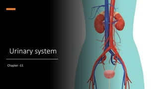
- 3. Anatomy and physiology of urinary system • The urinary system is one of the excretory systems of the body. • It consists of the following structures: • 2 kidneys, which secrete urine • 2 ureters, which convey the urine from the kidneys to the urinary bladder • Urinary bladder, where urine collects and is temporarily stored • 1 urethra through which the urine is discharged from the urinary bladder to the exterior.
- 4. Functions of the urinary system Elimination of waste products filtering gallons of fluid from the bloodstream every day creating “filtrate” “filtrate” includes metabolic wastes, ionic salts, toxins, drugs Maintenance of blood Red blood cell production- by producing hormone erythropoietin to stimulate RBC production in bone marrow Blood pressure (vessel size)- by producing renin which causes vasoconstriction Blood volume (water balance)- ADH released from Anterior Pituitary targets the kidney to limit water loss when blood pressure decreases or changes in blood composition Blood composition (electrolyte balance)- water follows salt; aldosterone reclaims sodium to the blood Blood pH- regulates H+ ions and HCO3- ions
- 5. 1. Kidneys- Location Dimensions Reddish-brown, bean shaped 12cm long, 6cm wide, 3cm thick High on posterior abdominal wall at the level of T12 to L3- superior lumbar region Retroperitoneal & against the dorsal body wall The right kidney is slightly lower than the left ,convex laterally Attached to ureters, renal blood vessels, and nerves at renal hilus (medial indention) Atop each kidney is an adrenal gland
- 6. Coverings of the Kidneys Adipose capsule Surrounds the kidney Provides protection to the kidney Helps keep the kidney in its correct location against muscles of posterior trunk wall Ptosis-kidneys drop to a lower position due to rapid fat loss, creating problems with the ureters. Ptosis can lead to hydronephrosis, a condition where urine backs up the ureters and exerts pressure on the kidney tissue. Renal capsule Surrounds each kidney
- 7. Organs associated with the kidneys • As the kidneys lie on either side of the vertebral column each is associated with a different group of structures. • Right kidney • Superiorly — the right adrenal gland • Anteriorly —the right lobe of the liver, the duodenum and the hepatic flexure of the colon • Posteriorly —the diaphragm, and muscles of the posterior abdominal wall • Left kidney • Superiorly — the left adrenal gland • Anteriorly —the spleen, stomach, pancreas, jejunum and splenic flexure of the colon • Posteriorly —the diaphragm and muscles of the posterior abdominal wall
- 9. Regions of the Kidney Three regions of kidneys Renal cortex – outer region, forms an outer shell Renal columns – extensions of cortex- material inward Renal medulla – inside the cortex, contains medullary (renal) pyramids Medullary pyramids – triangular regions of tissue in the medulla, appear striated Renal pelvis – inner collecting tube, divides into major and minor calyces Calyces – cup-shaped structures enclosing the tips of the pyramids that collect and funnel urine towards the renal pelvis
- 11. Blood Flow in the Kidneys Rich blood supply to filter blood and adjust blood composition ~¼ of blood supply passes through the kidneys each minute Blood enters the kidneys under extremely high pressure Renal artery arises from abdominal aorta, divides into Segmental artery at hilus Inside renal pelvis, Segmental artery divides into Lobar artery, which branch into Interlobar artery travelling thru the renal column to reach the renal cortex At the medulla-cortex junction, the Interlobar artery curves over the medullary pyramids as the Arcuate artery. Small Interlobular arterioles branch off of the Arcuate artery and move away from the renal cortex and into the Nephron of the kidney
- 12. The final branches of the interlobular arteries are called afferent arterioles. Afferent arterioles lead to the glomerulus, a network of capillaries that are involved in filtration. Leading away from the glomerulus, blood less filtrate travels through the efferent arterioles and into the peritubular capillaries. From there, blood moves through similar veins that parallel the arteries at their respective locations.
- 13. Microscopic structure of the kidney • The kidney is composed of about 1 million functional units, the nephrons, and a smaller number of collecting tubules. • The collecting tubules transport urine through the pyramids to the renal pelvis giving them their striped appearance. • The tubules are supported by a small amount of connective tissue, containing blood vessels, nerves and lymph vessels
- 14. The nephron • The nephron consists of a tubule closed at one end, the other end opening into a collecting tubule. • The closed or blind end is indented to form the cup- shaped glomerular capsule (Bowman's capsule) which almost completely encloses a network of arterial capillaries, the glomerulus. • Continuing from the glomerular capsule the remainder of the nephron is about 3 cm long and is described in three parts: • The proximal convoluted tubule • The medullary loop (loop of henle) • The distal convoluted tubule, leading into a collecting duct.
- 15. Glomerulus A specialized capillary bed fed and drained by arterioles. Glomerular capillaries filter fluid from the blood into the renal tubule GC is attached to arterioles on both sides in order to maintain high pressure Large afferent arteriole-arises from interlobular artery (feeder vessel); large in diameter, high resistance vessels that force fluid & solutes (filtrate) out of the blood into the glomerular capsule. 99% of the filtrate will be reclaimed by the renal tubule cells and returned to the blood in the peritubular capillary beds(blood vessels surrounding renal tubule) . Narrow efferent arteriole-merges to become the interlobular vein; draining vessel.
- 16. Glomerulus Glomerular capillaries are covered with podocytes from the inner (visceral) layer of the glomerular capsule. Podocytes have long, branching processes called pedicels that intertwine with one another and cling to the glomerular capillaries. Filtration slits between the pedicels form a porous membrane around the glomerular capillaries. The glomerular capillaries sit within a glomerular capsule (Bowman’s capsule) Expansion of renal tubule Receives filtered fluid Renal tubule coils into the PCT, then the dLOH, aLOH, DCT and finally, the CD. Along the PCT, much of the filtrate is reclaimed
- 17. Renal Tubule Glomerular (Bowman’s) capsule enlarged beginning of renal tubule Proximal convoluted tubule- lumen surface (surface exposed to filtrate) is covered with dense microvilli to increase surface area. The descending limb of the nephron - Loop of Henle The ascending limb of the nephron coils tightly again into the distal convoluted tubule Many DCT’s merge in renal cortex to form a collecting duct Collecting ducts not a part of nephron Collecting ducts receive urine from nephrons and deliver it to the major calyx and renal pelvis. CD run downward through the medullary pyramids, giving them their striped appearance.
- 19. Blood Supply of a Nephron Peritubular capillary Efferent arteriole braches into a second capillary bed Blood under low pressure Capillaries adapted for reabsorption instead of filtration. Attached to a venule and eventually lead to the interlobular veins to drain blood from the glomerulus Cling close to the renal tubule where they receive solutes and water from the renal tubule cells as these substances from the filtrate are reabsorbed into the blood.
- 20. Juxtaglomerular apparatus At origin of the DCT it contacts afferent and efferent arterioles Epithelial cells of DCT narrow and densely packed, called macula densa Together with smooth muscle cells, comprise the juxtaglomerular apparatus Control renin secretion & indirectly, aldosterone secretion
- 21. Types of Nephrons Cortical nephrons Located entirely in the cortex Includes most nephrons Juxtamedullary nephrons Found at the boundary of the cortex and medulla and their LOH dip deep into the medulla.
- 22. • The collecting ducts unite, forming larger ducts that empty into the minor calyces. • After entering the kidney at the hilum, the renal artery divides into smaller arteries and arterioles. • In the cortex an arteriole, the afferent arteriole, enters each glomerular capsule then subdivides into a cluster of capillaries, forming the glomerulus. • Between the capillary loops there are connective tissue phagocytic mesangial cells, which are part of the reticuloendothelial system • The blood vessel leading away from the glomerulus is the efferent arteriole; it breaks up into a second capillary network to supply oxygen and nutrients to the remainder of the nephron. • Venous blood drained from this capillary bed eventually leaves the kidney in the renal vein which empties into the inferior vena cava
- 23. • The blood pressure in the glomerulus is higher than in other capillaries because the diameter of the afferent arteriole is greater than that of the efferent arteriole. • The walls of the glomerulus and the glomerular capsule consist of a single layer of flattened epithelial cells. • The glomerular walls are more permeable than those of other capillaries. • The remainder of the nephron and the collecting tubule are formed by a single layer of highly specialized cells. • The nerve supply to the blood vessels of the kidney consists of sympathetic and parasympathetic nerves. • The presence of both branches of the autonomic nervous system permits control of renal blood vessel diameter and renal blood flow independently of autoregulation.
- 24. 2. URETERS • The ureters are the tubes that convey urine from the kidneys to the urinary bladder. • They are about 25 to 30 cm long with a diameter of about 3 mm. • The ureter is continuous with the funnel-shaped renal pelvis. • It passes downwards through the abdominal cavity, behind the peritoneum in front of the psoas muscle into the pelvic cavity and passes obliquely through the posterior wall of the bladder.
- 25. • Because of this arrangement, when urine accumulates and the pressure in the bladder rises, the ureters are compressed, and the openings occluded. • This prevents reflux of urine into the ureters (towards the kidneys) as the bladder fills and during micturition, when pressure increases as the muscular bladder wall contracts. • Structure • The ureters consist of three layers of tissue: • An outer covering of fibrous tissue, continuous with the fibrous capsule of the kidney • A middle muscular layer consisting of interlacing smooth muscle fibers that form a syncytium spiraling round the ureter, some in clockwise and some in anticlockwise directions and an additional outer longitudinal layer in the lower third • An inner layer, the mucosa, lined with transitional epithelium
- 26. Function • The ureters propel the urine from the kidneys into the bladder by peristaltic contraction of the smooth muscle layer. • This is an intrinsic property of the smooth muscle and is not under autonomic nerve control. • The waves of contraction originate in a pacemaker in the minor calyces. • Peristaltic waves occur several times per minute, increasing in frequency with the volume of urine produced, and send little spurts of urine into the bladder
- 27. 3. URINARY BLADDER • The urinary bladder is a reservoir for urine. It lies in the pelvic cavity and its size and position vary, depending on the amount of urine it contains. When distended, the bladder rises into the abdominal cavity. • Structure • The bladder is roughly pear-shaped but becomes more oval as it fills with urine. • It has anterior, superior and posterior surfaces. • The posterior surface is the base. • The bladder opens into the urethra at its lowest point, the neck. • Coverings • The peritoneum covers only the superior surface before it turns upwards as the parietal peritoneum, lining the anterior abdominal wall. • Posteriorly it surrounds the uterus in the female and the rectum in the male
- 28. The pelvic organs associated with the bladder and the urethra in female
- 29. The pelvic organs associated with the bladder and the urethra in male.
- 30. Bladder walls • The bladder wall is composed of three layers: • The outer layer of loose connective tissue, containing blood and lymphatic vessels and nerves, covered on the upper surface by the peritoneum • The middle layer, consisting of a mass of interlacing smooth muscle fibres and elastic tissue loosely arranged in three layers. This is called the detrusor muscle and it empties the bladder when it contracts • The mucosa, lined with transitional epithelium • When the bladder is empty the inner lining is arranged in folds, or rugae, and these gradually disappear as the bladder fills. • The bladder is distensible but when it contains 300 to 400 ml the awareness of the desire to urinate is initiated.
- 31. • The total capacity is rarely more than about 600 ml. • The three orifices in the bladder wall form a triangle or trigone. • The upper two orifices on the posterior wall are the openings of the ureters. • The lower orifice is the point of origin of the urethra. • Where the urethra commences is a thickening of the smooth muscle layer forming the internal urethral sphincter. • This sphincter is not under voluntary control.
- 32. 4. URETHRA • The urethra is a canal extending from the neck of the bladder to the exterior, at the external urethral orifice. • Its length differs in the male and in the female. • The male urethra is associated with the urinary and the reproductive systems. • The female urethra is approximately 4 cm long. • It runs downwards and forwards behind the symphysis pubis and opens at the external urethral orifice just in front of the vagina. • The external urethral orifice is guarded by the external urethral sphincter which is under voluntary control. • Except during the passage of urine, the walls of the urethra are in close apposition.
- 33. Urethra walls • Its walls consist of three layers of tissue. • The muscle layer, continuous with that of the bladder. At its origin there is the internal urethral sphincter, consisting mainly of elastic tissue and smooth muscle fibres, under autonomic nerve control. Slow and continuous contraction of this sphincter keeps the urethra closed. In the middle third there is skeletal muscle surrounding the urethra, under voluntary nerve control, that forms the external urethral sphincter • The submucosa, a spongy layer containing blood vessels and nerves • The mucosa, which is continuous with that of the bladder in the upper part. In the lower part the lining consists of stratified squamous epithelium, continuous externally with the skin of the vulva.
- 34. Juxtaglomerular Apparatus • The juxtaglomerular apparatus is a specialized structure formed by the distal convoluted tubule and the glomerular afferent arteriole. • It is located near the vascular pole of the glomerulus and its main function is to regulate blood pressure and the filtration rate of the glomerulus. • The macula densa is a collection of specialized epithelial cells in the distal convoluted tubule that detect sodium concentration of the fluid in the tubule. • In response to elevated sodium, the macula densa cells trigger contraction of the afferent arteriole, reducing flow of blood to the glomerulus and the glomerular filtration rate. • The juxtaglomerular cells, derived from smooth muscle cells, of the afferent arteriole secrete renin when blood pressure in the arteriole falls. • Renin increases blood pressure via the renin-angiotensin-aldosterone system. • Lacis cells, also called extraglomerular mesangial cells, are flat and elongated cells located near the macula densa.
- 36. Formation of urine • The kidneys form urine which passes through the ureters to the bladder for storage prior to excretion. • The composition of urine reflects the activities of the nephrons in the maintenance of homeostasis. • Waste products of protein metabolism are excreted, electrolyte balance is maintained and the pH (acid-base balance) is maintained by the excretion of hydrogen ions. • There are three processes involved in the formation of urine: 1. Simple filtration 2. Selective reabsorption 3. Secretion.
- 37. 1. Simple filtration • Filtration takes place through the semipermeable walls of the glomerulus and glomerular capsule. • Water and a large number of small molecules pass through, although some are reabsorbed later. Blood cells, plasma proteins and other large molecules are unable to filter through and remain in the capillaries . • The filtrate in the glomerulus is very similar in composition to plasma with the important exception of plasma proteins. • Filtration is assisted by the difference between the blood pressure in the glomerulus and the pressure of the filtrate in the glomerular capsule. • Because the diameter of the efferent arteriole is less than that of the afferent arteriole, a capillary hydrostatic pressure of about 7.3 kPa (55 mmHg) builds up in the glomerulus.
- 38. The pressures that drive glomerular filtration
- 39. • This pressure is opposed by the osmotic pressure of the blood, about 4 kPa (30 mmHg), and by filtrate hydrostatic pressure of about 2 kPa (15 mmHg) in the glomerular capsule. • Net filtration pressure (NFP), the total pressure that promotes filtration, is determined as follows: • Net filtration pressure (NFP) =GBHP -CHP –BCOP • By substituting the values just given, normal NFP may be calculated: • NFP = 55 mmHg -15 mmHg- 30 mmHg =10 mmHg • The volume of filtrate formed by both kidneys each minute is called the glomerular filtration rate (GFR). • In a healthy adult the GFR is about 125 ml/min; i.e. 180 liters of dilute filtrate are formed each day by the two kidneys. • Most of the filtrate is reabsorbed with less than 1%, i.e. 1 to 1.5 litres, excreted as urine. • The difference in volume and concentration is due to selective reabsorption of some constituents of the filtrate and tubular secretion of others.
- 40. • Autoregulation of filtration. • Renal blood flow is protected by a mechanism called autoregulation whereby renal blood flow is maintained at a constant pressure across a wide range of systolic blood pressures (from 80 to 200 mmHg). • Autoregulation operates independently of nervous control; i.e. if the nerve supply to the renal blood vessels is interrupted, autoregulation continues to operate. • It is therefore a property inherent in renal blood vessels; it may be stimulated by changes in blood pressure in the renal arteries or by fluctuating levels of certain metabolites, e.g. prostaglandins. • In severe shock when the systolic blood pressure falls below 80 mmHg, autoregulation fails and renal blood flow and the hydrostatic pressure decrease, impairing filtration within the nephrons.
- 41. Regulation of Glomerular Filtration Rate
- 42. 2. Selective reabsorption • Selective reabsorption is the process by which the composition and volume of the glomerular filtrate are altered during its passage through the convoluted tubules, the medullary loop and the collecting tubule. • The general purpose of this process is to reabsorb into the blood those filtrate constituents needed by the body to maintain fluid and electrolyte balance and the pH of the blood. • Active transport is carried out at carrier sites in the epithelial membrane using chemical energy to transport substances against their concentration gradients. • Some constituents of glomerular filtrate (e.g. glucose, amino acids) do not normally appear in urine because they are completely reabsorbed unless they are present in blood in excessive quantities.
- 44. • The kidneys' maximum capacity for reabsorption of a substance is the transport maximum, or renal threshold, e.g. normal blood glucose level is 2.5 to 5.3 mmol/l (45 to 95 mg/100 ml). • If the level rises above the transport maximum of about 9 mmol/1 (160 mg/100 ml) glucose appears in the urine because all the carrier sites are occupied and the mechanism for active transfer out of the tubules is overloaded. • Other substances reabsorbed by active transport include amino acids and sodium, calcium, potassium, phosphate and chloride. Some ions, e.g. sodium and chloride, can be absorbed by both active and passive mechanisms depending on the site in the nephron.
- 45. • The transport maximum, or renal threshold, of some substances varies according to the body's need for them at the time, and in some cases reabsorption is regulated by hormones. • Parathyroid hormone from the parathyroid glands and calcitonin from the thyroid gland together regulate reabsorption of calcium and phosphate. • Antidiuretic hormone (ADH) from the posterior lobe of the pituitary gland increases the permeability of the distal convoluted tubules and collecting tubules, increasing water reabsorption.
- 46. • Aldosterone, secreted by the adrenal cortex, increases the reabsorption of sodium and excretion of potassium. • Nitrogenous waste products, such as urea and uric acid, are reabsorbed only to a slight extent. • Substances that are not normal blood constituents are not reabsorbed. • If the blood passes through the glomerulus too quickly for filtration to clear such substances from the blood, the tubules secrete them into the filtrate.
- 47. 3. Secretion Filtration occurs as the blood flows through the glomerulus. Substances not required and foreign materials, e.g. drugs including penicillin and aspirin, may not be cleared from the blood by filtration because of the short time it remains in the glomerulus. Such substances are cleared by secretion into the convoluted tubules and excreted from the body in the urine. Tubular secretion of hydrogen ( H+ ) ions is important in maintaining homeostasis of blood pH.
- 48. Relationship of a nephron’s structure to its three basic functions.
- 50. Summary of filtration, reabsorption, and secretion in the nephron and collecting duct
- 51. Composition of urine • Urine is clear and amber in color due to the presence of urobilin, a bile pigment altered in the intestine, reabsorbed then excreted by the kidneys. • The specific gravity is between 1020 and 1030, and the pH is around 6 (normal range of 4.5 to 8). • A healthy adult passes 1000 to 1500 ml per day. • The amount of urine produced and the specific gravity vary according to the fluid intake and the amount of solute excreted. • During sleep and muscular exercise urine production is decreased
- 52. Renin–Angiotensin–Aldosterone System • When blood volume and blood pressure decrease, the walls of the afferent arterioles are stretched less, and the juxtaglomerular cells secrete the enzyme renin into the blood. • Sympathetic stimulation also directly stimulates release of renin from juxtaglomerular cells. • Renin clips off a 10–amino acid peptide called angiotensin I from angiotensinogen, which is synthesized by hepatocytes. • By clipping off two more amino acids, angiotensin-converting enzyme (ACE) converts angiotensin I to angiotensin II, which is the active form of the hormone.
- 53. • Angiotensin II affects renal physiology in three main ways: 1. It decreases the glomerular filtration rate by causing vasoconstriction of the afferent arterioles. 2. It enhances reabsorption of Na, Cl, and water in the proximal convoluted tubule by stimulating the activity of Na–H antiporters. 3. It stimulates the adrenal cortex to release aldosterone, a hormone that in turn stimulates the principal cells in the collecting ducts to reabsorb more Na and Cl and secrete more K. • The osmotic consequence of reabsorbing more Na and Cl is that more water is reabsorbed, which causes an increase in blood volume and blood pressure
- 55. Electrolyte balance • Changes in the concentration of electrolytes in the body fluids may be due to changes in: • The body water content, or • Electrolyte levels. • There are several mechanisms that maintain the balance between water and electrolyte concentration. • Sodium and potassium concentration • Sodium is the most common cation (positively charged ion) in extracellular fluid and potassium is the most common intracellular cation. • Sodium is a constituent of almost all foods and it is often added to food during cooking. This means that intake is usually in excess of the body's needs. • It is excreted mainly in urine and sweat. Sodium is a normal constituent of urine and the amount excreted is regulated by the hormone aldosterone, secreted by the adrenal cortex. • Cells in the afferent arteriole of the nephron are stimulated to produce the enzyme renin by sympathetic stimulation, low blood volume or by low arterial blood pressure.
- 56. • Renin converts the plasma protein angiotensinogen, produced by the liver, to angiotensin 1. Angiotensin converting enzyme (ACE), formed in small quantities in the lungs, proximal convoluted tubules and other tissues, converts angiotensin 1 into angiotensin 2 which is a very potent vasoconstrictor and increases blood pressure. Renin and raised blood potassium levels also stimulate the adrenal gland to secrete aldosterone • Water is reabsorbed with sodium and together they increase the blood volume, leading to reduced renin secretion through the negative feedback mechanism. • When sodium reabsorption is increased potassium excretion is increased, indirectly reducing intracellular potassium. • The amount of sodium excreted in sweat is insignificant except when sweating is excessive. • This may occur when there is pyrexia, a high environmental temperature or during sustained physical exercise.
- 57. • Normally the renal mechanism described above maintains the concentration of sodium and potassium within physiological limits. • When excessive sweating is sustained, e.g. living in a hot climate or working in a hot environment, acclimatization occurs in about 7 to 10 days and the amount of electrolytes lost in sweat is reduced. • Sodium and potassium occur in high concentrations in digestive juices — sodium in gastric juice and potassium in pancreatic and intestinal juice. • Normally these ions are reabsorbed by the colon but following acute and prolonged diarrhea they may be excreted in large quantities with resultant electrolyte imbalance.
- 58. Acid-base balance • In order to maintain the normal pH (acid-base balance) of the blood, the cells of the proximal convoluted tubules secrete hydrogen ions. In the filtrate they combine with buffers : • Carbonic acid is converted to carbon dioxide (CO2) and water (H2O), and the CO2 is reabsorbed maintaining the buffering capacity of the blood. • Hydrogen ions are excreted in the urine as ammonium salts and hydrogen phosphate. • The normal pH of urine varies from 4.5 to 7.8 depending on diet, time of day and a number of other factors. • Individuals whose diet contains a large amount of animal proteins tend to produce more acidic urine (lower pH) than vegetarians
- 59. MICTURITION • The urinary bladder acts as a reservoir for urine. When 300 to 400 ml of urine have accumulated, afferent autonomic nerve fibers in the bladder wall sensitive to stretch are stimulated. • In the infant this initiates a spinal reflex action and micturition occurs. • Micturition occurs when autonomic efferent fibres convey impulses to the bladder causing contraction of the detrusor muscle and relaxation of the internal urethral sphincter. • when the nervous system is fully developed the micturition reflex is stimulated but sensory impulses pass upwards to the brain and there is an awareness of the desire to pass urine.
- 60. • By conscious effort, reflex contraction of the bladder wall and relaxation of the internal sphincter can be inhibited for a limited period of time. • In adults, micturition occurs when the detrusor muscle contracts, and there is reflex relaxation of the internal sphincter and voluntary relaxation of the external sphincter. • It can be assisted by increasing the pressure within the pelvic cavity, achieved by lowering the diaphragm and contracting the abdominal muscles (Valsalva's manoeuvre). • Over-distension of the bladder is extremely painful, and when this stage is reached there is a tendency for involuntary relaxation of the external sphincter to occur and a small amount of urine to escape, provided there is no mechanical obstruction.
- 61. EVALUATION OF KIDNEY FUNCTION • Routine assessment of kidney function involves evaluating both the quantity and quality of urine and the levels of wastes in the blood. • They include • Urinalysis – Volume, Colour, Turbidity, Ordor, pH, Specific gravity • Blood Tests - Blood urea nitrogen (BUN) test, Plasma creatinine • Renal Plasma Clearance / Renal Clearance tests • Renal Plasma Clearance • Even more useful than BUN and blood creatinine values in the diagnosis of kidney problems is an evaluation of how effectively the kidneys are removing a given substance from blood plasma.
- 62. • Renal plasma clearance is the volume of blood that is “cleaned” or cleared of a substance per unit of time, usually expressed in units of milliliters per minute • High renal plasma clearance indicates efficient excretion of a substance in the urine; low clearance indicates inefficient excretion. For example, the clearance of glucose normally is zero because it is completely reabsorbed; therefore, glucose is not excreted at all. • Knowing a drug’s clearance is essential for determining the correct dosage. If clearance is high (one example is penicillin), then the dosage must also be high, and the drug must be given several times a day to maintain an adequate therapeutic level in the blood. • The following equation is used to calculate clearance
- 63. • The clearance of a solute depends on the three basic processes of a nephron: glomerular filtration, tubular reabsorption, and tubular secretion. • Consider a substance that is filtered but neither reabsorbed nor secreted. • Its clearance equals the glomerular filtration rate because all the molecules that pass the filtration membrane appear in the urine. • This is the situation for the plant polysaccharide inulin; it easily passes the filter, it is not reabsorbed, and it is not secreted. • Typically, the clearance of inulin is about 125 mL/min, which equals the GFR. • Clinically, the clearance of inulin can be used to determine the GFR.
- 64. • The clearance of inulin is obtained in the following way: • Inulin is administered intravenously and then the concentrations of inulin in plasma and urine are measured along with the urine flow rate. • Although using the clearance of inulin is an accurate method for determining the GFR, it has a few drawbacks: Inulin is not produced by the body and it must be infused continuously while clearance measurements are being determined. • Measuring the creatinine clearance, however, is an easier way to assess the GFR because creatinine is a substance that is naturally produced by the body as an end product of muscle metabolism. • Once creatinine is filtered, it is not reabsorbed, and is secreted only to a very small extent. • Because there is a small amount of creatinine secretion, the creatinine clearance is only a close estimate of the GFR and is not as accurate as using the inulin clearance to determine the GFR. • The creatinine clearance is normally about 120–140 mL/min.
- 65. • The clearance of the organic anion para-aminohippuric acid is also of clinical importance. • After PAH is administered intravenously, it is filtered and secreted in a single pass through the kidneys. • Thus, the clearance of PAH is used to measure renal plasma flow, the amount of plasma that passes through the kidneys in one minute. • Typically, the renal plasma flow is 650 mL per minute, which is about 55% of the renal blood flow (1200 mL per minute)